Movie
Movie Controller

[English] 日本語
 Yorodumi
- EMDB-24666: Cryo-EM structure of Kip3 (AMPPNP) bound to Taxol-Stabilized Micr... -
+ Open data

- Basic information
| Entry |  | |||||||||
|---|---|---|---|---|---|---|---|---|---|---|
| Title | Cryo-EM structure of Kip3 (AMPPNP) bound to Taxol-Stabilized Microtubules | |||||||||
 Map data Map data | Cryo-EM structure of Kip3 (AMPPNP) bound to taxol-stabilized microtubules | |||||||||
 Sample Sample |
| |||||||||
 Keywords Keywords | kinesin-8 / microtubules / complex / MOTOR PROTEIN | |||||||||
| Function / homology |  Function and homology information Function and homology informationmotile cilium / structural constituent of cytoskeleton / microtubule cytoskeleton organization / neuron migration / mitotic cell cycle / Hydrolases; Acting on acid anhydrides; Acting on GTP to facilitate cellular and subcellular movement / microtubule / hydrolase activity / GTPase activity / GTP binding ...motile cilium / structural constituent of cytoskeleton / microtubule cytoskeleton organization / neuron migration / mitotic cell cycle / Hydrolases; Acting on acid anhydrides; Acting on GTP to facilitate cellular and subcellular movement / microtubule / hydrolase activity / GTPase activity / GTP binding / metal ion binding / cytoplasm Similarity search - Function | |||||||||
| Biological species |   | |||||||||
| Method | helical reconstruction / cryo EM / Resolution: 3.9 Å | |||||||||
 Authors Authors | Hernandez-Lopez RA / Leschziner AE | |||||||||
| Funding support | 1 items
| |||||||||
 Citation Citation | Journal: Biorxiv / Year: 2021 Title: Multimodal tubulin binding by the yeast kinesin-8, Kip3, underlies its motility and depolymerization Authors: Arellano-Santoyo H / Hernandez-Lopez RA / Stokasimov E / Wang RYR / Pellman D / Leschziner AE | |||||||||
| History |
|
- Structure visualization
| Supplemental images |
|---|

- Downloads & links
-EMDB archive
| Map data | emd_24666.map.gz | 10.5 MB | EMDB map data format | |
|---|---|---|---|---|
| Header (meta data) | emd-24666-v30.xmlemd-24666.xml | 19.9 KB 19.9 KB | Display Display | EMDB header |
| Images | 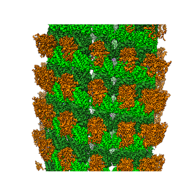 emd_24666.png emd_24666.png | 209.8 KB | ||
| Filedesc metadata | emd-24666.cif.gz | 8 KB | ||
| Archive directory |  http://ftp.pdbj.org/pub/emdb/structures/EMD-24666ftp://ftp.pdbj.org/pub/emdb/structures/EMD-24666 http://ftp.pdbj.org/pub/emdb/structures/EMD-24666ftp://ftp.pdbj.org/pub/emdb/structures/EMD-24666 | HTTPS FTP |
-Validation report
| Summary document | emd_24666_validation.pdf.gz | 461.9 KB | Display | EMDB validaton report |
|---|---|---|---|---|
| Full document | emd_24666_full_validation.pdf.gz | 461.5 KB | Display | |
| Data in XML | emd_24666_validation.xml.gz | 6.6 KB | Display | |
| Data in CIF | emd_24666_validation.cif.gz | 7.5 KB | Display | |
| Arichive directory | https://ftp.pdbj.org/pub/emdb/validation_reports/EMD-24666ftp://ftp.pdbj.org/pub/emdb/validation_reports/EMD-24666 | HTTPS FTP |
-Related structure data
| Related structure data |  7rs5MC  7rs6C C: citing same article ( M: atomic model generated by this map |
|---|---|
| Similar structure data |

-Links
| EMDB pages | EMDB (EBI/PDBe) / EMDataResource |
|---|---|
| Related items in Molecule of the Month |


-Map
| File | Download / File: emd_24666.map.gz / Format: CCP4 / Size: 103 MB / Type: IMAGE STORED AS FLOATING POINT NUMBER (4 BYTES) | ||||||||||||||||||||||||||||||||||||
|---|---|---|---|---|---|---|---|---|---|---|---|---|---|---|---|---|---|---|---|---|---|---|---|---|---|---|---|---|---|---|---|---|---|---|---|---|---|
| Annotation | Cryo-EM structure of Kip3 (AMPPNP) bound to taxol-stabilized microtubules | ||||||||||||||||||||||||||||||||||||
| Projections & slices | Image control
Images are generated by Spider. | ||||||||||||||||||||||||||||||||||||
| Voxel size | X=Y=Z: 1.04 Å | ||||||||||||||||||||||||||||||||||||
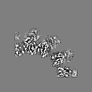
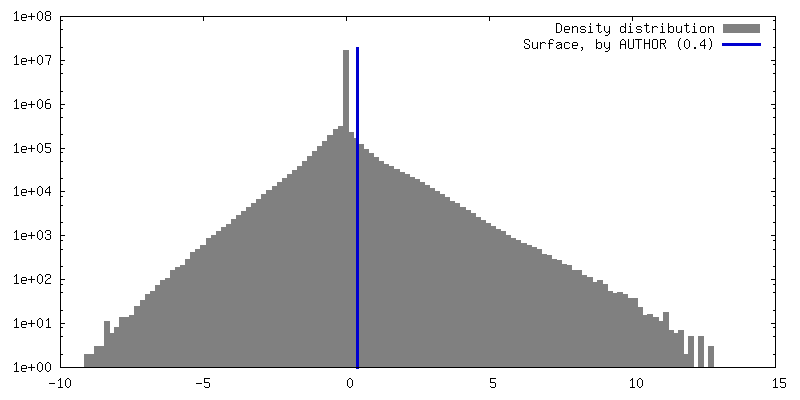
| Density |
| ||||||||||||||||||||||||||||||||||||
| Symmetry | Space group: 1 | ||||||||||||||||||||||||||||||||||||
| Details | EMDB XML:
|
 Z (Sec.)
 Y (Row.)
 X (Col.)


-Supplemental data
- Sample components
+Entire : Cryo-EM structure of Kip3 (AMPPNP) bound to Taxol-Stabilized Micr...
+Supramolecule #1: Cryo-EM structure of Kip3 (AMPPNP) bound to Taxol-Stabilized Micr...
+Supramolecule #2: Taxol-stabilized microtubules
+Supramolecule #3: yeast kinesin-8/ Kip3
+Macromolecule #1: Tubulin alpha-1A chain
+Macromolecule #2: Tubulin beta chain
+Macromolecule #3: yeast kinesin-8/ Kip3
+Macromolecule #4: GUANOSINE-5'-TRIPHOSPHATE

+Macromolecule #5: MAGNESIUM ION
+Macromolecule #6: GUANOSINE-5'-DIPHOSPHATE

+Macromolecule #7: TAXOL

+Macromolecule #8: PHOSPHOAMINOPHOSPHONIC ACID-ADENYLATE ESTER

-Experimental details
-Structure determination
| Method | cryo EM |
|---|---|
 Processing Processing | helical reconstruction |
| Aggregation state | helical array |
-Sample preparation
| Buffer | pH: 8 Details: cryoEM buffer (50 mM Tris-HCl, pH 8.0, 1 mM MgCl2, 1 mM EGTA, 1 mM DTT supplemented with 2mM AMPPNP) |
|---|---|
| Grid | Model: C-flat-1.2/1.3 / Material: COPPER / Pretreatment - Type: GLOW DISCHARGE / Pretreatment - Time: 20 sec. |
| Vitrification | Cryogen name: ETHANE / Chamber humidity: 100 % / Chamber temperature: 22 K / Instrument: FEI VITROBOT MARK IV |
| Details | Highly purified, glycerol-free tubulin (Cytoskeleton, Inc.) was resuspended in BRB80 buffer (80 mM PIPES-KOH, pH 6.8, 1 mM MgCl2, 1 mM EGTA, 1 mM DTT) to a concentration of 10 mg/mL. To prepare Taxol-stabilized microtubules, tubulin was polymerized with a stepwise addition of Taxol as follows: 20 uL of tubulin stock was thawed quickly and placed on ice. 10 uL of BRB80 supplemented with 3 mM GTP were added and the mixture was transferred to a 37 C water bath. After 15, 30, and 45 minutes, additions of 0.5, 0.5, and 1.0 uL of 2 mM Taxol were added by gentle swirling. The mixture was then incubated for an additional 1 h at 37C. Purified Kip3 438 protein was buffer exchanged to cryoEM buffer (50 mM Tris-HCl, pH 8.0, 1 mM MgCl2, 1 mM EGTA, 1 mM DTT supplemented with 2 mM AMPPNP) and desalted using a ZEBA spin desalting column. The protein was recovered by centrifugation at 15,000 rcf for 2 min. A final spin at 30,000 x g in a TLA 100 rotor (Beckman) for 10 min at 4 C was carried out to remove big aggregates. |
- Electron microscopy
| Microscope | FEI TITAN KRIOS |
|---|---|
| Details | Images were recorded using a semi-automated acquisition program Serial EM with a defocus range from 1.5 to 3.5 um. |
| Image recording | Film or detector model: GATAN K2 SUMMIT (4k x 4k) / Detector mode: SUPER-RESOLUTION / Number grids imaged: 1 / Number real images: 1194 / Average exposure time: 4.0 sec. / Average electron dose: 40.0 e/Å2 Details: Final accumulated electron doses were 40 electrons/A2. Images were collected in super-resolution mode. The total exposure time was 4 seconds, fractionated into 20 subframes, each with an exposure time of 0.2 s. |
| Electron beam | Acceleration voltage: 300 kV / Electron source:  FIELD EMISSION GUN FIELD EMISSION GUN |
| Electron optics | Illumination mode: FLOOD BEAM / Imaging mode: BRIGHT FIELD |
| Sample stage | Specimen holder model: FEI TITAN KRIOS AUTOGRID HOLDER |
| Experimental equipment |  Model: Titan Krios / Image courtesy: FEI Company |
-Image processing
| Final reconstruction | Applied symmetry - Helical parameters - Δz: 8.55 Å Applied symmetry - Helical parameters - Δ&Phi: -25.76 ° Applied symmetry - Helical parameters - Axial symmetry: C14 (14 fold cyclic) Resolution.type: BY AUTHOR / Resolution: 3.9 Å / Resolution method: FSC 0.143 CUT-OFF / Software - Name: FREALIGN Details: Pseudo-helical symmetry was applied during the reconstruction step Number images used: 14934 |
|---|---|
| Segment selection | Number selected: 67040 / Software - Name: Appion Details: Inspection, defocus estimation, microtubule picking, and stack creation were performed within the Appion processing environment (Lander et al., 2009). Images were selected for processing on ...Details: Inspection, defocus estimation, microtubule picking, and stack creation were performed within the Appion processing environment (Lander et al., 2009). Images were selected for processing on the basis of high decoration, straight MTs, and the absence of crystalline ice. |
| Startup model | Type of model: INSILICO MODEL |
| Final angle assignment | Type: NOT APPLICABLE / Software - Name: FREALIGN |
-Atomic model buiding 1
| Initial model |
| ||||||||
|---|---|---|---|---|---|---|---|---|---|
| Details | Multiple rounds of refinement were carried out against one half map (training map), and the other half map (validation map) was used to monitor overfitting based on the procedure described in Wang et al. elife, 2016. It is to note that the molecular interactions of ligand-protein were restrained to the initial poses adapted from the high-resolution structures during structure refinement. | ||||||||
| Refinement | Space: REAL / Protocol: OTHER / Overall B value: 100 Target criteria: Overall correlation of the residues to the map | ||||||||
| Output model | PDB-7rs5: |