Movie
Movie Controller


+ Open data

- Basic information
| Entry | Database: EMDB / ID: EMD-23037 | ||||||||||||
|---|---|---|---|---|---|---|---|---|---|---|---|---|---|
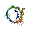
| Title | MicroED structure of mVDAC | ||||||||||||
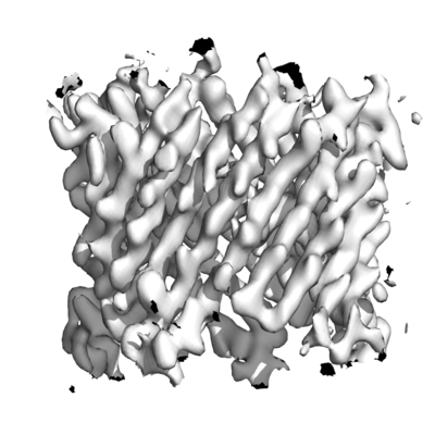
 Map data Map data | 2Fo-Fc map of mVDAC determined by MicroED | ||||||||||||
 Sample Sample |
| ||||||||||||
 Keywords Keywords | Channel / mammalian / voltage dependent / TRANSPORT PROTEIN | ||||||||||||
| Function / homology |  Function and homology information Function and homology informationnegative regulation of calcium import into the mitochondrion / positive regulation of parkin-mediated stimulation of mitophagy in response to mitochondrial depolarization / Pyruvate metabolism / voltage-gated monoatomic anion channel activity / PINK1-PRKN Mediated Mitophagy / neuron-neuron synaptic transmission / mitochondrial calcium ion transmembrane transport / ceramide binding / acetyl-CoA biosynthetic process from pyruvate / monoatomic anion channel activity ...negative regulation of calcium import into the mitochondrion / positive regulation of parkin-mediated stimulation of mitophagy in response to mitochondrial depolarization / Pyruvate metabolism / voltage-gated monoatomic anion channel activity / PINK1-PRKN Mediated Mitophagy / neuron-neuron synaptic transmission / mitochondrial calcium ion transmembrane transport / ceramide binding / acetyl-CoA biosynthetic process from pyruvate / monoatomic anion channel activity / regulation of mitophagy / mitochondrial permeability transition pore complex / phosphatidylcholine binding / Ub-specific processing proteases / oxysterol binding / porin activity / cholesterol binding / mitochondrial nucleoid / negative regulation of reactive oxygen species metabolic process / behavioral fear response / presynaptic active zone membrane / epithelial cell differentiation / learning / postsynaptic density membrane / synaptic vesicle / myelin sheath / chemical synaptic transmission / mitochondrial outer membrane / transmembrane transporter binding / mitochondrial inner membrane / membrane raft / nucleotide binding / apoptotic process / protein-containing complex binding / negative regulation of apoptotic process / protein kinase binding / protein-containing complex / mitochondrion / identical protein binding / membrane / plasma membrane Similarity search - Function | ||||||||||||
| Biological species |  | ||||||||||||
| Method | electron crystallography / cryo EM / Resolution: 3.12 Å | ||||||||||||
 Authors Authors | Martynowycz MW / Khan F | ||||||||||||
| Funding support |  United States, 3 items United States, 3 items
| ||||||||||||
 Citation Citation | Journal: Proc Natl Acad Sci U S A / Year: 2020 Title: MicroED structure of lipid-embedded mammalian mitochondrial voltage-dependent anion channel. Authors: Michael W Martynowycz / Farha Khan / Johan Hattne / Jeff Abramson / Tamir Gonen / Abstract: A structure of the murine voltage-dependent anion channel (VDAC) was determined by microcrystal electron diffraction (MicroED). Microcrystals of an essential mutant of VDAC grew in a viscous bicelle ...A structure of the murine voltage-dependent anion channel (VDAC) was determined by microcrystal electron diffraction (MicroED). Microcrystals of an essential mutant of VDAC grew in a viscous bicelle suspension, making it unsuitable for conventional X-ray crystallography. Thin, plate-like crystals were identified using scanning-electron microscopy (SEM). Crystals were milled into thin lamellae using a focused-ion beam (FIB). MicroED data were collected from three crystal lamellae and merged for completeness. The refined structure revealed unmodeled densities between protein monomers, indicative of lipids that likely mediate contacts between the proteins in the crystal. This body of work demonstrates the effectiveness of milling membrane protein microcrystals grown in viscous media using a focused ion beam for subsequent structure determination by MicroED. This approach is well suited for samples that are intractable by X-ray crystallography. To our knowledge, the presented structure is a previously undescribed mutant of the membrane protein VDAC, crystallized in a lipid bicelle matrix and solved by MicroED. | ||||||||||||
| History |
|
- Structure visualization
| Movie |
Movie viewer |
|---|---|
| Structure viewer | EM map: SurfViewMolmilJmol/JSmol |
| Supplemental images |
- Downloads & links
-EMDB archive
| Map data | emd_23037.map.gz | 1.8 MB | EMDB map data format | |
|---|---|---|---|---|
| Header (meta data) | emd-23037-v30.xmlemd-23037.xml | 15.1 KB 15.1 KB | Display Display | EMDB header |
| Images |  emd_23037.png emd_23037.png | 107 KB | ||
| Filedesc metadata | emd-23037.cif.gz | 5.8 KB | ||
| Filedesc structureFactors | emd_23037_sf.cif.gz | 146.2 KB | ||
| Archive directory |  http://ftp.pdbj.org/pub/emdb/structures/EMD-23037ftp://ftp.pdbj.org/pub/emdb/structures/EMD-23037 http://ftp.pdbj.org/pub/emdb/structures/EMD-23037ftp://ftp.pdbj.org/pub/emdb/structures/EMD-23037 | HTTPS FTP |
-Validation report
| Summary document | emd_23037_validation.pdf.gz | 529.3 KB | Display | EMDB validaton report |
|---|---|---|---|---|
| Full document | emd_23037_full_validation.pdf.gz | 528.9 KB | Display | |
| Data in XML | emd_23037_validation.xml.gz | 4.5 KB | Display | |
| Data in CIF | emd_23037_validation.cif.gz | 5 KB | Display | |
| Arichive directory | https://ftp.pdbj.org/pub/emdb/validation_reports/EMD-23037ftp://ftp.pdbj.org/pub/emdb/validation_reports/EMD-23037 | HTTPS FTP |
-Related structure data
| Related structure data |  7kuhMC M: atomic model generated by this map C: citing same article ( |
|---|---|
| Similar structure data |
-Links
| EMDB pages | EMDB (EBI/PDBe) / EMDataResource |
|---|---|
| Related items in Molecule of the Month |


-Map
| File | Download / File: emd_23037.map.gz / Format: CCP4 / Size: 2.2 MB / Type: IMAGE STORED AS FLOATING POINT NUMBER (4 BYTES) | ||||||||||||||||||||||||||||||||||||||||||||||||||||||||||||||||||||
|---|---|---|---|---|---|---|---|---|---|---|---|---|---|---|---|---|---|---|---|---|---|---|---|---|---|---|---|---|---|---|---|---|---|---|---|---|---|---|---|---|---|---|---|---|---|---|---|---|---|---|---|---|---|---|---|---|---|---|---|---|---|---|---|---|---|---|---|---|---|
| Annotation | 2Fo-Fc map of mVDAC determined by MicroED | ||||||||||||||||||||||||||||||||||||||||||||||||||||||||||||||||||||
| Voxel size | X: 0.77266 Å / Y: 0.77613 Å / Z: 0.77267 Å | ||||||||||||||||||||||||||||||||||||||||||||||||||||||||||||||||||||
| Density |
| ||||||||||||||||||||||||||||||||||||||||||||||||||||||||||||||||||||
| Symmetry | Space group: 5 | ||||||||||||||||||||||||||||||||||||||||||||||||||||||||||||||||||||
| Details | EMDB XML:
CCP4 map header:
| ||||||||||||||||||||||||||||||||||||||||||||||||||||||||||||||||||||
-Supplemental data
- Sample components
-Entire : Murine voltage-dependent anion channel mutant
| Entire | Name: Murine voltage-dependent anion channel mutant |
|---|---|
| Components |
|
-Supramolecule #1: Murine voltage-dependent anion channel mutant
| Supramolecule | Name: Murine voltage-dependent anion channel mutant / type: complex / ID: 1 / Parent: 0 / Macromolecule list: all |
|---|---|
| Source (natural) | Organism: |
| Molecular weight | Theoretical: 32.87 KDa |
-Macromolecule #1: Voltage-dependent anion-selective channel protein 1
| Macromolecule | Name: Voltage-dependent anion-selective channel protein 1 / type: protein_or_peptide / ID: 1 / Number of copies: 1 / Enantiomer: LEVO |
|---|---|
| Source (natural) | Organism: |
| Molecular weight | Theoretical: 32.195879 KDa |
| Recombinant expression | Organism:  |
| Sequence | String: MRGSHHHHHH GSMAVPPTYA DLGESARDVF TKGYGFGLIK LDLKTKSENG LEFTSSGSAN TETTKVNGSL ETKYRWTEYG LTFTEKWNT DNTLGTEITV EDQLARGLKL TFDSSFSPNT GKKNAKIKTG YKREHINLGC DVDFDIAGPS IRGALVLGYE G WLAGYQMN ...String: MRGSHHHHHH GSMAVPPTYA DLGESARDVF TKGYGFGLIK LDLKTKSENG LEFTSSGSAN TETTKVNGSL ETKYRWTEYG LTFTEKWNT DNTLGTEITV EDQLARGLKL TFDSSFSPNT GKKNAKIKTG YKREHINLGC DVDFDIAGPS IRGALVLGYE G WLAGYQMN FETSKSRVTQ SNFAVGYKTD EFQLHTNVND GTEFGGSIYQ KVNKKLETAV NLAWTAGNSN TRFGIAAKYQ VD PDACFSA KVNNSSLIGL GYTQTLKPGI KLTLSALLDG KNVNAGGHKL GLGLEFQA UniProtKB: Voltage-dependent anion-selective channel protein 1 |
-Experimental details
-Structure determination
| Method | cryo EM |
|---|---|
 Processing Processing | electron crystallography |
| Aggregation state | 3D array |
-Sample preparation
| Concentration | 12 mg/mL |
|---|---|
| Buffer | pH: 8.5 |
| Grid | Model: Quantifoil R2/2 / Material: COPPER / Mesh: 200 / Support film - Material: CARBON / Support film - topology: HOLEY ARRAY / Support film - Film thickness: 12 / Pretreatment - Type: GLOW DISCHARGE / Pretreatment - Time: 30 sec. |
| Vitrification | Cryogen name: ETHANE / Chamber humidity: 90 % / Chamber temperature: 277 K / Instrument: LEICA EM GP |
| Details | Monodisperse crystal embedded in lipid bicelles |
- Electron microscopy
| Microscope | FEI TITAN KRIOS |
|---|---|
| Temperature | Min: 77.0 K / Max: 90.0 K |
| Image recording | Film or detector model: FEI CETA (4k x 4k) / Digitization - Dimensions - Width: 2048 pixel / Digitization - Dimensions - Height: 2048 pixel / Number grids imaged: 2 / Number real images: 150 / Number diffraction images: 180 / Average exposure time: 5.0 sec. / Average electron dose: 0.01 e/Å2 |
| Electron beam | Acceleration voltage: 300 kV / Electron source:  FIELD EMISSION GUN FIELD EMISSION GUN |
| Electron optics | C2 aperture diameter: 100.0 µm / Illumination mode: FLOOD BEAM / Imaging mode: DIFFRACTION / Cs: 2.7 mm / Nominal defocus max: 0.0 µm / Nominal defocus min: 0.0 µm / Camera length: 3000 mm |
| Sample stage | Specimen holder model: FEI TITAN KRIOS AUTOGRID HOLDER / Cooling holder cryogen: NITROGEN / Tilt angle: -60.0, 68.0 |
| Experimental equipment |  Model: Titan Krios / Image courtesy: FEI Company |
+Image processing
-Atomic model buiding 1
| Initial model | PDB ID: Chain - Source name: PDB / Chain - Initial model type: experimental model |
|---|---|
| Details | Standard refinement with electron scattering factors |
| Refinement | Space: RECIPROCAL / Protocol: RIGID BODY FIT / Overall B value: 90 / Target criteria: Maximum liklihood |
| Output model | PDB-7kuh: |