[English] 日本語
Yorodumi
- EMDB-22357: Structural Basis of the Activation of Heterotrimeric Gs-protein b... -

+
Open data


ID or keywords:

Loading...

-
Basic information

Entry
Database: EMDB / ID: EMD-22357
TitleStructural Basis of the Activation of Heterotrimeric Gs-protein by Isoproterenol-bound Beta1-Adrenergic Receptor
Map dataActivation of Heterotrimeric Gs-protein by Isoproterenol-bound Beta1-Adrenergic Receptor
Sample
  • Complex: Isoproterenol bound beta1 adrenergic receptor in complex with heterotrimeric Gs protein
    • Complex: Guanine nucleotide-binding protein G(I)/G(S)/G(T) subunit beta-1, Guanine nucleotide-binding protein G(I)/G(S)/G(O) subunit gamma-2
      • Protein or peptide: Guanine nucleotide-binding protein G(I)/G(S)/G(T) subunit beta-1
      • Protein or peptide: Guanine nucleotide-binding protein G(I)/G(S)/G(O) subunit gamma-2
    • Complex: Nanobody 35
      • Protein or peptide: Nanobody 35
    • Complex: Guanine nucleotide-binding protein G(s) subunit alpha isoforms short
      • Protein or peptide: Guanine nucleotide-binding protein G(s) subunit alpha isoforms short
    • Complex: Beta1-Adrenergic Receptor
      • Protein or peptide: Beta1-Adrenergic Receptor
  • Ligand: ISOPRENALINE
Function / homology
Function and homology information


beta1-adrenergic receptor activity / positive regulation of heart rate by epinephrine-norepinephrine / Olfactory Signaling Pathway / Sensory perception of sweet, bitter, and umami (glutamate) taste / Synthesis, secretion, and inactivation of Glucagon-like Peptide-1 (GLP-1) / regulation of circadian sleep/wake cycle, sleep / sensory perception of chemical stimulus / Activation of the phototransduction cascade / Activation of G protein gated Potassium channels / G-protein activation ...beta1-adrenergic receptor activity / positive regulation of heart rate by epinephrine-norepinephrine / Olfactory Signaling Pathway / Sensory perception of sweet, bitter, and umami (glutamate) taste / Synthesis, secretion, and inactivation of Glucagon-like Peptide-1 (GLP-1) / regulation of circadian sleep/wake cycle, sleep / sensory perception of chemical stimulus / Activation of the phototransduction cascade / Activation of G protein gated Potassium channels / G-protein activation / G beta:gamma signalling through PI3Kgamma / Prostacyclin signalling through prostacyclin receptor / G beta:gamma signalling through PLC beta / ADP signalling through P2Y purinoceptor 1 / Thromboxane signalling through TP receptor / Presynaptic function of Kainate receptors / G beta:gamma signalling through CDC42 / Inhibition of voltage gated Ca2+ channels via Gbeta/gamma subunits / Glucagon-type ligand receptors / Adrenaline,noradrenaline inhibits insulin secretion / G alpha (12/13) signalling events / G beta:gamma signalling through BTK / ADP signalling through P2Y purinoceptor 12 / norepinephrine-epinephrine-mediated vasodilation involved in regulation of systemic arterial blood pressure / Cooperation of PDCL (PhLP1) and TRiC/CCT in G-protein beta folding / Thrombin signalling through proteinase activated receptors (PARs) / Ca2+ pathway / G alpha (z) signalling events / Extra-nuclear estrogen signaling / G alpha (s) signalling events / G alpha (q) signalling events / G alpha (i) signalling events / Glucagon-like Peptide-1 (GLP1) regulates insulin secretion / Vasopressin regulates renal water homeostasis via Aquaporins / mu-type opioid receptor binding / corticotropin-releasing hormone receptor 1 binding / D1 dopamine receptor binding / beta-2 adrenergic receptor binding / adenylate cyclase-activating adrenergic receptor signaling pathway / adenylate cyclase activator activity / insulin-like growth factor receptor binding / ionotropic glutamate receptor binding / G-protein beta/gamma-subunit complex binding / adenylate cyclase-activating G protein-coupled receptor signaling pathway / photoreceptor disc membrane / cellular response to catecholamine stimulus / adenylate cyclase-activating dopamine receptor signaling pathway / cellular response to prostaglandin E stimulus / G-protein beta-subunit binding / heterotrimeric G-protein complex / signaling receptor complex adaptor activity / early endosome / G protein-coupled receptor signaling pathway / GTPase activity / GTP binding / membrane / identical protein binding / metal ion binding / plasma membrane / cytoplasm
Similarity search - Function
Beta 1 adrenoceptor / Adrenoceptor family / G-protein alpha subunit, group S / Serpentine type 7TM GPCR chemoreceptor Srsx / G-alpha domain profile. / Guanine nucleotide binding protein (G-protein), alpha subunit / G protein alpha subunit, helical insertion / G-protein alpha subunit / G protein alpha subunit / G-protein, gamma subunit ...Beta 1 adrenoceptor / Adrenoceptor family / G-protein alpha subunit, group S / Serpentine type 7TM GPCR chemoreceptor Srsx / G-alpha domain profile. / Guanine nucleotide binding protein (G-protein), alpha subunit / G protein alpha subunit, helical insertion / G-protein alpha subunit / G protein alpha subunit / G-protein, gamma subunit / G-protein gamma subunit domain profile. / GGL domain / G-protein gamma-like domain superfamily / G-protein gamma-like domain / GGL domain / G protein gamma subunit-like motifs / Guanine nucleotide-binding protein, beta subunit / G-protein, beta subunit / G-protein coupled receptors family 1 signature. / G protein-coupled receptor, rhodopsin-like / GPCR, rhodopsin-like, 7TM / G-protein coupled receptors family 1 profile. / 7 transmembrane receptor (rhodopsin family) / G-protein beta WD-40 repeat / WD40 repeat, conserved site / Trp-Asp (WD) repeats signature. / Trp-Asp (WD) repeats profile. / Trp-Asp (WD) repeats circular profile. / WD domain, G-beta repeat / WD40 repeats / WD40 repeat / WD40-repeat-containing domain superfamily / WD40/YVTN repeat-like-containing domain superfamily / P-loop containing nucleoside triphosphate hydrolase
Similarity search - Domain/homology
Guanine nucleotide-binding protein G(s) subunit alpha isoforms short / Beta-1 adrenergic receptor / Guanine nucleotide-binding protein G(I)/G(S)/G(T) subunit beta-1 / Guanine nucleotide-binding protein G(I)/G(S)/G(O) subunit gamma-2
Similarity search - Component
Biological speciesBos taurus (cattle) / Lama glama (llama) / Meleagris gallopavo (turkey)
Methodsingle particle reconstruction / cryo EM / Resolution: 2.6 Å
AuthorsSu M / Zhu L / Zhang Y / Paknejad N / Dey R / Huang J / Lee MY / Williams D / Jordan KD / Eng ET ...Su M / Zhu L / Zhang Y / Paknejad N / Dey R / Huang J / Lee MY / Williams D / Jordan KD / Eng ET / Ernst OP / Meyerson JR / Hite RK / Walz T / Liu W / Huang XY
Funding support United States, 1 items
OrganizationGrant numberCountry
National Institutes of Health/National Heart, Lung, and Blood Institute (NIH/NHLBI)HL130478 United States
Citation
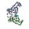
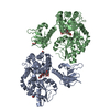
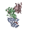
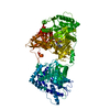
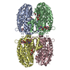
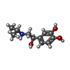
Journal: Mol Cell / Year: 2020
Title: Structural Basis of the Activation of Heterotrimeric Gs-Protein by Isoproterenol-Bound β-Adrenergic Receptor.
Authors: Minfei Su / Lan Zhu / Yixiao Zhang / Navid Paknejad / Raja Dey / Jianyun Huang / Ming-Yue Lee / Dewight Williams / Kelsey D Jordan / Edward T Eng / Oliver P Ernst / Joel R Meyerson / Richard ...Authors: Minfei Su / Lan Zhu / Yixiao Zhang / Navid Paknejad / Raja Dey / Jianyun Huang / Ming-Yue Lee / Dewight Williams / Kelsey D Jordan / Edward T Eng / Oliver P Ernst / Joel R Meyerson / Richard K Hite / Thomas Walz / Wei Liu / Xin-Yun Huang /
Abstract: Cardiac disease remains the leading cause of morbidity and mortality worldwide. The β-adrenergic receptor (β-AR) is a major regulator of cardiac functions and is downregulated in the majority of ...Cardiac disease remains the leading cause of morbidity and mortality worldwide. The β-adrenergic receptor (β-AR) is a major regulator of cardiac functions and is downregulated in the majority of heart failure cases. A key physiological process is the activation of heterotrimeric G-protein Gs by β-ARs, leading to increased heart rate and contractility. Here, we use cryo-electron microscopy and functional studies to investigate the molecular mechanism by which β-AR activates Gs. We find that the tilting of α5-helix breaks a hydrogen bond between the sidechain of His373 in the C-terminal α5-helix and the backbone carbonyl of Arg38 in the N-terminal αN-helix of Gα. Together with the disruption of another interacting network involving Gln59 in the α1-helix, Ala352 in the β6-α5 loop, and Thr355 in the α5-helix, these conformational changes might lead to the deformation of the GDP-binding pocket. Our data provide molecular insights into the activation of G-proteins by G-protein-coupled receptors.
History
DepositionJul 27, 2020-
Header (metadata) releaseSep 2, 2020-
Map releaseSep 2, 2020-
UpdateOct 14, 2020-
Current statusOct 14, 2020Processing site: RCSB / Status: Released

-
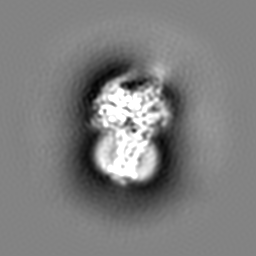
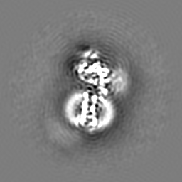
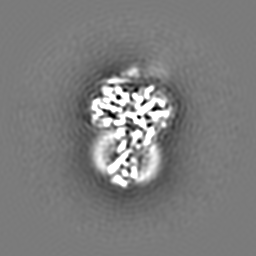
Structure visualization
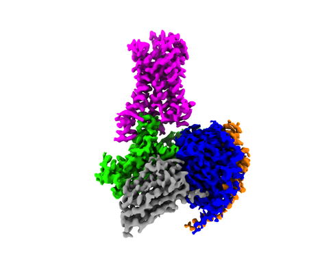
Movie
  • Surface view with section colored by density value
  • Surface level: 3
  • Imaged by UCSF Chimera
  • Download
  • Surface view colored by height
  • Surface level: 3
  • Imaged by UCSF Chimera
  • Download
  • Surface view with fitted model
  • Atomic models: PDB-7jjo
  • Surface level: 3
  • Imaged by UCSF Chimera
  • Download
Movie viewer
Structure viewerEM map:
SurfViewMolmilJmol/JSmol
Supplemental images

Downloads & links

-
Map

FileDownload / File: emd_22357.map.gz / Format: CCP4 / Size: 512 MB / Type: IMAGE STORED AS FLOATING POINT NUMBER (4 BYTES)
AnnotationActivation of Heterotrimeric Gs-protein by Isoproterenol-bound Beta1-Adrenergic Receptor
Voxel sizeX=Y=Z: 0.532 Å
Density
Contour LevelBy AUTHOR: 3 / Movie #1: 3
Minimum - Maximum-81.77604 - 188.34462
Average (Standard dev.)-0.0011023238 (±1.248299)
SymmetrySpace group: 1
Details

EMDB XML:

Map geometry
Axis orderZYX
Origin000
Dimensions512512512
Spacing512512512
CellA=B=C: 272.384 Å
α=β=γ: 90.0 °

CCP4 map header:

modeImage stored as Reals
Å/pix. X/Y/Z0.5320.5320.532
M x/y/z512512512
origin x/y/z0.0000.0000.000
length x/y/z272.384272.384272.384
α/β/γ90.00090.00090.000
start NX/NY/NZ000
NX/NY/NZ512512512
MAP C/R/S321
start NC/NR/NS000
NC/NR/NS512512512
D min/max/mean-81.776188.345-0.001

-
Supplemental data

-
Additional map: low-pass filtered to 6 A map

Fileemd_22357_additional.map
Annotationlow-pass filtered to 6 A map
Projections & Slices
AxesZYX

Projections

Slices (1/2)
Density Histograms

-
Sample components

+
Entire : Isoproterenol bound beta1 adrenergic receptor in complex with het...

EntireName: Isoproterenol bound beta1 adrenergic receptor in complex with heterotrimeric Gs protein
Components
  • Complex: Isoproterenol bound beta1 adrenergic receptor in complex with heterotrimeric Gs protein
    • Complex: Guanine nucleotide-binding protein G(I)/G(S)/G(T) subunit beta-1, Guanine nucleotide-binding protein G(I)/G(S)/G(O) subunit gamma-2
      • Protein or peptide: Guanine nucleotide-binding protein G(I)/G(S)/G(T) subunit beta-1
      • Protein or peptide: Guanine nucleotide-binding protein G(I)/G(S)/G(O) subunit gamma-2
    • Complex: Nanobody 35
      • Protein or peptide: Nanobody 35
    • Complex: Guanine nucleotide-binding protein G(s) subunit alpha isoforms short
      • Protein or peptide: Guanine nucleotide-binding protein G(s) subunit alpha isoforms short
    • Complex: Beta1-Adrenergic Receptor
      • Protein or peptide: Beta1-Adrenergic Receptor
  • Ligand: ISOPRENALINE

+
Supramolecule #1: Isoproterenol bound beta1 adrenergic receptor in complex with het...

SupramoleculeName: Isoproterenol bound beta1 adrenergic receptor in complex with heterotrimeric Gs protein
type: complex / ID: 1 / Parent: 0 / Macromolecule list: #1-#5
Molecular weightTheoretical: 160 KDa

+
Supramolecule #2: Guanine nucleotide-binding protein G(I)/G(S)/G(T) subunit beta-1,...

SupramoleculeName: Guanine nucleotide-binding protein G(I)/G(S)/G(T) subunit beta-1, Guanine nucleotide-binding protein G(I)/G(S)/G(O) subunit gamma-2
type: complex / ID: 2 / Parent: 1 / Macromolecule list: #1, #4
Source (natural)Organism: Bos taurus (cattle)
Recombinant expressionOrganism: Spodoptera frugiperda (fall armyworm)

+
Supramolecule #3: Nanobody 35

SupramoleculeName: Nanobody 35 / type: complex / ID: 3 / Parent: 1 / Macromolecule list: #2
Source (natural)Organism: Lama glama (llama)
Recombinant expressionOrganism: Escherichia coli (E. coli)

+
Supramolecule #4: Guanine nucleotide-binding protein G(s) subunit alpha isoforms short

SupramoleculeName: Guanine nucleotide-binding protein G(s) subunit alpha isoforms short
type: complex / ID: 4 / Parent: 1 / Macromolecule list: #3
Source (natural)Organism: Bos taurus (cattle)
Recombinant expressionOrganism: Escherichia coli (E. coli)

+
Supramolecule #5: Beta1-Adrenergic Receptor

SupramoleculeName: Beta1-Adrenergic Receptor / type: complex / ID: 5 / Parent: 1 / Macromolecule list: #5
Source (natural)Organism: Meleagris gallopavo (turkey)
Recombinant expressionOrganism: Spodoptera frugiperda (fall armyworm)

+
Macromolecule #1: Guanine nucleotide-binding protein G(I)/G(S)/G(T) subunit beta-1

MacromoleculeName: Guanine nucleotide-binding protein G(I)/G(S)/G(T) subunit beta-1
type: protein_or_peptide / ID: 1 / Number of copies: 1 / Enantiomer: LEVO
Source (natural)Organism: Bos taurus (cattle)
Molecular weightTheoretical: 37.285734 KDa
Recombinant expressionOrganism: Spodoptera frugiperda (fall armyworm)
SequenceString: SELDQLRQEA EQLKNQIRDA RKACADATLS QITNNIDPVG RIQMRTRRTL RGHLAKIYAM HWGTDSRLLV SASQDGKLII WDSYTTNKV HAIPLRSSWV MTCAYAPSGN YVACGGLDNI CSIYNLKTRE GNVRVSRELA GHTGYLSCCR FLDDNQIVTS S GDTTCALW ...String:
SELDQLRQEA EQLKNQIRDA RKACADATLS QITNNIDPVG RIQMRTRRTL RGHLAKIYAM HWGTDSRLLV SASQDGKLII WDSYTTNKV HAIPLRSSWV MTCAYAPSGN YVACGGLDNI CSIYNLKTRE GNVRVSRELA GHTGYLSCCR FLDDNQIVTS S GDTTCALW DIETGQQTTT FTGHTGDVMS LSLAPDTRLF VSGACDASAK LWDVREGMCR QTFTGHESDI NAICFFPNGN AF ATGSDDA TCRLFDLRAD QELMTYSHDN IICGITSVSF SKSGRLLLAG YDDFNCNVWD ALKADRAGVL AGHDNRVSCL GVT DDGMAV ATGSWDSFLK IWN

+
Macromolecule #2: Nanobody 35

MacromoleculeName: Nanobody 35 / type: protein_or_peptide / ID: 2 / Number of copies: 1 / Enantiomer: LEVO
Source (natural)Organism: Lama glama (llama)
Molecular weightTheoretical: 15.140742 KDa
Recombinant expressionOrganism: Escherichia coli (E. coli)
SequenceString:
QVQLQESGGG LVQPGGSLRL SCAASGFTFS NYKMNWVRQA PGKGLEWVSD ISQSGASISY TGSVKGRFTI SRDNAKNTLY LQMNSLKPE DTAVYYCARC PAPFTRDCFD VTSTTYAYRG QGTQVTVSSH HHHHHEPEA

+
Macromolecule #3: Guanine nucleotide-binding protein G(s) subunit alpha isoforms short

MacromoleculeName: Guanine nucleotide-binding protein G(s) subunit alpha isoforms short
type: protein_or_peptide / ID: 3 / Number of copies: 1 / Enantiomer: LEVO
Source (natural)Organism: Bos taurus (cattle)
Molecular weightTheoretical: 44.694551 KDa
Recombinant expressionOrganism: Escherichia coli (E. coli)
SequenceString: GPLAMGCLGN SKTEDQRNEE KGQREANKKI EKQLQKDKQV YRATHRLLLL GAGESGKSTI VKQMRILHVN GFNGDSEKAT KVQDIKNNL KEAIETIVAA MSNLVPPVEL ANPENQFRVD YILSVMNVPD FDFPPEFYEH AKALWEDEGV RACYERSNEY Q LIDCAQYF ...String:
GPLAMGCLGN SKTEDQRNEE KGQREANKKI EKQLQKDKQV YRATHRLLLL GAGESGKSTI VKQMRILHVN GFNGDSEKAT KVQDIKNNL KEAIETIVAA MSNLVPPVEL ANPENQFRVD YILSVMNVPD FDFPPEFYEH AKALWEDEGV RACYERSNEY Q LIDCAQYF LDKIDVIKQD DYVPSDQDLL RCRVLTSGIF ETKFQVDKVN FHMFDVGGQR DERRKWIQCF NDVTAIIFVV AS SSYNMVI REDNQTNRLQ EALNLFKSIW NNRWLRTISV ILFLNKQDLL AEKVLAGKSK IEDYFPEFAR YTTPEDATPE PGE DPRVTR AKYFIRDEFL RISTASGDGR HYCYPHFTCA VDTENIRRVF NDCRDIIQRM HLRQYELL

+
Macromolecule #4: Guanine nucleotide-binding protein G(I)/G(S)/G(O) subunit gamma-2

MacromoleculeName: Guanine nucleotide-binding protein G(I)/G(S)/G(O) subunit gamma-2
type: protein_or_peptide / ID: 4 / Number of copies: 1 / Enantiomer: LEVO
Source (natural)Organism: Bos taurus (cattle)
Molecular weightTheoretical: 7.845078 KDa
Recombinant expressionOrganism: Spodoptera frugiperda (fall armyworm)
SequenceString:
MASNNTASIA QARKLVEQLK MEANIDRIKV SKAAADLMAY CEAHAKEDPL LTPVPASENP FREKKFFSAI L

+
Macromolecule #5: Beta1-Adrenergic Receptor

MacromoleculeName: Beta1-Adrenergic Receptor / type: protein_or_peptide / ID: 5 / Number of copies: 1 / Enantiomer: LEVO
Source (natural)Organism: Meleagris gallopavo (turkey)
Molecular weightTheoretical: 57.958668 KDa
Recombinant expressionOrganism: Spodoptera frugiperda (fall armyworm)
SequenceString: MKTIIALSYI FCLVFADYKD DDDKLEVLFQ GPNIFEMLRI DEGLRLKIYK DTEGYYTIGI GHLLTKSPSL NAAKSELDKA IGRNTNGVI TKDEAEKLFN QDVDAAVRGI LRNAKLKPVY DSLDAVRRAA LINMVFQMGE TGVAGFTNSL RMLQQKRWDE A AVNLAKSR ...String:
MKTIIALSYI FCLVFADYKD DDDKLEVLFQ GPNIFEMLRI DEGLRLKIYK DTEGYYTIGI GHLLTKSPSL NAAKSELDKA IGRNTNGVI TKDEAEKLFN QDVDAAVRGI LRNAKLKPVY DSLDAVRRAA LINMVFQMGE TGVAGFTNSL RMLQQKRWDE A AVNLAKSR WYNQTPNRAK RVITTFRTGT WDAYAAGAEL LSQQWEAGMS LLMALVVLLI VAGNVLVIAA IGSTQRLQTL TN LFITSLA CADLVVGLLV VPFGATLVVR GTWLWGSFLC ELWTSLDVLC VTASIETLCV IAIDRYLAIT SPFRYQSLMT RAR AKVIIC TVWAISALVS FLPIMMHWWR DEDPQALKCY QDPGCCDFVT NRAYAIASSI ISFYIPLLIM IFVYLRVYRE AKEQ IRKID RCEGRFREHK ALKTLGIIMG VFTLCWLPFF LVNIVNVFNR DLVPDWLFVF FNWLGYANSA FNPIIYCRSP DFRKA FKRL LCFPRKADRR LEVLFQGPHH HHHH

+
Macromolecule #6: ISOPRENALINE

MacromoleculeName: ISOPRENALINE / type: ligand / ID: 6 / Number of copies: 1 / Formula: 5FW
Molecular weightTheoretical: 211.258 Da
Chemical component information

ChemComp-5FW:
ISOPRENALINE / medication, agonist*YM

-
Experimental details

-
Structure determination

Methodcryo EM
Processingsingle particle reconstruction
Aggregation stateparticle

-
Sample preparation

Concentration1.5 mg/mL
BufferpH: 7
GridModel: Quantifoil / Material: GOLD / Mesh: 400 / Support film - Material: CARBON / Support film - topology: HOLEY
VitrificationCryogen name: ETHANE / Chamber humidity: 100 % / Chamber temperature: 277 K / Instrument: FEI VITROBOT MARK IV

-
Electron microscopy

MicroscopeFEI TITAN KRIOS
Image recordingFilm or detector model: GATAN K3 (6k x 4k) / Average electron dose: 46.0 e/Å2
Electron beamAcceleration voltage: 300 kV / Electron source: FIELD EMISSION GUN
Electron opticsIllumination mode: FLOOD BEAM / Imaging mode: BRIGHT FIELD
Experimental equipment
Model: Titan Krios / Image courtesy: FEI Company

-
Image processing

Final reconstructionResolution.type: BY AUTHOR / Resolution: 2.6 Å / Resolution method: FSC 0.143 CUT-OFF / Number images used: 452312
Initial angle assignmentType: MAXIMUM LIKELIHOOD / Software - Name: cryoSPARC (ver. v2) / Details: ab initio
Final angle assignmentType: MAXIMUM LIKELIHOOD / Software - Name: cryoSPARC (ver. v2) / Details: non-uniform refinement

+
About Yorodumi

-
News

-
Feb 9, 2022. New format data for meta-information of EMDB entries

New format data for meta-information of EMDB entries

  • Version 3 of the EMDB header file is now the official format.
  • The previous official version 1.9 will be removed from the archive.

Related info.:EMDB header

External links:wwPDB to switch to version 3 of the EMDB data model

-
Aug 12, 2020. Covid-19 info

Covid-19 info

URL: https://pdbjlvh1.pdbj.org/emnavi/covid19.php

New page: Covid-19 featured information page in EM Navigator.

Related info.:Covid-19 info / Mar 5, 2020. Novel coronavirus structure data

+
Mar 5, 2020. Novel coronavirus structure data

Novel coronavirus structure data

Related info.:Yorodumi Speices / Aug 12, 2020. Covid-19 info

External links:COVID-19 featured content - PDBj / Molecule of the Month (242):Coronavirus Proteases

+
Jan 31, 2019. EMDB accession codes are about to change! (news from PDBe EMDB page)

EMDB accession codes are about to change! (news from PDBe EMDB page)

  • The allocation of 4 digits for EMDB accession codes will soon come to an end. Whilst these codes will remain in use, new EMDB accession codes will include an additional digit and will expand incrementally as the available range of codes is exhausted. The current 4-digit format prefixed with “EMD-” (i.e. EMD-XXXX) will advance to a 5-digit format (i.e. EMD-XXXXX), and so on. It is currently estimated that the 4-digit codes will be depleted around Spring 2019, at which point the 5-digit format will come into force.
  • The EM Navigator/Yorodumi systems omit the EMD- prefix.

Related info.:Q: What is EMD? / ID/Accession-code notation in Yorodumi/EM Navigator

External links:EMDB Accession Codes are Changing Soon! / Contact to PDBj

+
Jul 12, 2017. Major update of PDB

Major update of PDB

  • wwPDB released updated PDB data conforming to the new PDBx/mmCIF dictionary.
  • This is a major update changing the version number from 4 to 5, and with Remediation, in which all the entries are updated.
  • In this update, many items about electron microscopy experimental information are reorganized (e.g. em_software).
  • Now, EM Navigator and Yorodumi are based on the updated data.

External links:wwPDB Remediation / Enriched Model Files Conforming to OneDep Data Standards Now Available in the PDB FTP Archive

-
Yorodumi

Thousand views of thousand structures

  • Yorodumi is a browser for structure data from EMDB, PDB, SASBDB, etc.
  • This page is also the successor to EM Navigator detail page, and also detail information page/front-end page for Omokage search.
  • The word "yorodu" (or yorozu) is an old Japanese word meaning "ten thousand". "mi" (miru) is to see.

Related info.:EMDB / PDB / SASBDB / Comparison of 3 databanks / Yorodumi Search / Aug 31, 2016. New EM Navigator & Yorodumi / Yorodumi Papers / Jmol/JSmol / Function and homology information / Changes in new EM Navigator and Yorodumi

Read more