regulation of amino acid metabolic process / negative regulation of glucose mediated signaling pathway / translational readthrough / mTORC1-mediated signalling / Protein hydroxylation / positive regulation of protein kinase activity / ribosome-associated ubiquitin-dependent protein catabolic process / pre-mRNA 5'-splice site binding / GDP-dissociation inhibitor activity / cytosolic large ribosomal subunit assembly ...regulation of amino acid metabolic process / negative regulation of glucose mediated signaling pathway / translational readthrough / mTORC1-mediated signalling / Protein hydroxylation / positive regulation of protein kinase activity / ribosome-associated ubiquitin-dependent protein catabolic process / pre-mRNA 5'-splice site binding / GDP-dissociation inhibitor activity / cytosolic large ribosomal subunit assembly / nonfunctional rRNA decay / Formation of the ternary complex, and subsequently, the 43S complex / Translation initiation complex formation / response to cycloheximide / Ribosomal scanning and start codon recognition / cleavage in ITS2 between 5.8S rRNA and LSU-rRNA of tricistronic rRNA transcript (SSU-rRNA, 5.8S rRNA, LSU-rRNA) / Major pathway of rRNA processing in the nucleolus and cytosol / mRNA destabilization / SRP-dependent cotranslational protein targeting to membrane / GTP hydrolysis and joining of the 60S ribosomal subunit / negative regulation of mRNA splicing, via spliceosome / preribosome, large subunit precursor / Formation of a pool of free 40S subunits / Nonsense Mediated Decay (NMD) independent of the Exon Junction Complex (EJC) / Nonsense Mediated Decay (NMD) enhanced by the Exon Junction Complex (EJC) / L13a-mediated translational silencing of Ceruloplasmin expression / negative regulation of translational frameshifting / ribosomal large subunit export from nucleus / 90S preribosome / G-protein alpha-subunit binding / endonucleolytic cleavage to generate mature 3'-end of SSU-rRNA from (SSU-rRNA, 5.8S rRNA, LSU-rRNA) / ribosomal subunit export from nucleus / regulation of translational fidelity / protein-RNA complex assembly / maturation of LSU-rRNA / translation regulator activity / endonucleolytic cleavage in ITS1 to separate SSU-rRNA from 5.8S rRNA and LSU-rRNA from tricistronic rRNA transcript (SSU-rRNA, 5.8S rRNA, LSU-rRNA) / rescue of stalled cytosolic ribosome / cellular response to amino acid starvation / protein kinase C binding / ribosomal large subunit biogenesis / maturation of LSU-rRNA from tricistronic rRNA transcript (SSU-rRNA, 5.8S rRNA, LSU-rRNA) / maturation of SSU-rRNA from tricistronic rRNA transcript (SSU-rRNA, 5.8S rRNA, LSU-rRNA) / positive regulation of apoptotic signaling pathway / macroautophagy / maturation of SSU-rRNA / translational initiation / small-subunit processome / maintenance of translational fidelity / modification-dependent protein catabolic process / cytoplasmic stress granule / protein tag activity / rRNA processing / ribosomal small subunit assembly / ribosome biogenesis / ribosome binding / ribosomal small subunit biogenesis / 5S rRNA binding / ribosomal large subunit assembly / small ribosomal subunit / small ribosomal subunit rRNA binding / cytosolic small ribosomal subunit / large ribosomal subunit rRNA binding / cytosolic large ribosomal subunit / cytoplasmic translation / negative regulation of translation / rRNA binding / structural constituent of ribosome / protein ubiquitination / ribosome / translation / G protein-coupled receptor signaling pathway / negative regulation of gene expression / response to antibiotic / mRNA binding / ubiquitin protein ligase binding / nucleolus / mitochondrion / RNA binding / zinc ion binding / nucleoplasm / metal ion binding / nucleus / cytoplasm / cytosol Similarity search - Function
: / 60S acidic ribosomal protein P0 / 50S ribosomal protein L10, insertion domain superfamily / : / 60S ribosomal protein L10P, insertion domain / Insertion domain in 60S ribosomal protein L10P / Ribosomal protein L1, conserved site / Ribosomal protein L1 signature. / Ribosomal protein L1 / : ...: / 60S acidic ribosomal protein P0 / 50S ribosomal protein L10, insertion domain superfamily / : / 60S ribosomal protein L10P, insertion domain / Insertion domain in 60S ribosomal protein L10P / Ribosomal protein L1, conserved site / Ribosomal protein L1 signature. / Ribosomal protein L1 / : / Ribosomal protein S26e signature. / Ribosomal protein L1, 3-layer alpha/beta-sandwich / Ribosomal protein L41 / Ribosomal protein L41 / Ribosomal protein S21e, conserved site / Ribosomal protein S21e signature. / Ribosomal protein L13e, conserved site / Ribosomal protein L13e signature. / Ribosomal protein S26e / Ribosomal protein S26e superfamily / Ribosomal protein S26e / Ribosomal protein L29e / Ribosomal L29e protein family / Ribosomal protein L1-like / Ribosomal protein L1/ribosomal biogenesis protein / Ribosomal protein L1p/L10e family / Ribosomal protein L27e, conserved site / Ribosomal protein L27e signature. / Ribosomal protein L22e / Ribosomal protein L22e superfamily / Ribosomal L22e protein family / Small (40S) ribosomal subunit Asc1/RACK1 / Ribosomal protein L13e / Ribosomal protein S5, eukaryotic/archaeal / Ribosomal protein L13e / Ribosomal protein S21e / Ribosomal protein L38e / Ribosomal protein L38e superfamily / Ribosomal protein S21e superfamily / Ribosomal protein S21e / Ribosomal L38e protein family / Ribosomal protein S19e, conserved site / Ribosomal protein S19e signature. / Ribosomal protein S2, eukaryotic / Ribosomal protein L19, eukaryotic / : / Ribosomal protein L10e, conserved site / Ribosomal protein L6e signature. / Ribosomal protein L10e signature. / Ribosomal protein L19/L19e conserved site / Ribosomal protein L19e signature. / Ribosomal protein L44e signature. / 60S ribosomal protein L18a/ L20, eukaryotes / 40S Ribosomal protein S10 / Ribosomal protein L10e / Ribosomal protein L24e, conserved site / Ribosomal protein L24e signature. / Ribosomal protein L18/L18-A/B/e, conserved site / Ribosomal protein L18e signature. / Ribosomal protein L34e, conserved site / Ribosomal protein L34e signature. / Plectin/S10, N-terminal / Plectin/S10 domain / Ribosomal protein L5 eukaryotic, C-terminal / Ribosomal L18 C-terminal region / Ribosomal protein L23/L25, N-terminal / Ribosomal protein L23, N-terminal domain / : / Ribosomal L40e family / Ribosomal protein L36e signature. / Ribosomal protein S30 / Ribosomal protein S30 / Ribosomal protein L30e signature 1. / Ribosomal protein L44e / Ribosomal protein S10, eukaryotic/archaeal / Ribosomal protein L44 / Ribosomal protein S25 / 50S ribosomal protein L18Ae/60S ribosomal protein L20 and L18a / Ribosomal protein S8e subdomain, eukaryotes / S25 ribosomal protein / : / Ribosomal protein L35Ae, conserved site / Ribosomal protein 50S-L18Ae/60S-L20/60S-L18A / Ribosomal proteins 50S-L18Ae/60S-L20/60S-L18A / Ribosomal protein 60S L18 and 50S L18e / Ribosomal protein L35Ae signature. / Eukaryotic Ribosomal Protein L27, KOW domain / Ribosomal protein S7e signature. / Ribosomal_L40e / Ribosomal protein L40e / Ribosomal protein L40e superfamily / Ribosomal protein L27e / Ribosomal protein L27e superfamily / Ribosomal L27e protein family / Ribosomal protein S17e, conserved site / Ribosomal Protein L6, KOW domain / Ribosomal protein S17e signature. / Ribosomal protein S2, eukaryotic/archaeal / Ribosomal protein L39e, conserved site / Ribosomal protein L39e signature. Similarity search - Domain/homology
Rps5p / Small ribosomal subunit protein eS1 / RPS22A isoform 1 / 60S acidic ribosomal protein P0 / RPL38 isoform 1 / RPL10 isoform 1 / RPS29A isoform 1 / RPS20 isoform 1 / RPS2 isoform 1 / 60S ribosomal protein L29 ...Rps5p / Small ribosomal subunit protein eS1 / RPS22A isoform 1 / 60S acidic ribosomal protein P0 / RPL38 isoform 1 / RPL10 isoform 1 / RPS29A isoform 1 / RPS20 isoform 1 / RPS2 isoform 1 / 60S ribosomal protein L29 / 60S ribosomal protein L8 / RPL41A isoform 1 / RPL24A isoform 1 / RPL11B isoform 1 / 40S ribosomal protein S25 / RPL9A isoform 1 / 40S ribosomal protein S26 / RPL5 isoform 1 / 40S ribosomal protein S8 / RPL32 isoform 1 / RPL12A isoform 1 / Ribosomal protein / 40S ribosomal protein S3 / RPL4A isoform 1 / Large ribosomal subunit protein uL3 / RPS15 isoform 1 / RPS28A isoform 1 / Small ribosomal subunit protein uS4A / Large ribosomal subunit protein uL15 / Large ribosomal subunit protein uL23 / Large ribosomal subunit protein eL39 / Large ribosomal subunit protein uL30A / Large ribosomal subunit protein uL22A / Large ribosomal subunit protein uL24A / Large ribosomal subunit protein eL33A / Large ribosomal subunit protein eL36A / Large ribosomal subunit protein eL15A / Large ribosomal subunit protein eL22A / Small ribosomal subunit protein uS15 / Small ribosomal subunit protein eS19A / Small ribosomal subunit protein eS21A / Large ribosomal subunit protein eL27A / Large ribosomal subunit protein eL31A / Ubiquitin-ribosomal protein eL40A fusion protein / Large ribosomal subunit protein eL20A / Large ribosomal subunit protein eL43A / Large ribosomal subunit protein eL42A / Small ribosomal subunit protein uS12A / Small ribosomal subunit protein eS24A / Small ribosomal subunit protein eS30A / Small ribosomal subunit protein eS4A / Small ribosomal subunit protein eS6A / Large ribosomal subunit protein uL14A / Large ribosomal subunit protein uL2A / Small ribosomal subunit protein uS17A / Large ribosomal subunit protein eL18A / Small ribosomal subunit protein uS9A / Small ribosomal subunit protein uS13A / Large ribosomal subunit protein eL19A / Large ribosomal subunit protein uL29A / Large ribosomal subunit protein eL30 / Small ribosomal subunit protein eS17B / Large ribosomal subunit protein uL13A / Small ribosomal subunit protein eS7A / Small ribosomal subunit protein uS2A / Small ribosomal subunit protein eS27A / Large ribosomal subunit protein eL14A / Small ribosomal subunit protein RACK1 / Small ribosomal subunit protein uS11B / Large ribosomal subunit protein eL37A / Large ribosomal subunit protein eL34A / Large ribosomal subunit protein eL6A / Large ribosomal subunit protein eL21A / Small ribosomal subunit protein eS10A / Large ribosomal subunit protein eL13A Similarity search - Component
Biological species
Saccharomyces cerevisiae (brewer's yeast)
Method
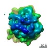
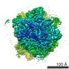
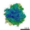
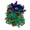
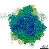
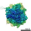
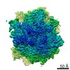
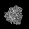
single particle reconstruction / cryo EM / Resolution: 4.2 Å
National Institutes of Health/National Institute of Environmental Health Sciences (NIH/NIEHS)
5R00ES025835-05
United States
Citation
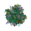
Journal: Proc Natl Acad Sci U S A / Year: 2020 Title: Structural impact of K63 ubiquitin on yeast translocating ribosomes under oxidative stress. Authors: Ye Zhou / Panagiotis L Kastritis / Shannon E Dougherty / Jonathan Bouvette / Allen L Hsu / Laura Burbaum / Shyamal Mosalaganti / Stefan Pfeffer / Wim J H Hagen / Friedrich Förster / Mario J ...Authors: Ye Zhou / Panagiotis L Kastritis / Shannon E Dougherty / Jonathan Bouvette / Allen L Hsu / Laura Burbaum / Shyamal Mosalaganti / Stefan Pfeffer / Wim J H Hagen / Friedrich Förster / Mario J Borgnia / Christine Vogel / Martin Beck / Alberto Bartesaghi / Gustavo M Silva / Abstract: Subpopulations of ribosomes are responsible for fine tuning the control of protein synthesis in dynamic environments. K63 ubiquitination of ribosomes has emerged as a new posttranslational ...Subpopulations of ribosomes are responsible for fine tuning the control of protein synthesis in dynamic environments. K63 ubiquitination of ribosomes has emerged as a new posttranslational modification that regulates protein synthesis during cellular response to oxidative stress. K63 ubiquitin, a type of ubiquitin chain that functions independently of the proteasome, modifies several sites at the surface of the ribosome, however, we lack a molecular understanding on how this modification affects ribosome structure and function. Using cryoelectron microscopy (cryo-EM), we resolved the first three-dimensional (3D) structures of K63 ubiquitinated ribosomes from oxidatively stressed yeast cells at 3.5-3.2 Å resolution. We found that K63 ubiquitinated ribosomes are also present in a polysome arrangement, similar to that observed in yeast polysomes, which we determined using cryoelectron tomography (cryo-ET). We further showed that K63 ubiquitinated ribosomes are captured uniquely at the rotated pretranslocation stage of translation elongation. In contrast, cryo-EM structures of ribosomes from mutant cells lacking K63 ubiquitin resolved at 4.4-2.7 Å showed 80S ribosomes represented in multiple states of translation, suggesting that K63 ubiquitin regulates protein synthesis at a selective stage of elongation. Among the observed structural changes, ubiquitin mediates the destabilization of proteins in the 60S P-stalk and in the 40S beak, two binding regions of the eukaryotic elongation factor eEF2. These changes would impact eEF2 function, thus, inhibiting translocation. Our findings help uncover the molecular effects of K63 ubiquitination on ribosomes, providing a model of translation control during oxidative stress, which supports elongation halt at pretranslocation.
History
Deposition
Jun 21, 2020
-
Header (metadata) release
Aug 26, 2020
-
Map release
Aug 26, 2020
-
Update
Oct 23, 2024
-
Current status
Oct 23, 2024
Processing site: RCSB / Status: Released
-
Structure visualization
Movie
Surface view with section colored by density value
In the structure databanks used in Yorodumi, some data are registered as the other names, "COVID-19 virus" and "2019-nCoV". Here are the details of the virus and the list of structure data.
Jan 31, 2019. EMDB accession codes are about to change! (news from PDBe EMDB page)
EMDB accession codes are about to change! (news from PDBe EMDB page)
The allocation of 4 digits for EMDB accession codes will soon come to an end. Whilst these codes will remain in use, new EMDB accession codes will include an additional digit and will expand incrementally as the available range of codes is exhausted. The current 4-digit format prefixed with “EMD-” (i.e. EMD-XXXX) will advance to a 5-digit format (i.e. EMD-XXXXX), and so on. It is currently estimated that the 4-digit codes will be depleted around Spring 2019, at which point the 5-digit format will come into force.
The EM Navigator/Yorodumi systems omit the EMD- prefix.
Related info.:Q: What is EMD? / ID/Accession-code notation in Yorodumi/EM Navigator
Yorodumi is a browser for structure data from EMDB, PDB, SASBDB, etc.
This page is also the successor to EM Navigator detail page, and also detail information page/front-end page for Omokage search.
The word "yorodu" (or yorozu) is an old Japanese word meaning "ten thousand". "mi" (miru) is to see.
Related info.:EMDB / PDB / SASBDB / Comparison of 3 databanks / Yorodumi Search / Aug 31, 2016. New EM Navigator & Yorodumi / Yorodumi Papers / Jmol/JSmol / Function and homology information / Changes in new EM Navigator and Yorodumi
 Movie
Movie Controller

 Yorodumi
 Open data
 Basic information
 Map data
 Sample
 Keywords
 Function and homology information
 Authors
 United States, 1 items
  Citation

 Structure visualization
 Downloads & links
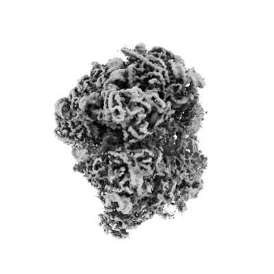 emd_22196.png
 http://ftp.pdbj.org/pub/emdb/structures/EMD-22196


 X (Sec.)
 Y (Row.)
 Z (Col.)


 Sample components
 Processing
 Electron microscopy
 FIELD EMISSION GUN