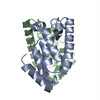
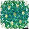
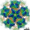
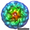
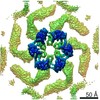
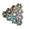
Journal: Nature / Year: 2012 Title: Structure of the immature retroviral capsid at 8 Å resolution by cryo-electron microscopy. Authors: Tanmay A M Bharat / Norman E Davey / Pavel Ulbrich / James D Riches / Alex de Marco / Michaela Rumlova / Carsten Sachse / Tomas Ruml / John A G Briggs / Abstract: The assembly of retroviruses such as HIV-1 is driven by oligomerization of their major structural protein, Gag. Gag is a multidomain polyprotein including three conserved folded domains: MA (matrix), ...The assembly of retroviruses such as HIV-1 is driven by oligomerization of their major structural protein, Gag. Gag is a multidomain polyprotein including three conserved folded domains: MA (matrix), CA (capsid) and NC (nucleocapsid). Assembly of an infectious virion proceeds in two stages. In the first stage, Gag oligomerization into a hexameric protein lattice leads to the formation of an incomplete, roughly spherical protein shell that buds through the plasma membrane of the infected cell to release an enveloped immature virus particle. In the second stage, cleavage of Gag by the viral protease leads to rearrangement of the particle interior, converting the non-infectious immature virus particle into a mature infectious virion. The immature Gag shell acts as the pivotal intermediate in assembly and is a potential target for anti-retroviral drugs both in inhibiting virus assembly and in disrupting virus maturation. However, detailed structural information on the immature Gag shell has not previously been available. For this reason it is unclear what protein conformations and interfaces mediate the interactions between domains and therefore the assembly of retrovirus particles, and what structural transitions are associated with retrovirus maturation. Here we solve the structure of the immature retroviral Gag shell from Mason-Pfizer monkey virus by combining cryo-electron microscopy and tomography. The 8-Å resolution structure permits the derivation of a pseudo-atomic model of CA in the immature retrovirus, which defines the protein interfaces mediating retrovirus assembly. We show that transition of an immature retrovirus into its mature infectious form involves marked rotations and translations of CA domains, that the roles of the amino-terminal and carboxy-terminal domains of CA in assembling the immature and mature hexameric lattices are exchanged, and that the CA interactions that stabilize the immature and mature viruses are almost completely distinct.
History
Deposition
Apr 23, 2012
-
Header (metadata) release
May 17, 2012
-
Map release
May 25, 2012
-
Update
Aug 12, 2015
-
Current status
Aug 12, 2015
Processing site: PDBe / Status: Released
-
Structure visualization
Movie
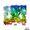
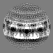
Surface view with section colored by density value
Category: FILM / Film or detector model: KODAK SO-163 FILM / Digitization - Scanner: ZEISS SCAI / Digitization - Sampling interval: 7 µm / Number real images: 46 / Average electron dose: 20 e/Å2
Electron beam
Acceleration voltage: 300 kV / Electron source: FIELD EMISSION GUN
Reconstruction carried out using real-space helical reconstruction followed by 3D averaging of the asymmetric unit from assemblies with different helical symmetry parameters.
Final reconstruction
Algorithm: OTHER / Resolution.type: BY AUTHOR / Resolution: 8.0 Å / Resolution method: OTHER / Software - Name: Spider, AV3 / Details: Helical reconstructions were averaged in 3D
CTF correction
Details: Division by 3D CTF^2
+
About Yorodumi
-
News
-
Feb 9, 2022. New format data for meta-information of EMDB entries
New format data for meta-information of EMDB entries
Version 3 of the EMDB header file is now the official format.
The previous official version 1.9 will be removed from the archive.
In the structure databanks used in Yorodumi, some data are registered as the other names, "COVID-19 virus" and "2019-nCoV". Here are the details of the virus and the list of structure data.
Jan 31, 2019. EMDB accession codes are about to change! (news from PDBe EMDB page)
EMDB accession codes are about to change! (news from PDBe EMDB page)
The allocation of 4 digits for EMDB accession codes will soon come to an end. Whilst these codes will remain in use, new EMDB accession codes will include an additional digit and will expand incrementally as the available range of codes is exhausted. The current 4-digit format prefixed with “EMD-” (i.e. EMD-XXXX) will advance to a 5-digit format (i.e. EMD-XXXXX), and so on. It is currently estimated that the 4-digit codes will be depleted around Spring 2019, at which point the 5-digit format will come into force.
The EM Navigator/Yorodumi systems omit the EMD- prefix.
Related info.:Q: What is EMD? / ID/Accession-code notation in Yorodumi/EM Navigator
Yorodumi is a browser for structure data from EMDB, PDB, SASBDB, etc.
This page is also the successor to EM Navigator detail page, and also detail information page/front-end page for Omokage search.
The word "yorodu" (or yorozu) is an old Japanese word meaning "ten thousand". "mi" (miru) is to see.
Related info.:EMDB / PDB / SASBDB / Comparison of 3 databanks / Yorodumi Search / Aug 31, 2016. New EM Navigator & Yorodumi / Yorodumi Papers / Jmol/JSmol / Function and homology information / Changes in new EM Navigator and Yorodumi
 Movie
Movie Controller

 Yorodumi
 Open data
 Basic information
 Map data
 Sample
 Keywords
 Function and homology information
 Mason-Pfizer monkey virus
 Authors
 Citation
 Structure visualization
 Downloads & links
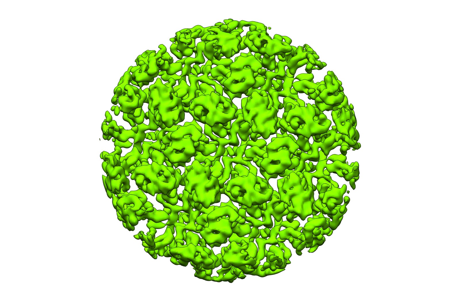 emd_2089.jpg
 http://ftp.pdbj.org/pub/emdb/structures/EMD-2089


 Z (Sec.)
 Y (Row.)
 X (Col.)


 Sample components
 Processing
 Electron microscopy
 FIELD EMISSION GUN
