 Movie
Movie Controller

 Yorodumi
+ Open data

- Basic information
| Entry |  | |||||||||
|---|---|---|---|---|---|---|---|---|---|---|
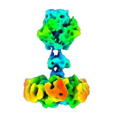
| Title | Receptor binding protein of bacteriophage JBD30 computed with C3 symmetry | |||||||||
 Map data Map data | main map | |||||||||
 Sample Sample |
| |||||||||
 Keywords Keywords | bacteriophage / virion / receptor binding protein / VIRUS | |||||||||
| Function / homology | DUF2793 domain-containing protein Function and homology information Function and homology information | |||||||||
| Biological species |  Pseudomonas phage JBD30 (virus) Pseudomonas phage JBD30 (virus) | |||||||||
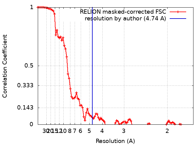
| Method | single particle reconstruction / cryo EM / Resolution: 4.74 Å | |||||||||
 Authors Authors | Valentova L / Fuzik T / Plevka P | |||||||||
| Funding support |  Czech Republic, European Union, 2 items Czech Republic, European Union, 2 items
| |||||||||
 Citation Citation | Journal: Embo J. / Year: 2024 Title: Structure and replication of Pseudomonas aeruginosa phage JBD30 Authors: Valentova L / Plevka P / Fuzik T / Novacek J / Pospisil J | |||||||||
| History |
|
- Structure visualization
| Supplemental images |
|---|
- Downloads & links
-EMDB archive
| Map data | emd_19259.map.gz | 3.7 MB | EMDB map data format | |
|---|---|---|---|---|
| Header (meta data) | emd-19259-v30.xmlemd-19259.xml | 19.2 KB 19.2 KB | Display Display | EMDB header |
| FSC (resolution estimation) | emd_19259_fsc.xml | 9.2 KB | Display | FSC data file |
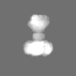
| Images |  emd_19259.png emd_19259.png | 69.6 KB | ||
| Masks | emd_19259_msk_1.map | 64 MB | Mask map | |
| Filedesc metadata | emd-19259.cif.gz | 6.3 KB | ||
| Others | emd_19259_half_map_1.map.gzemd_19259_half_map_2.map.gz | 49.7 MB 49.6 MB | ||
| Archive directory |  http://ftp.pdbj.org/pub/emdb/structures/EMD-19259ftp://ftp.pdbj.org/pub/emdb/structures/EMD-19259 http://ftp.pdbj.org/pub/emdb/structures/EMD-19259ftp://ftp.pdbj.org/pub/emdb/structures/EMD-19259 | HTTPS FTP |
-Related structure data
| Related structure data | 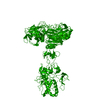 8rk4MC 19256 19257 19258 19260 19261 19262 19263 19264 19265 19266 19267 19268 19269 19270 19271 19272 19273 19274 19280 19281 19285 19439 19561  8rk3C 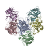 8rk5C  8rk6C 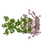 8rk7C  8rk8C 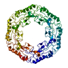 8rk9C 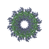 8rkaC 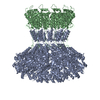 8rkbC  8rkcC  8rknC  8rkoC 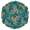 8rkxC 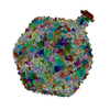 8rqeC M: atomic model generated by this map C: citing same article ( |
|---|---|
| Similar structure data |
-Links
| EMDB pages | EMDB (EBI/PDBe) / EMDataResource |
|---|
-Map
| File | Download / File: emd_19259.map.gz / Format: CCP4 / Size: 64 MB / Type: IMAGE STORED AS FLOATING POINT NUMBER (4 BYTES) | ||||||||||||||||||||
|---|---|---|---|---|---|---|---|---|---|---|---|---|---|---|---|---|---|---|---|---|---|
| Annotation | main map | ||||||||||||||||||||
| Voxel size | X=Y=Z: 0.8336 Å | ||||||||||||||||||||
| Density |
| ||||||||||||||||||||
| Symmetry | Space group: 1 | ||||||||||||||||||||
| Details | EMDB XML:
|
-Supplemental data
-Mask #1
| File | emd_19259_msk_1.map | ||||||||||||
|---|---|---|---|---|---|---|---|---|---|---|---|---|---|
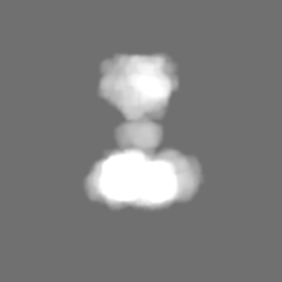
| Projections & Slices |
| ||||||||||||
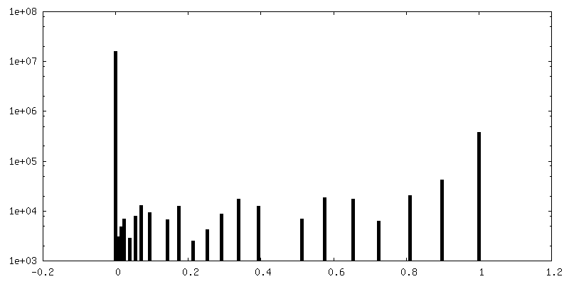
| Density Histograms |
 Z
Z Y
Y X
X


- Sample components
-Entire : Pseudomonas phage JBD30
| Entire | Name: Pseudomonas phage JBD30 (virus) |
|---|---|
| Components |
|
-Supramolecule #1: Pseudomonas phage JBD30
| Supramolecule | Name: Pseudomonas phage JBD30 / type: virus / ID: 1 / Parent: 0 / Macromolecule list: all Details: Phage JBD30 was propagated in P. aeruginosa strain BAA-28 and purified using CsCl gradient. NCBI-ID: 1223260 / Sci species name: Pseudomonas phage JBD30 / Virus type: VIRION / Virus isolate: STRAIN / Virus enveloped: No / Virus empty: No |
|---|---|
| Host (natural) | Organism:   Pseudomonas aeruginosa (bacteria) / Strain: BAA-28 Pseudomonas aeruginosa (bacteria) / Strain: BAA-28 |
| Molecular weight | Theoretical: 137 KDa |
| Virus shell | Shell ID: 1 / Name: JBD30 capsid / Diameter: 640.0 Å / T number (triangulation number): 7 |
-Macromolecule #1: DUF2793 domain-containing protein
| Macromolecule | Name: DUF2793 domain-containing protein / type: protein_or_peptide / ID: 1 / Details: Receptor binding protein / Number of copies: 1 / Enantiomer: LEVO |
|---|---|
| Source (natural) | Organism: Pseudomonas phage JBD30 (virus) |
| Molecular weight | Theoretical: 40.578426 KDa |
| Sequence | String: MTLYMGPNTG LLINGLPGEG HYSDLIRMWR WDDFLRQPVV KGRVATLPTT GQAEGDTYIF TGSGSNQNRL ARWWATGATT AIWEYMPPR LGWRVQVANE TTPSGQVKTY EYSGTAWVEL VGGMSDAPSD GKAYARESGA WTELGSAAKS ALNVLPFMNL M PDMGRFAG ...String: MTLYMGPNTG LLINGLPGEG HYSDLIRMWR WDDFLRQPVV KGRVATLPTT GQAEGDTYIF TGSGSNQNRL ARWWATGATT AIWEYMPPR LGWRVQVANE TTPSGQVKTY EYSGTAWVEL VGGMSDAPSD GKAYARESGA WTELGSAAKS ALNVLPFMNL M PDMGRFAG TAANPLATMF TTSWTPSSFL NGWNGATVAD GGKFAFDNST NGGAGPALNA RVQALLAAMG RTWTSVSRYG VE FFTAVLT AGSQTTTGSA GADGVTRYLC CSNGSKTVFN AGGWATVVMW LRVESGSAHI SSAPYTTHRL WINGAVAAPG VVL PANQWV HLRFSMQSYN GYDNACPYIY ASAGAQIAFA CPAWFGGLVD PGIHVAPILT INGASA UniProtKB: DUF2793 domain-containing protein |
-Experimental details
-Structure determination
| Method | cryo EM |
|---|---|
 Processing Processing | single particle reconstruction |
| Aggregation state | particle |
-Sample preparation
| Buffer | pH: 8 Component:
Details: 10 mM MgSO4, 10 mM NaCl, 50 mM Tris pH 8 | ||||||||||||
|---|---|---|---|---|---|---|---|---|---|---|---|---|---|
| Grid | Model: Quantifoil R2/1 / Material: COPPER / Mesh: 300 / Pretreatment - Type: GLOW DISCHARGE / Pretreatment - Time: 15 sec. / Pretreatment - Atmosphere: OTHER / Details: Gatan Solarus II | ||||||||||||
| Vitrification | Cryogen name: ETHANE / Chamber humidity: 100 % / Chamber temperature: 277.15 K / Instrument: FEI VITROBOT MARK IV Details: blotting force 0, blotting time 2 s, waiting time 15 s. | ||||||||||||
| Details | phage titer 10^11 PFU |
- Electron microscopy
| Microscope | FEI TITAN KRIOS |
|---|---|
| Specialist optics | Energy filter - Name: GIF Bioquantum / Energy filter - Slit width: 10 eV |
| Image recording | Film or detector model: GATAN K3 (6k x 4k) / Digitization - Dimensions - Width: 5760 pixel / Digitization - Dimensions - Height: 4092 pixel / Number grids imaged: 1 / Number real images: 12356 / Average exposure time: 2.0 sec. / Average electron dose: 34.0 e/Å2 |
| Electron beam | Acceleration voltage: 300 kV / Electron source:  FIELD EMISSION GUN FIELD EMISSION GUN |
| Electron optics | C2 aperture diameter: 50.0 µm / Illumination mode: FLOOD BEAM / Imaging mode: BRIGHT FIELD / Cs: 2.7 mm / Nominal defocus max: 1.6 µm / Nominal defocus min: 0.6 µm / Nominal magnification: 105000 |
| Sample stage | Specimen holder model: FEI TITAN KRIOS AUTOGRID HOLDER / Cooling holder cryogen: NITROGEN |
| Experimental equipment |  Model: Titan Krios / Image courtesy: FEI Company |
+Image processing
-Atomic model buiding 1
| Refinement | Space: REAL / Protocol: AB INITIO MODEL |
|---|---|
| Output model | PDB-8rk4: |
