ムービー
ムービー コントローラー


+ データを開く

- 基本情報
基本情報
| 登録情報 |  | |||||||||
|---|---|---|---|---|---|---|---|---|---|---|
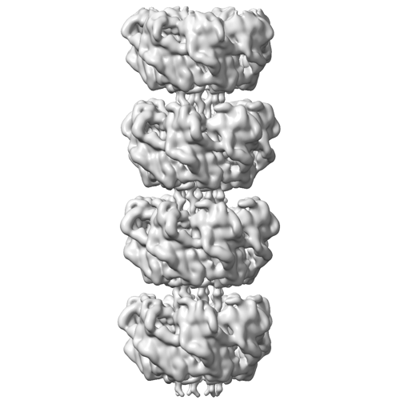
| タイトル | Recombinant Ena3A L-Type endospore appendages | |||||||||
 マップデータ マップデータ | ||||||||||
 試料 試料 |
| |||||||||
 キーワード キーワード | Endospore / Pili / Protein / Fiber / Spore / Appendage / Bacillus paranthracis / PROTEIN FIBRIL / recombinant | |||||||||
| 機能・相同性 | Endospore appendages core / Endospore appendages / Endospore appendages core domain-containing protein 機能・相同性情報 機能・相同性情報 | |||||||||
| 生物種 |  | |||||||||
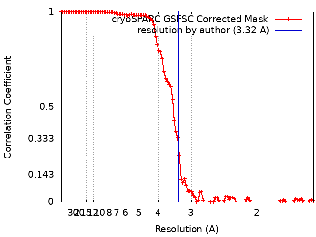
| 手法 | らせん対称体再構成法 / クライオ電子顕微鏡法 / 解像度: 3.32 Å | |||||||||
 データ登録者 データ登録者 | Sleutel M / Remaut H | |||||||||
| 資金援助 |  ベルギー, 1件 ベルギー, 1件
| |||||||||
 引用 引用 | ジャーナル: Nat Commun / 年: 2024 タイトル: Helical ultrastructure of the L-ENA spore aggregation factor of a Bacillus paranthracis foodborne outbreak strain. 著者: Mike Sleutel / Ephrem Debebe Zegeye / Ann-Katrin Llarena / Brajabandhu Pradhan / Marcus Fislage / Kristin O'Sullivan / Nani Van Gerven / Marina Aspholm / Han Remaut /  要旨: In pathogenic Bacillota, spores can form an infectious particle and can take up a central role in the environmental persistence and dissemination of disease. A poorly understood aspect of spore- ...In pathogenic Bacillota, spores can form an infectious particle and can take up a central role in the environmental persistence and dissemination of disease. A poorly understood aspect of spore-mediated infection is the fibrous structures or 'endospore appendages' (ENAs) that have been seen to decorate the spores of pathogenic Bacilli and Clostridia. Current methodological approaches are opening a window on these long enigmatic structures. Using cryoID, Alphafold modelling and genetic approaches we identify a sub-class of robust ENAs in a Bacillus paranthracis foodborne outbreak strain. We demonstrate that L-ENA are encoded by a rare three-gene cluster (ena3) that contains all components for the self-assembly of ladder-like protein nanofibers of stacked heptameric rings, their anchoring to the exosporium, and their termination in a trimeric 'ruffle' made of a complement C1Q-like BclA paralogue. The role of ENA fibers in spore-spore interaction and the distribution of L-ENA operon as mobile genetic elements in B. cereus s.l. strains suggest that L-ENA fibers may increase the survival, spread and virulence of these strains. #1: ジャーナル: Biorxiv / 年: 2023タイトル: A novel class of ultra-stable endospore appendages decorated with collagen-like tip fibrillae 著者: Sleutel M / Zegeye ED / Llarena AK / Pradhan B / Fislage M / O'Sullivan K / Aspholm M / Remaut H | |||||||||
| 履歴 |
|
- 構造の表示
構造の表示
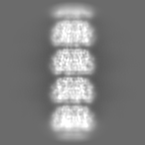
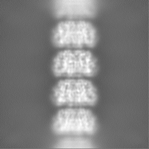
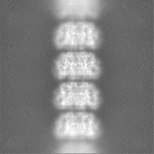
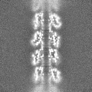
| 添付画像 |
|---|
- ダウンロードとリンク
-EMDBアーカイブ
| マップデータ | emd_17627.map.gz | 50.5 MB | EMDBマップデータ形式 | |
|---|---|---|---|---|
| ヘッダ (付随情報) | emd-17627-v30.xmlemd-17627.xml | 19.2 KB 19.2 KB | 表示 表示 | EMDBヘッダ |
| FSC (解像度算出) | emd_17627_fsc.xml | 9.9 KB | 表示 | FSCデータファイル |
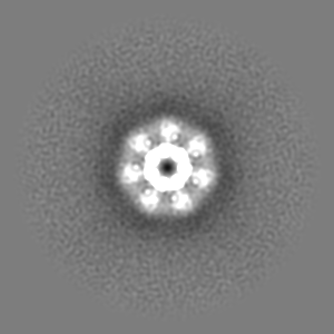
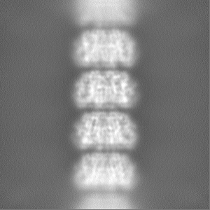
| 画像 |  emd_17627.png emd_17627.png | 71.8 KB | ||
| マスクデータ | emd_17627_msk_1.map | 103 MB | マスクマップ | |
| Filedesc metadata | emd-17627.cif.gz | 6.4 KB | ||
| その他 | emd_17627_half_map_1.map.gzemd_17627_half_map_2.map.gz | 95.6 MB 95.6 MB | ||
| アーカイブディレクトリ |  http://ftp.pdbj.org/pub/emdb/structures/EMD-17627ftp://ftp.pdbj.org/pub/emdb/structures/EMD-17627 http://ftp.pdbj.org/pub/emdb/structures/EMD-17627ftp://ftp.pdbj.org/pub/emdb/structures/EMD-17627 | HTTPS FTP |
-関連構造データ

-リンク
| EMDBのページ | EMDB (EBI/PDBe) / EMDataResource |
|---|
-マップ
| ファイル | ダウンロード / ファイル: emd_17627.map.gz / 形式: CCP4 / 大きさ: 103 MB / タイプ: IMAGE STORED AS FLOATING POINT NUMBER (4 BYTES) | ||||||||||||||||||||||||||||||||||||
|---|---|---|---|---|---|---|---|---|---|---|---|---|---|---|---|---|---|---|---|---|---|---|---|---|---|---|---|---|---|---|---|---|---|---|---|---|---|
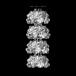
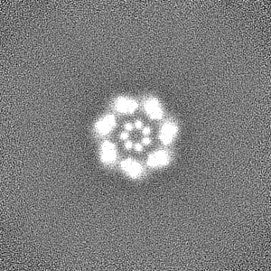
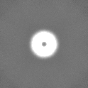
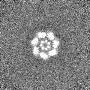
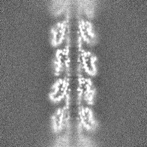
| 投影像・断面図 | 画像のコントロール
画像は Spider により作成 | ||||||||||||||||||||||||||||||||||||
| ボクセルのサイズ | X=Y=Z: 0.764 Å | ||||||||||||||||||||||||||||||||||||
| 密度 |
| ||||||||||||||||||||||||||||||||||||
| 対称性 | 空間群: 1 | ||||||||||||||||||||||||||||||||||||
| 詳細 | EMDB XML:
|
 Z (Sec.)
 Y (Row.)
 X (Col.)


-添付データ
-マスク #1
| ファイル | emd_17627_msk_1.map | ||||||||||||
|---|---|---|---|---|---|---|---|---|---|---|---|---|---|
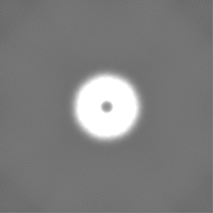
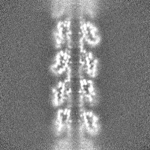
| 投影像・断面図 |
| ||||||||||||
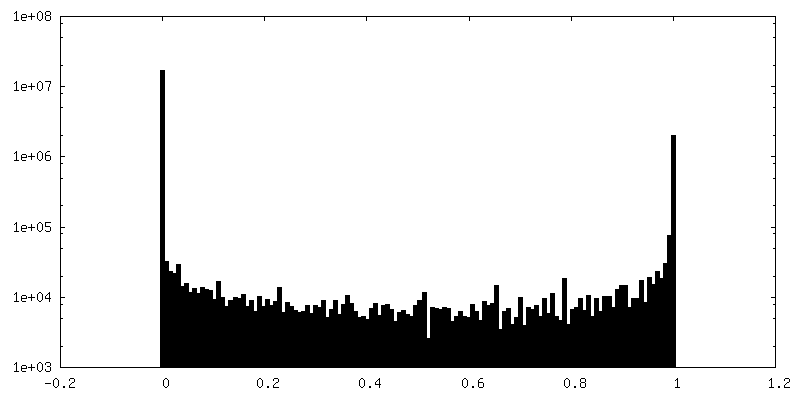
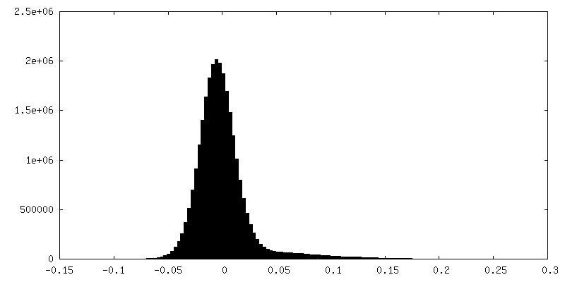
| 密度ヒストグラム |


-ハーフマップ: #2
| ファイル | emd_17627_half_map_1.map | ||||||||||||
|---|---|---|---|---|---|---|---|---|---|---|---|---|---|
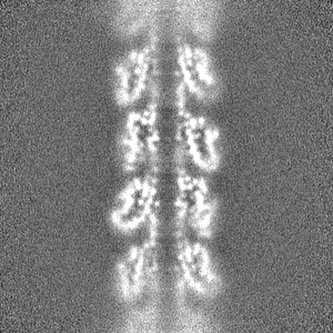
| 投影像・断面図 |
| ||||||||||||
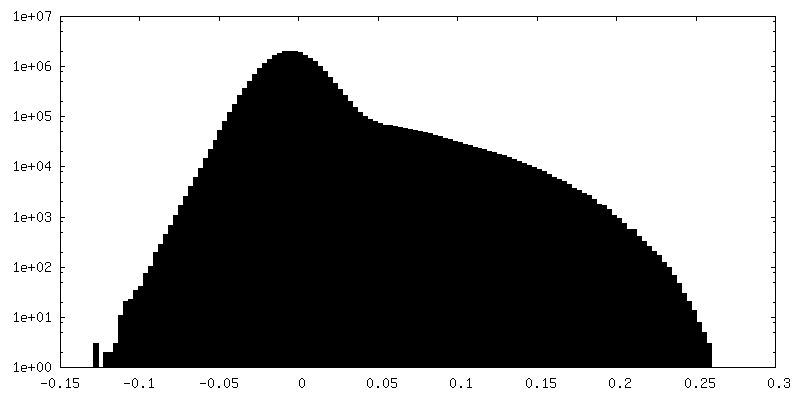
| 密度ヒストグラム |


-ハーフマップ: #1
| ファイル | emd_17627_half_map_2.map | ||||||||||||
|---|---|---|---|---|---|---|---|---|---|---|---|---|---|
| 投影像・断面図 |
| ||||||||||||
| 密度ヒストグラム |
- 試料の構成要素
-全体 : Recombinant Ena3A L-type endospore appendage
| 全体 | 名称: Recombinant Ena3A L-type endospore appendage |
|---|---|
| 要素 |
|
-超分子 #1: Recombinant Ena3A L-type endospore appendage
| 超分子 | 名称: Recombinant Ena3A L-type endospore appendage / タイプ: organelle_or_cellular_component / ID: 1 / 親要素: 0 / 含まれる分子: all |
|---|---|
| 由来(天然) | 生物種: |
| 分子量 | 理論値: 18.03 kDa/nm |
-分子 #1: DUF3992 domain-containing protein
| 分子 | 名称: DUF3992 domain-containing protein / タイプ: protein_or_peptide / ID: 1 / コピー数: 28 / 光学異性体: LEVO |
|---|---|
| 由来(天然) | 生物種: |
| 分子量 | 理論値: 11.58715 KDa |
| 組換発現 | 生物種: |
| 配列 | 文字列: MAQIGNCCTE QLCCVNDAVC CTIILDDTGG TALPIWDDAT TFVINGTIMV ENNGTVGVGP TAALTVNGTA VGGFVVAPGE CRSITMNDI NSIAIVGAGT GTSSVKISFS INYKF UniProtKB: Endospore appendages core domain-containing protein |
-実験情報
-構造解析
| 手法 | クライオ電子顕微鏡法 |
|---|---|
 解析 解析 | らせん対称体再構成法 |
| 試料の集合状態 | filament |
-試料調製
| 緩衝液 | pH: 7 / 構成要素 - 式: miliQ |
|---|---|
| 凍結 | 凍結剤: ETHANE |
- 電子顕微鏡法
| 顕微鏡 | JEOL CRYO ARM 300 |
|---|---|
| 撮影 | フィルム・検出器のモデル: GATAN K2 SUMMIT (4k x 4k) 実像数: 8528 / 平均電子線量: 62.5 e/Å2 |
| 電子線 | 加速電圧: 300 kV / 電子線源:  FIELD EMISSION GUN FIELD EMISSION GUN |
| 電子光学系 | 照射モード: SPOT SCAN / 撮影モード: BRIGHT FIELD / 最大 デフォーカス(公称値): 3.5 µm / 最小 デフォーカス(公称値): 0.5 µm / 倍率(公称値): 60000 |