establishment of RNA localization to telomere / positive regulation of telomerase catalytic core complex assembly / cellular response to nitrosative stress / negative regulation of telomere capping / establishment of protein-containing complex localization to telomere / Sensing of DNA Double Strand Breaks / peptidyl-serine autophosphorylation / positive regulation of telomere maintenance via telomere lengthening / meiotic telomere clustering / DNA-dependent protein kinase activity ...establishment of RNA localization to telomere / positive regulation of telomerase catalytic core complex assembly / cellular response to nitrosative stress / negative regulation of telomere capping / establishment of protein-containing complex localization to telomere / Sensing of DNA Double Strand Breaks / peptidyl-serine autophosphorylation / positive regulation of telomere maintenance via telomere lengthening / meiotic telomere clustering / DNA-dependent protein kinase activity / extrinsic component of synaptic vesicle membrane / pre-B cell allelic exclusion / histone mRNA catabolic process / female meiotic nuclear division / regulation of telomere maintenance via telomerase / histone H2AXS139 kinase activity / male meiotic nuclear division / lipoprotein catabolic process / DNA double-strand break processing / V(D)J recombination / cellular response to X-ray / regulation of autophagosome assembly / pexophagy / negative regulation of helicase activity / Loss of function of TP53 in cancer due to loss of tetramerization ability / Impaired BRCA2 binding to PALB2 / Regulation of TP53 Expression / signal transduction by p53 class mediator / negative regulation of G1 to G0 transition / negative regulation of glucose catabolic process to lactate via pyruvate / Transcriptional activation of cell cycle inhibitor p21 / regulation of intrinsic apoptotic signaling pathway by p53 class mediator / negative regulation of pentose-phosphate shunt / Activation of NOXA and translocation to mitochondria / ATP-dependent DNA/DNA annealing activity / regulation of cell cycle G2/M phase transition / oligodendrocyte apoptotic process / negative regulation of miRNA processing / intrinsic apoptotic signaling pathway in response to hypoxia / oocyte development / oxidative stress-induced premature senescence / regulation of tissue remodeling / positive regulation of thymocyte apoptotic process / DNA repair complex / positive regulation of mitochondrial membrane permeability / germ cell nucleus / regulation of fibroblast apoptotic process / bone marrow development / circadian behavior / cellular response to actinomycin D / histone deacetylase regulator activity / positive regulation of programmed necrotic cell death / : / regulation of mitochondrial membrane permeability involved in apoptotic process / RUNX3 regulates CDKN1A transcription / T cell proliferation involved in immune response / TP53 Regulates Transcription of Death Receptors and Ligands / reciprocal meiotic recombination / Activation of PUMA and translocation to mitochondria / TP53 regulates transcription of additional cell cycle genes whose exact role in the p53 pathway remain uncertain / mRNA transcription / negative regulation of glial cell proliferation / positive regulation of DNA damage response, signal transduction by p53 class mediator / regulation of DNA damage response, signal transduction by p53 class mediator / Regulation of TP53 Activity through Association with Co-factors / negative regulation of neuroblast proliferation / Formation of Senescence-Associated Heterochromatin Foci (SAHF) / 1-phosphatidylinositol-3-kinase activity / mitochondrial DNA repair / T cell lineage commitment / thymocyte apoptotic process / ER overload response / TP53 Regulates Transcription of Caspase Activators and Caspases / cardiac septum morphogenesis / HDR through Single Strand Annealing (SSA) / B cell lineage commitment / negative regulation of B cell proliferation / mitotic spindle assembly checkpoint signaling / cellular response to stress / entrainment of circadian clock by photoperiod / response to ionizing radiation / positive regulation of double-strand break repair / Homologous DNA Pairing and Strand Exchange / Defective homologous recombination repair (HRR) due to BRCA1 loss of function / Defective HDR through Homologous Recombination Repair (HRR) due to PALB2 loss of BRCA1 binding function / Defective HDR through Homologous Recombination Repair (HRR) due to PALB2 loss of BRCA2/RAD51/RAD51C binding function / Resolution of D-loop Structures through Synthesis-Dependent Strand Annealing (SDSA) / negative regulation of DNA replication / Resolution of D-loop Structures through Holliday Junction Intermediates / Zygotic genome activation (ZGA) / negative regulation of mitophagy / TP53 Regulates Transcription of Genes Involved in Cytochrome C Release / necroptotic process / mitotic G2 DNA damage checkpoint signaling / PI5P Regulates TP53 Acetylation / Association of TriC/CCT with target proteins during biosynthesis / positive regulation of release of cytochrome c from mitochondria / negative regulation of telomere maintenance via telomerase / SUMOylation of transcription factors / TP53 regulates transcription of several additional cell death genes whose specific roles in p53-dependent apoptosis remain uncertain Similarity search - Function
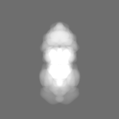
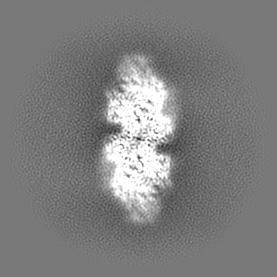
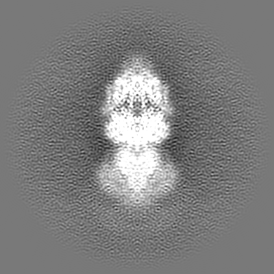
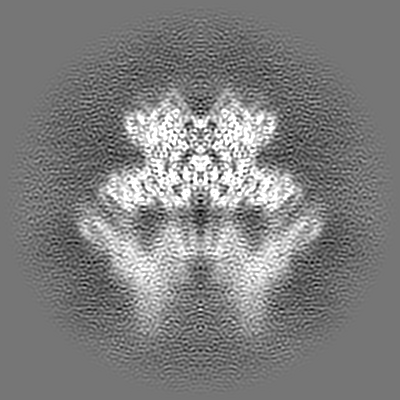
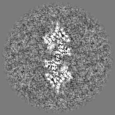
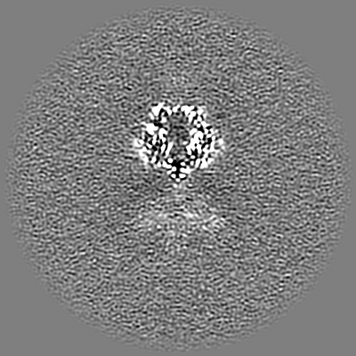
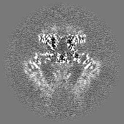
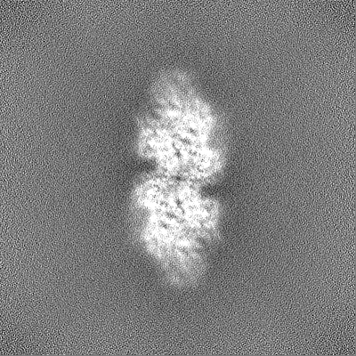
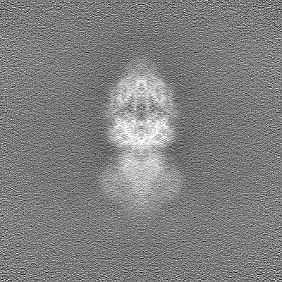
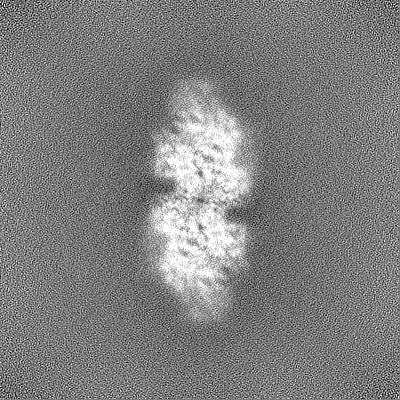
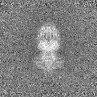
Journal: Sci Adv / Year: 2023 Title: Structural insights into the activation of ataxia-telangiectasia mutated by oxidative stress. Authors: Anna C Howes / Olga Perisic / Roger L Williams / Abstract: Ataxia-telangiectasia mutated (ATM) is a master kinase regulating DNA damage response that is activated by DNA double-strand breaks. However, ATM is also directly activated by reactive oxygen ...Ataxia-telangiectasia mutated (ATM) is a master kinase regulating DNA damage response that is activated by DNA double-strand breaks. However, ATM is also directly activated by reactive oxygen species, but how oxidative activation is achieved remains unknown. We determined the cryo-EM structure of an HO-activated ATM and showed that under oxidizing conditions, ATM formed an intramolecular disulfide bridge between two protomers that are rotated relative to each other when compared to the basal state. This rotation is accompanied by release of the substrate-blocking PRD region and twisting of the N-lobe relative to the C-lobe, which greatly optimizes catalysis. This active site remodeling enabled us to capture a substrate (p53) bound to the enzyme. This provides the first structural insights into how ATM is activated during oxidative stress.
In the structure databanks used in Yorodumi, some data are registered as the other names, "COVID-19 virus" and "2019-nCoV". Here are the details of the virus and the list of structure data.
Jan 31, 2019. EMDB accession codes are about to change! (news from PDBe EMDB page)
EMDB accession codes are about to change! (news from PDBe EMDB page)
The allocation of 4 digits for EMDB accession codes will soon come to an end. Whilst these codes will remain in use, new EMDB accession codes will include an additional digit and will expand incrementally as the available range of codes is exhausted. The current 4-digit format prefixed with “EMD-” (i.e. EMD-XXXX) will advance to a 5-digit format (i.e. EMD-XXXXX), and so on. It is currently estimated that the 4-digit codes will be depleted around Spring 2019, at which point the 5-digit format will come into force.
The EM Navigator/Yorodumi systems omit the EMD- prefix.
Related info.:Q: What is EMD? / ID/Accession-code notation in Yorodumi/EM Navigator
Yorodumi is a browser for structure data from EMDB, PDB, SASBDB, etc.
This page is also the successor to EM Navigator detail page, and also detail information page/front-end page for Omokage search.
The word "yorodu" (or yorozu) is an old Japanese word meaning "ten thousand". "mi" (miru) is to see.
Related info.:EMDB / PDB / SASBDB / Comparison of 3 databanks / Yorodumi Search / Aug 31, 2016. New EM Navigator & Yorodumi / Yorodumi Papers / Jmol/JSmol / Function and homology information / Changes in new EM Navigator and Yorodumi
 Movie
Movie Controller

 Yorodumi
 Open data
 Basic information
 Map data
 Sample
 Keywords
 Function and homology information
 Homo sapiens (human)
 Authors
 United Kingdom, 3 items
  Citation
 Structure visualization
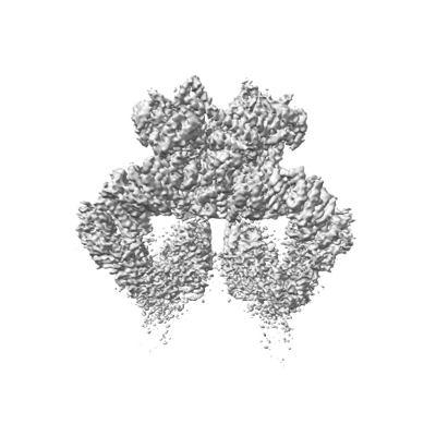
 Downloads & links
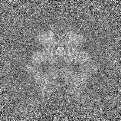
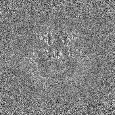
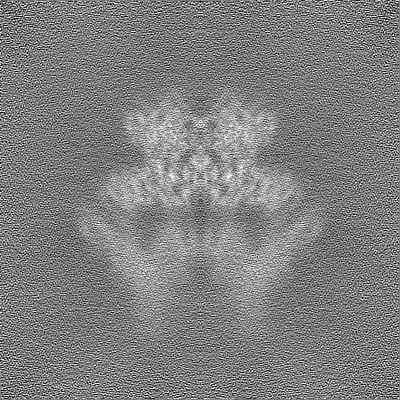
 emd_17265.png
 http://ftp.pdbj.org/pub/emdb/structures/EMD-17265


 Z (Sec.)
 Y (Row.)
 X (Col.)


 Sample components
 Processing
 Electron microscopy
 FIELD EMISSION GUN