[English] 日本語
 Yorodumi
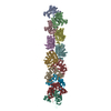
- EMDB-1674: The cryo-EM structure of actin filament in the presence of phosphate -
+ Open data
- Basic information
| Entry | Database: EMDB / ID: EMD-1674 | |||||||||
|---|---|---|---|---|---|---|---|---|---|---|
| Title | The cryo-EM structure of actin filament in the presence of phosphate | |||||||||
 Map data Map data | This is an image of a surface with B-factor correction. | |||||||||
 Sample Sample |
| |||||||||
 Keywords Keywords | actin / cytoskeleton / cell adhesion / cellular signaling / cytokinesis / muscle | |||||||||
| Function / homology |  Function and homology information Function and homology informationcytoskeletal motor activator activity / myosin heavy chain binding / tropomyosin binding / actin filament bundle / troponin I binding / filamentous actin / mesenchyme migration / skeletal muscle myofibril / actin filament bundle assembly / striated muscle thin filament ...cytoskeletal motor activator activity / myosin heavy chain binding / tropomyosin binding / actin filament bundle / troponin I binding / filamentous actin / mesenchyme migration / skeletal muscle myofibril / actin filament bundle assembly / striated muscle thin filament / skeletal muscle thin filament assembly / actin monomer binding / skeletal muscle fiber development / stress fiber / titin binding / actin filament polymerization / filopodium / actin filament / Hydrolases; Acting on acid anhydrides; Acting on acid anhydrides to facilitate cellular and subcellular movement / calcium-dependent protein binding / lamellipodium / cell body / hydrolase activity / protein domain specific binding / calcium ion binding / positive regulation of gene expression / magnesium ion binding / ATP binding / identical protein binding / cytoplasm Similarity search - Function | |||||||||
| Biological species |  | |||||||||
| Method | single particle reconstruction / cryo EM / Resolution: 6.0 Å | |||||||||
 Authors Authors | Murakami K / Yasunaga T / Noguchi TQ / Uyeda TQ / Wakabayashi T | |||||||||
 Citation Citation |  Journal: Cell / Year: 2010 Journal: Cell / Year: 2010Title: Structural basis for actin assembly, activation of ATP hydrolysis, and delayed phosphate release. Authors: Kenji Murakami / Takuo Yasunaga / Taro Q P Noguchi / Yuki Gomibuchi / Kien X Ngo / Taro Q P Uyeda / Takeyuki Wakabayashi /  Abstract: Assembled actin filaments support cellular signaling, intracellular trafficking, and cytokinesis. ATP hydrolysis triggered by actin assembly provides the structural cues for filament turnover in vivo. ...Assembled actin filaments support cellular signaling, intracellular trafficking, and cytokinesis. ATP hydrolysis triggered by actin assembly provides the structural cues for filament turnover in vivo. Here, we present the cryo-electron microscopic (cryo-EM) structure of filamentous actin (F-actin) in the presence of phosphate, with the visualization of some α-helical backbones and large side chains. A complete atomic model based on the EM map identified intermolecular interactions mediated by bound magnesium and phosphate ions. Comparison of the F-actin model with G-actin monomer crystal structures reveals a critical role for bending of the conserved proline-rich loop in triggering phosphate release following ATP hydrolysis. Crystal structures of G-actin show that mutations in this loop trap the catalytic site in two intermediate states of the ATPase cycle. The combined structural information allows us to propose a detailed molecular mechanism for the biochemical events, including actin polymerization and ATPase activation, critical for actin filament dynamics. | |||||||||
| History |
|
- Structure visualization
| Movie |
 Movie viewer Movie viewer |
|---|---|
| Structure viewer | EM map:  SurfView SurfView Molmil Molmil Jmol/JSmol Jmol/JSmol |
| Supplemental images |
- Downloads & links
-EMDB archive
| Map data |  emd_1674.map.gz emd_1674.map.gz | 2 MB |  EMDB map data format EMDB map data format | |
|---|---|---|---|---|
| Header (meta data) |  emd-1674-v30.xml emd-1674-v30.xml emd-1674.xml emd-1674.xml | 10.4 KB 10.4 KB | Display Display |  EMDB header EMDB header |
| Images |  1674.png 1674.png | 345 KB | ||
| Archive directory |  http://ftp.pdbj.org/pub/emdb/structures/EMD-1674 http://ftp.pdbj.org/pub/emdb/structures/EMD-1674 ftp://ftp.pdbj.org/pub/emdb/structures/EMD-1674 ftp://ftp.pdbj.org/pub/emdb/structures/EMD-1674 | HTTPS FTP |
-Validation report
| Summary document |  emd_1674_validation.pdf.gz emd_1674_validation.pdf.gz | 401.5 KB | Display |  EMDB validaton report EMDB validaton report |
|---|---|---|---|---|
| Full document |  emd_1674_full_validation.pdf.gz emd_1674_full_validation.pdf.gz | 401.1 KB | Display | |
| Data in XML |  emd_1674_validation.xml.gz emd_1674_validation.xml.gz | 5.4 KB | Display | |
| Arichive directory |  https://ftp.pdbj.org/pub/emdb/validation_reports/EMD-1674 https://ftp.pdbj.org/pub/emdb/validation_reports/EMD-1674 ftp://ftp.pdbj.org/pub/emdb/validation_reports/EMD-1674 ftp://ftp.pdbj.org/pub/emdb/validation_reports/EMD-1674 | HTTPS FTP |
-Related structure data
| Related structure data |  3g37MC 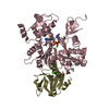 3a5lC  3a5mC  3a5nC  3a5oC M: atomic model generated by this map C: citing same article ( |
|---|---|
| Similar structure data |
- Links
Links
| EMDB pages |  EMDB (EBI/PDBe) / EMDB (EBI/PDBe) /  EMDataResource EMDataResource |
|---|---|
| Related items in Molecule of the Month |
- Map
Map
| File |  Download / File: emd_1674.map.gz / Format: CCP4 / Size: 6.6 MB / Type: IMAGE STORED AS FLOATING POINT NUMBER (4 BYTES) Download / File: emd_1674.map.gz / Format: CCP4 / Size: 6.6 MB / Type: IMAGE STORED AS FLOATING POINT NUMBER (4 BYTES) | ||||||||||||||||||||||||||||||||||||||||||||||||||||||||||||||||||||
|---|---|---|---|---|---|---|---|---|---|---|---|---|---|---|---|---|---|---|---|---|---|---|---|---|---|---|---|---|---|---|---|---|---|---|---|---|---|---|---|---|---|---|---|---|---|---|---|---|---|---|---|---|---|---|---|---|---|---|---|---|---|---|---|---|---|---|---|---|---|
| Annotation | This is an image of a surface with B-factor correction. | ||||||||||||||||||||||||||||||||||||||||||||||||||||||||||||||||||||
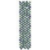
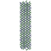
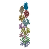
| Projections & slices | Image control
Images are generated by Spider. | ||||||||||||||||||||||||||||||||||||||||||||||||||||||||||||||||||||
| Voxel size | X=Y=Z: 2.275 Å | ||||||||||||||||||||||||||||||||||||||||||||||||||||||||||||||||||||
| Density |
| ||||||||||||||||||||||||||||||||||||||||||||||||||||||||||||||||||||
| Symmetry | Space group: 1 | ||||||||||||||||||||||||||||||||||||||||||||||||||||||||||||||||||||
| Details | EMDB XML:
CCP4 map header:
| ||||||||||||||||||||||||||||||||||||||||||||||||||||||||||||||||||||
-Supplemental data
- Sample components
-Entire : Actin filament in the presence of phosphate
| Entire | Name: Actin filament in the presence of phosphate |
|---|---|
| Components |
|
-Supramolecule #1000: Actin filament in the presence of phosphate
| Supramolecule | Name: Actin filament in the presence of phosphate / type: sample / ID: 1000 / Oligomeric state: Filament / Number unique components: 1 |
|---|---|
| Molecular weight | Theoretical: 1.17 MDa |
-Macromolecule #1: Actin
| Macromolecule | Name: Actin / type: protein_or_peptide / ID: 1 / Name.synonym: Actin / Number of copies: 26 / Oligomeric state: Filament / Recombinant expression: No / Database: NCBI |
|---|---|
| Source (natural) | Organism:  |
| Molecular weight | Theoretical: 45 KDa |
-Experimental details
-Structure determination
| Method | cryo EM |
|---|---|
 Processing Processing | single particle reconstruction |
| Aggregation state | particle |
- Sample preparation
| Buffer | pH: 7.4 Details: 50 mM NaCl, 5 mM MgCl2, 0.025 mM ATP, 10 mM sodium phosphate (pH 7.4), 0.05 %NaN3, and 0.7 mM DTT |
|---|---|
| Vitrification | Cryogen name: ETHANE / Chamber humidity: 90 % / Chamber temperature: 77 K / Instrument: OTHER |
- Electron microscopy
| Microscope | HITACHI EF2000 |
|---|---|
| Temperature | Min: 77 K / Max: 77 K / Average: 77 K |
| Specialist optics | Energy filter - Name: HITACH |
| Image recording | Category: CCD / Film or detector model: GENERIC CCD / Digitization - Sampling interval: 2.275 µm / Average electron dose: 15 e/Å2 |
| Tilt angle min | 0 |
| Tilt angle max | 0 |
| Electron beam | Acceleration voltage: 200 kV / Electron source:  FIELD EMISSION GUN FIELD EMISSION GUN |
| Electron optics | Illumination mode: SPOT SCAN / Imaging mode: BRIGHT FIELD / Nominal defocus max: 1.5 µm / Nominal defocus min: 1.0 µm / Nominal magnification: 100000 |
| Sample stage | Specimen holder: Side entry liquid nitrogen-cooled cryo specimen holder Specimen holder model: GATAN LIQUID NITROGEN |
- Image processing
| CTF correction | Details: Each particle |
|---|---|
| Final reconstruction | Algorithm: OTHER / Resolution.type: BY AUTHOR / Resolution: 6.0 Å / Resolution method: FSC 0.143 CUT-OFF / Software - Name: EOS / Number images used: 8000 |
| Final two d classification | Number classes: 72 |
-Atomic model buiding 1
| Software | Name: O, NAMD, EOS |
|---|---|
| Details | Protocol: Real-space refinement. The initial model was docked manually and refined using O, NAMD, and EOS. |
| Refinement | Space: REAL / Protocol: RIGID BODY FIT / Target criteria: R-factor |
| Output model |  PDB-3g37: |
 Movie
Movie Controller







 Z (Sec.)
 X (Row.)
 Y (Col.)


