+ データを開く
- 基本情報
基本情報
| 登録情報 |  | |||||||||
|---|---|---|---|---|---|---|---|---|---|---|
| タイトル | Cyanide dihydratase from Bacillus pumilus C1 variant - Q86R,H305K,H308K,H323K | |||||||||
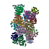
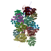
 マップデータ マップデータ | Map of cyanide dihydratase from Bacillus pumilus C1 variant (Q86R/H305K/H308K/H323K) | |||||||||
 試料 試料 |
| |||||||||
 キーワード キーワード | Helical / homo-oligomeric / cyanide dihydratase / HYDROLASE | |||||||||
| 機能・相同性 |  機能・相同性情報 機能・相同性情報 | |||||||||
| 生物種 |  | |||||||||
| 手法 | らせん対称体再構成法 / クライオ電子顕微鏡法 / 解像度: 3.15 Å | |||||||||
 データ登録者 データ登録者 | Mulelu AE / Reitz J / van Rooyen J / Scheffer M / Frangakis AS / Dlamini LS / Woodward JD / Benedik MJ / Sewell BT | |||||||||
| 資金援助 |  南アフリカ, 1件 南アフリカ, 1件
| |||||||||
 引用 引用 |  ジャーナル: To Be Published ジャーナル: To Be Publishedタイトル: The Role of Histidine Residues in the Oligomerization of Cyanide Dihydratase from Bacillus pumilus C1 著者: Mulelu AE / Reitz J / van Rooyen J / Scheffer M / Frangakis AS / Dlamini LS / Woodward JD / Benedik MJ / Sewell BT | |||||||||
| 履歴 |
|
- 構造の表示
構造の表示
| 添付画像 |
|---|
- ダウンロードとリンク
-EMDBアーカイブ
| マップデータ |  emd_16437.map.gz emd_16437.map.gz | 95.1 MB |  EMDBマップデータ形式 EMDBマップデータ形式 | |
|---|---|---|---|---|
| ヘッダ (付随情報) |  emd-16437-v30.xml emd-16437-v30.xml emd-16437.xml emd-16437.xml | 13 KB 13 KB | 表示 表示 |  EMDBヘッダ EMDBヘッダ |
| 画像 |  emd_16437.png emd_16437.png | 39.3 KB | ||
| Filedesc metadata |  emd-16437.cif.gz emd-16437.cif.gz | 6.3 KB | ||
| アーカイブディレクトリ |  http://ftp.pdbj.org/pub/emdb/structures/EMD-16437 http://ftp.pdbj.org/pub/emdb/structures/EMD-16437 ftp://ftp.pdbj.org/pub/emdb/structures/EMD-16437 ftp://ftp.pdbj.org/pub/emdb/structures/EMD-16437 | HTTPS FTP |
-検証レポート
| 文書・要旨 |  emd_16437_validation.pdf.gz emd_16437_validation.pdf.gz | 662.3 KB | 表示 |  EMDB検証レポート EMDB検証レポート |
|---|---|---|---|---|
| 文書・詳細版 |  emd_16437_full_validation.pdf.gz emd_16437_full_validation.pdf.gz | 661.9 KB | 表示 | |
| XML形式データ |  emd_16437_validation.xml.gz emd_16437_validation.xml.gz | 6.2 KB | 表示 | |
| CIF形式データ |  emd_16437_validation.cif.gz emd_16437_validation.cif.gz | 7.1 KB | 表示 | |
| アーカイブディレクトリ |  https://ftp.pdbj.org/pub/emdb/validation_reports/EMD-16437 https://ftp.pdbj.org/pub/emdb/validation_reports/EMD-16437 ftp://ftp.pdbj.org/pub/emdb/validation_reports/EMD-16437 ftp://ftp.pdbj.org/pub/emdb/validation_reports/EMD-16437 | HTTPS FTP |
-関連構造データ
| 関連構造データ |  8c5iMC  8p4iC M: このマップから作成された原子モデル C: 同じ文献を引用 ( |
|---|---|
| 類似構造データ | 類似検索 - 機能・相同性  F&H 検索 F&H 検索 |
- リンク
リンク
| EMDBのページ |  EMDB (EBI/PDBe) / EMDB (EBI/PDBe) /  EMDataResource EMDataResource |
|---|
- マップ
マップ
| ファイル |  ダウンロード / ファイル: emd_16437.map.gz / 形式: CCP4 / 大きさ: 103 MB / タイプ: IMAGE STORED AS FLOATING POINT NUMBER (4 BYTES) ダウンロード / ファイル: emd_16437.map.gz / 形式: CCP4 / 大きさ: 103 MB / タイプ: IMAGE STORED AS FLOATING POINT NUMBER (4 BYTES) | ||||||||||||||||||||||||||||||||||||
|---|---|---|---|---|---|---|---|---|---|---|---|---|---|---|---|---|---|---|---|---|---|---|---|---|---|---|---|---|---|---|---|---|---|---|---|---|---|
| 注釈 | Map of cyanide dihydratase from Bacillus pumilus C1 variant (Q86R/H305K/H308K/H323K) | ||||||||||||||||||||||||||||||||||||
| 投影像・断面図 | 画像のコントロール
画像は Spider により作成 | ||||||||||||||||||||||||||||||||||||
| ボクセルのサイズ | X=Y=Z: 1.05 Å | ||||||||||||||||||||||||||||||||||||
| 密度 |
| ||||||||||||||||||||||||||||||||||||
| 対称性 | 空間群: 1 | ||||||||||||||||||||||||||||||||||||
| 詳細 | EMDB XML:
|
-添付データ
- 試料の構成要素
-全体 : Active helical nitrilase homo-oligomer of cyanide dihydratase fro...
| 全体 | 名称: Active helical nitrilase homo-oligomer of cyanide dihydratase from Bacillus pumilus C1 variant (Q86R/H305K/H308K/H323K) |
|---|---|
| 要素 |
|
-超分子 #1: Active helical nitrilase homo-oligomer of cyanide dihydratase fro...
| 超分子 | 名称: Active helical nitrilase homo-oligomer of cyanide dihydratase from Bacillus pumilus C1 variant (Q86R/H305K/H308K/H323K) タイプ: complex / ID: 1 / 親要素: 0 / 含まれる分子: all 詳細: Cyanide dihydratase from Bacillus pumilus C1 variant generated by site-directed mutagenesis. |
|---|---|
| 由来(天然) | 生物種:  |
-分子 #1: Cyanide dihydratase
| 分子 | 名称: Cyanide dihydratase / タイプ: protein_or_peptide / ID: 1 / コピー数: 18 / 光学異性体: LEVO |
|---|---|
| 由来(天然) | 生物種:  |
| 分子量 | 理論値: 37.503363 KDa |
| 組換発現 | 生物種:  |
| 配列 | 文字列: MTSIYPKFRA AAVQAAPIYL NLEASVEKSC ELIDEAASNG AKLVAFPEAF LPGYPWFAFI GHPEYTRKFY HELYKNAVEI PSLAIRKIS EAAKRNETYV CISCSEKDGG SLYLAQLWFN PNGDLIGKHR KMRASVAERL IWGDGSGSMM PVFQTEIGNL G GLMCWEHQ ...文字列: MTSIYPKFRA AAVQAAPIYL NLEASVEKSC ELIDEAASNG AKLVAFPEAF LPGYPWFAFI GHPEYTRKFY HELYKNAVEI PSLAIRKIS EAAKRNETYV CISCSEKDGG SLYLAQLWFN PNGDLIGKHR KMRASVAERL IWGDGSGSMM PVFQTEIGNL G GLMCWEHQ VPLDLMAMNA QNEQVHVASW PGYFDDEISS RYYAIATQTF VLMTSSIYTE EMKEMICLTQ EQRDYFETFK SG HTCIYGP DGEPISDMVP AETEGIAYAE IDVERVIDYK YYIDPAGHYS NQSLSMNFNQ QPTPVVKKLN KQKNEVFTYE DIQ YQKGIL EEKV UniProtKB: Cyanide dihydratase |
-実験情報
-構造解析
| 手法 | クライオ電子顕微鏡法 |
|---|---|
 解析 解析 | らせん対称体再構成法 |
| 試料の集合状態 | filament |
- 試料調製
試料調製
| 濃度 | 0.2 mg/mL | |||||||||
|---|---|---|---|---|---|---|---|---|---|---|
| 緩衝液 | pH: 5.4 構成要素:
詳細: 150 mM NaCl, 50 mM Tris-HCl pH 5.4 | |||||||||
| グリッド | モデル: Quantifoil R2/2 / 材質: COPPER / 支持フィルム - 材質: CARBON / 支持フィルム - トポロジー: HOLEY / 支持フィルム - Film thickness: 12 / 前処理 - タイプ: GLOW DISCHARGE / 前処理 - 時間: 30 sec. / 前処理 - 雰囲気: OTHER / 前処理 - 気圧: 20.0 kPa | |||||||||
| 凍結 | 凍結剤: ETHANE / チャンバー内湿度: 100 % / チャンバー内温度: 277.15 K / 装置: FEI VITROBOT MARK IV 詳細: A 2.5 microlitre sample was applied onto a glow-discharged grid, blotted and plunged without incubation.. | |||||||||
| 詳細 | Homogeneous protein sample. |
- 電子顕微鏡法
| 顕微鏡 | FEI TITAN KRIOS |
|---|---|
| 撮影 | フィルム・検出器のモデル: GATAN K2 SUMMIT (4k x 4k) 検出モード: COUNTING / 平均電子線量: 54.0 e/Å2 |
| 電子線 | 加速電圧: 300 kV / 電子線源:  FIELD EMISSION GUN FIELD EMISSION GUN |
| 電子光学系 | 照射モード: OTHER / 撮影モード: BRIGHT FIELD / 最大 デフォーカス(公称値): 3.0 µm / 最小 デフォーカス(公称値): 0.6 µm / 倍率(公称値): 130000 |
| 試料ステージ | 試料ホルダーモデル: FEI TITAN KRIOS AUTOGRID HOLDER ホルダー冷却材: NITROGEN |
| 実験機器 |  モデル: Titan Krios / 画像提供: FEI Company |
- 画像解析
画像解析
| 最終 再構成 | 使用したクラス数: 92000 想定した対称性 - らせんパラメータ - Δz: 16.7 Å 想定した対称性 - らせんパラメータ - ΔΦ: -77 ° 想定した対称性 - らせんパラメータ - 軸対称性: C2 (2回回転対称) アルゴリズム: FOURIER SPACE / 解像度のタイプ: BY AUTHOR / 解像度: 3.15 Å / 解像度の算出法: FSC 0.5 CUT-OFF / ソフトウェア - 名称: RELION (ver. 2.1) / 使用した粒子像数: 103000 |
|---|---|
| 初期モデル | モデルのタイプ: INSILICO MODEL / In silico モデル: Featureless cylinder |
| 最終 角度割当 | タイプ: NOT APPLICABLE |
-原子モデル構築 1
| 精密化 | 空間: REAL / プロトコル: AB INITIO MODEL |
|---|---|
| 得られたモデル |  PDB-8c5i: |
 ムービー
ムービー コントローラー


 Z (Sec.)
 Y (Row.)
 X (Col.)


