[English] 日本語
 Yorodumi
- EMDB-16031: Elongating E. coli 70S ribosome containing acylated tRNA(iMet) in... -
+ Open data
- Basic information
| Entry |  | |||||||||
|---|---|---|---|---|---|---|---|---|---|---|
| Title | Elongating E. coli 70S ribosome containing acylated tRNA(iMet) in the P-site and AAA mRNA codon in the A-site after uncompleted di-peptide formation | |||||||||
 Map data Map data | ||||||||||
 Sample Sample |
| |||||||||
 Keywords Keywords | m6A / mRNA modification / tRNA recognition / TRANSLATION | |||||||||
| Function / homology |  Function and homology information Function and homology informationnegative regulation of cytoplasmic translational initiation / ornithine decarboxylase inhibitor activity / transcription antitermination factor activity, RNA binding / misfolded RNA binding / Group I intron splicing / RNA folding / transcriptional attenuation / endoribonuclease inhibitor activity / positive regulation of ribosome biogenesis / RNA-binding transcription regulator activity ...negative regulation of cytoplasmic translational initiation / ornithine decarboxylase inhibitor activity / transcription antitermination factor activity, RNA binding / misfolded RNA binding / Group I intron splicing / RNA folding / transcriptional attenuation / endoribonuclease inhibitor activity / positive regulation of ribosome biogenesis / RNA-binding transcription regulator activity / negative regulation of cytoplasmic translation / four-way junction DNA binding / DnaA-L2 complex / translation repressor activity / negative regulation of translational initiation / regulation of mRNA stability / negative regulation of DNA-templated DNA replication initiation / mRNA regulatory element binding translation repressor activity / assembly of large subunit precursor of preribosome / positive regulation of RNA splicing / ribosome assembly / transcription elongation factor complex / regulation of DNA-templated transcription elongation / cytosolic ribosome assembly / response to reactive oxygen species / DNA endonuclease activity / transcription antitermination / translational initiation / regulation of cell growth / DNA-templated transcription termination / response to radiation / maintenance of translational fidelity / mRNA 5'-UTR binding / ribosome biogenesis / large ribosomal subunit / regulation of translation / transferase activity / ribosome binding / ribosomal small subunit biogenesis / ribosomal small subunit assembly / small ribosomal subunit / 5S rRNA binding / small ribosomal subunit rRNA binding / ribosomal large subunit assembly / cytosolic small ribosomal subunit / large ribosomal subunit rRNA binding / cytosolic large ribosomal subunit / cytoplasmic translation / tRNA binding / negative regulation of translation / rRNA binding / structural constituent of ribosome / ribosome / translation / response to antibiotic / negative regulation of DNA-templated transcription / mRNA binding / DNA binding / RNA binding / zinc ion binding / membrane / cytosol / cytoplasm Similarity search - Function | |||||||||
| Biological species |  | |||||||||
| Method | single particle reconstruction / cryo EM / Resolution: 2.88 Å | |||||||||
 Authors Authors | Koziej L / Glatt S | |||||||||
| Funding support | European Union, 1 items
| |||||||||
 Citation Citation |  Journal: Nat Commun / Year: 2023 Journal: Nat Commun / Year: 2023Title: Modulation of translational decoding by mA modification of mRNA. Authors: Sakshi Jain / Lukasz Koziej / Panagiotis Poulis / Igor Kaczmarczyk / Monika Gaik / Michal Rawski / Namit Ranjan / Sebastian Glatt / Marina V Rodnina /   Abstract: N-methyladenosine (mA) is an abundant, dynamic mRNA modification that regulates key steps of cellular mRNA metabolism. mA in the mRNA coding regions inhibits translation elongation. Here, we show how ...N-methyladenosine (mA) is an abundant, dynamic mRNA modification that regulates key steps of cellular mRNA metabolism. mA in the mRNA coding regions inhibits translation elongation. Here, we show how mA modulates decoding in the bacterial translation system using a combination of rapid kinetics, smFRET and single-particle cryo-EM. We show that, while the modification does not impair the initial binding of aminoacyl-tRNA to the ribosome, in the presence of mA fewer ribosomes complete the decoding process due to the lower stability of the complexes and enhanced tRNA drop-off. The mRNA codon adopts a π-stacked codon conformation that is remodeled upon aminoacyl-tRNA binding. mA does not exclude canonical codon-anticodon geometry, but favors alternative more dynamic conformations that are rejected by the ribosome. These results highlight how modifications outside the Watson-Crick edge can still interfere with codon-anticodon base pairing and complex recognition by the ribosome, thereby modulating the translational efficiency of modified mRNAs. | |||||||||
| History |
|
- Structure visualization
| Supplemental images |
|---|
- Downloads & links
-EMDB archive
| Map data |  emd_16031.map.gz emd_16031.map.gz | 434.1 MB |  EMDB map data format EMDB map data format | |
|---|---|---|---|---|
| Header (meta data) |  emd-16031-v30.xml emd-16031-v30.xml emd-16031.xml emd-16031.xml | 88.7 KB 88.7 KB | Display Display |  EMDB header EMDB header |
| FSC (resolution estimation) |  emd_16031_fsc.xml emd_16031_fsc.xml | 16.9 KB | Display |  FSC data file FSC data file |
| Images |  emd_16031.png emd_16031.png | 155.1 KB | ||
| Masks |  emd_16031_msk_1.map emd_16031_msk_1.map | 512 MB |  Mask map Mask map | |
| Others |  emd_16031_additional_1.map.gz emd_16031_additional_1.map.gz emd_16031_half_map_1.map.gz emd_16031_half_map_1.map.gz emd_16031_half_map_2.map.gz emd_16031_half_map_2.map.gz | 257 MB 474.4 MB 474.4 MB | ||
| Archive directory |  http://ftp.pdbj.org/pub/emdb/structures/EMD-16031 http://ftp.pdbj.org/pub/emdb/structures/EMD-16031 ftp://ftp.pdbj.org/pub/emdb/structures/EMD-16031 ftp://ftp.pdbj.org/pub/emdb/structures/EMD-16031 | HTTPS FTP |
-Validation report
| Summary document |  emd_16031_validation.pdf.gz emd_16031_validation.pdf.gz | 1.2 MB | Display |  EMDB validaton report EMDB validaton report |
|---|---|---|---|---|
| Full document |  emd_16031_full_validation.pdf.gz emd_16031_full_validation.pdf.gz | 1.2 MB | Display | |
| Data in XML |  emd_16031_validation.xml.gz emd_16031_validation.xml.gz | 26.6 KB | Display | |
| Data in CIF |  emd_16031_validation.cif.gz emd_16031_validation.cif.gz | 34.9 KB | Display | |
| Arichive directory |  https://ftp.pdbj.org/pub/emdb/validation_reports/EMD-16031 https://ftp.pdbj.org/pub/emdb/validation_reports/EMD-16031 ftp://ftp.pdbj.org/pub/emdb/validation_reports/EMD-16031 ftp://ftp.pdbj.org/pub/emdb/validation_reports/EMD-16031 | HTTPS FTP |
-Related structure data
| Related structure data | 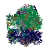 8bghMC  8bf7C 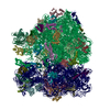 8bgeC  8bh4C  8bhjC 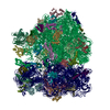 8bhlC  8bhnC 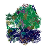 8bhpC 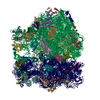 8bilC 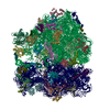 8bimC C: citing same article ( M: atomic model generated by this map |
|---|---|
| Similar structure data | Similarity search - Function & homology  F&H Search F&H Search |
- Links
Links
| EMDB pages |  EMDB (EBI/PDBe) / EMDB (EBI/PDBe) /  EMDataResource EMDataResource |
|---|---|
| Related items in Molecule of the Month |
- Map
Map
| File |  Download / File: emd_16031.map.gz / Format: CCP4 / Size: 512 MB / Type: IMAGE STORED AS FLOATING POINT NUMBER (4 BYTES) Download / File: emd_16031.map.gz / Format: CCP4 / Size: 512 MB / Type: IMAGE STORED AS FLOATING POINT NUMBER (4 BYTES) | ||||||||||||||||||||||||||||||||||||
|---|---|---|---|---|---|---|---|---|---|---|---|---|---|---|---|---|---|---|---|---|---|---|---|---|---|---|---|---|---|---|---|---|---|---|---|---|---|
| Projections & slices | Image control
Images are generated by Spider. | ||||||||||||||||||||||||||||||||||||
| Voxel size | X=Y=Z: 0.86 Å | ||||||||||||||||||||||||||||||||||||
| Density |
| ||||||||||||||||||||||||||||||||||||
| Symmetry | Space group: 1 | ||||||||||||||||||||||||||||||||||||
| Details | EMDB XML:
|
-Supplemental data
-Mask #1
| File |  emd_16031_msk_1.map emd_16031_msk_1.map | ||||||||||||
|---|---|---|---|---|---|---|---|---|---|---|---|---|---|
| Projections & Slices |
| ||||||||||||
| Density Histograms |
-Additional map: Map combined from half maps used for generation...
| File | emd_16031_additional_1.map | ||||||||||||
|---|---|---|---|---|---|---|---|---|---|---|---|---|---|
| Annotation | Map combined from half maps used for generation of the final high-resolution DeepEM enhanced map. | ||||||||||||
| Projections & Slices |
| ||||||||||||
| Density Histograms |
-Half map: #2
| File | emd_16031_half_map_1.map | ||||||||||||
|---|---|---|---|---|---|---|---|---|---|---|---|---|---|
| Projections & Slices |
| ||||||||||||
| Density Histograms |
-Half map: #1
| File | emd_16031_half_map_2.map | ||||||||||||
|---|---|---|---|---|---|---|---|---|---|---|---|---|---|
| Projections & Slices |
| ||||||||||||
| Density Histograms |
- Sample components
+Entire : Elongating E. coli 70S ribosome containing acylated tRNA(iMet) in...
+Supramolecule #1: Elongating E. coli 70S ribosome containing acylated tRNA(iMet) in...
+Supramolecule #2: 50S subunit
+Supramolecule #3: 30S subunit
+Supramolecule #4: 23S and 5S rRNA
+Supramolecule #5: 16S rRNA
+Supramolecule #6: mRNA and tRNA
+Macromolecule #1: 50S ribosomal protein L2
+Macromolecule #2: 50S ribosomal protein L3
+Macromolecule #3: 50S ribosomal protein L4
+Macromolecule #4: 50S ribosomal protein L5
+Macromolecule #5: 50S ribosomal protein L6
+Macromolecule #6: 50S ribosomal protein L9
+Macromolecule #7: 50S ribosomal protein L13
+Macromolecule #8: 50S ribosomal protein L14
+Macromolecule #9: 50S ribosomal protein L15
+Macromolecule #10: 50S ribosomal protein L16
+Macromolecule #11: 50S ribosomal protein L17
+Macromolecule #12: 50S ribosomal protein L18
+Macromolecule #13: 50S ribosomal protein L19
+Macromolecule #14: 50S ribosomal protein L20
+Macromolecule #15: 50S ribosomal protein L21
+Macromolecule #16: 50S ribosomal protein L22
+Macromolecule #17: 50S ribosomal protein L23
+Macromolecule #18: 50S ribosomal protein L24
+Macromolecule #19: 50S ribosomal protein L25
+Macromolecule #20: 50S ribosomal protein L27
+Macromolecule #21: 50S ribosomal protein L28
+Macromolecule #22: 50S ribosomal protein L29
+Macromolecule #23: 50S ribosomal protein L30
+Macromolecule #24: 50S ribosomal protein L31
+Macromolecule #25: 50S ribosomal protein L32
+Macromolecule #26: 50S ribosomal protein L33
+Macromolecule #27: 50S ribosomal protein L34
+Macromolecule #28: 50S ribosomal protein L35
+Macromolecule #29: 50S ribosomal protein L36
+Macromolecule #30: 30S ribosomal protein S2
+Macromolecule #31: 30S ribosomal protein S3
+Macromolecule #32: 30S ribosomal protein S4
+Macromolecule #33: 30S ribosomal protein S5
+Macromolecule #34: 30S ribosomal protein S6
+Macromolecule #35: 30S ribosomal protein S7
+Macromolecule #36: 30S ribosomal protein S8
+Macromolecule #37: 30S ribosomal protein S9
+Macromolecule #38: 30S ribosomal protein S10
+Macromolecule #39: 30S ribosomal protein S11
+Macromolecule #40: 30S ribosomal protein S12
+Macromolecule #41: 30S ribosomal protein S13
+Macromolecule #42: 30S ribosomal protein S14
+Macromolecule #43: 30S ribosomal protein S15
+Macromolecule #44: 30S ribosomal protein S16
+Macromolecule #45: 30S ribosomal protein S17
+Macromolecule #46: 30S ribosomal protein S18
+Macromolecule #47: 30S ribosomal protein S19
+Macromolecule #48: 30S ribosomal protein S20
+Macromolecule #49: 30S ribosomal protein S21
+Macromolecule #50: 23S rRNA RRLG-RRNA
+Macromolecule #51: 5S rRNA
+Macromolecule #52: 16S rRNA RRSB-RRNA
+Macromolecule #53: mRNA
+Macromolecule #54: Acylated P-site tRNA(fMet)
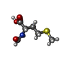
+Macromolecule #55: N-FORMYLMETHIONINE
-Experimental details
-Structure determination
| Method | cryo EM |
|---|---|
 Processing Processing | single particle reconstruction |
| Aggregation state | particle |
- Sample preparation
| Concentration | 10 mg/mL | |||||||||||||||
|---|---|---|---|---|---|---|---|---|---|---|---|---|---|---|---|---|
| Buffer | pH: 7.5 Component:
Details: 1x TAKM7 buffer | |||||||||||||||
| Grid | Model: Quantifoil R2/1 / Material: COPPER / Mesh: 200 / Support film - Material: CARBON / Support film - topology: HOLEY / Pretreatment - Type: GLOW DISCHARGE / Pretreatment - Time: 60 sec. / Pretreatment - Atmosphere: AIR | |||||||||||||||
| Vitrification | Cryogen name: ETHANE / Chamber humidity: 100 % / Chamber temperature: 277 K / Instrument: FEI VITROBOT MARK IV | |||||||||||||||
| Details | Initiation complex containing: 1x TAKM7 buffer 1 mM DTT (dithiothreitol) 1 mM GTP (guanosine 5'-triphosphate) 2.0 uM 70S ribosomes 3.0 uM IF1, IF2, IF3 mix 6.0 uM A-site AAA mRNA 3.0 uM fMet-tRNAfMet Mixed 1:1 with a ternary complex containing: 1x TAKM7 buffer 3 mM PEP (phosphoenolpyruvate) 1 mM GTP (guanosine 5'-triphosphate) 1 mM DTT (dithiothreitol) 1% PK (pyruvate kinase) 0.02 uM EF-Ts 1.5 uM EF-Tu 0.3 uM Lys-tRNALys Incubated on ice for 60 seconds Final concentration of the sample: 20 A260 (absorbance at 260 nm, 10 mm path), 10 A280 (absorbance at 280 nm, 10 mm path) |
- Electron microscopy
| Microscope | TFS KRIOS |
|---|---|
| Specialist optics | Energy filter - Name: GIF Bioquantum / Energy filter - Slit width: 20 eV |
| Image recording | Film or detector model: GATAN K3 BIOQUANTUM (6k x 4k) / Digitization - Dimensions - Width: 5760 pixel / Digitization - Dimensions - Height: 4092 pixel / Number grids imaged: 1 / Number real images: 8435 / Average exposure time: 1.82 sec. / Average electron dose: 40.0 e/Å2 |
| Electron beam | Acceleration voltage: 300 kV / Electron source:  FIELD EMISSION GUN FIELD EMISSION GUN |
| Electron optics | C2 aperture diameter: 50.0 µm / Calibrated magnification: 105000 / Illumination mode: FLOOD BEAM / Imaging mode: BRIGHT FIELD / Cs: 2.7 mm / Nominal defocus max: 2.1 µm / Nominal defocus min: 0.9 µm |
| Sample stage | Specimen holder model: FEI TITAN KRIOS AUTOGRID HOLDER / Cooling holder cryogen: NITROGEN |
| Experimental equipment |  Model: Titan Krios / Image courtesy: FEI Company |
+ Image processing
-Atomic model buiding 1
| Details | Following rigid body fitting we used online Namdiantor tool to perform initial flexible fitting: https://namdinator.au.dk/namdinator/ |
|---|---|
| Refinement | Space: REAL / Protocol: RIGID BODY FIT |
| Output model |  PDB-8bgh: |
-Atomic model buiding 2
| Refinement | Space: REAL / Protocol: FLEXIBLE FIT |
|---|---|
| Output model |  PDB-8bgh: |
 Movie
Movie Controller


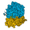


 Z (Sec.)
 Y (Row.)
 X (Col.)


