 Movie
Movie Controller

[English] 日本語
 Yorodumi
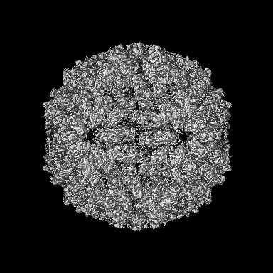
- EMDB-15189: Capsid structure of the L-A helper virus from native viral communities -
+ Open data

- Basic information
| Entry |  | ||||||||||||||||||
|---|---|---|---|---|---|---|---|---|---|---|---|---|---|---|---|---|---|---|---|
| Title | Capsid structure of the L-A helper virus from native viral communities | ||||||||||||||||||
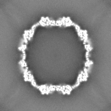
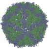
 Map data Map data | Icosahedral symmetrized map for the viral capsid of the Saccharomyces cerevisiae L-A helper virus | ||||||||||||||||||
 Sample Sample |
| ||||||||||||||||||
 Keywords Keywords | Capsid structure ScVLA / viral particle / wildtype / endogenous / VIRUS | ||||||||||||||||||
| Function / homology | Major coat protein, L-A virus / L-A virus major coat protein superfamily / L-A virus, major coat protein / viral capsid / viral translational frameshifting / Major capsid protein Function and homology information Function and homology information | ||||||||||||||||||
| Biological species |   Saccharomyces cerevisiae virus L-A Saccharomyces cerevisiae virus L-A | ||||||||||||||||||
| Method | single particle reconstruction / cryo EM / Resolution: 3.78 Å | ||||||||||||||||||
 Authors Authors | Schmidt L / Tueting C / Kyrilis F / Hamdi F / Semchonok DA / Kastritis PL | ||||||||||||||||||
| Funding support |  Germany, European Union, 5 items Germany, European Union, 5 items
| ||||||||||||||||||
 Citation Citation | Journal: Commun Biol / Year: 2024 Title: Delineating organizational principles of the endogenous L-A virus by cryo-EM and computational analysis of native cell extracts. Authors: Lisa Schmidt / Christian Tüting / Fotis L Kyrilis / Farzad Hamdi / Dmitry A Semchonok / Gerd Hause / Annette Meister / Christian Ihling / Milton T Stubbs / Andrea Sinz / Panagiotis L Kastritis /  Abstract: The high abundance of most viruses in infected host cells benefits their structural characterization. However, endogenous viruses are present in low copy numbers and are therefore challenging to ...The high abundance of most viruses in infected host cells benefits their structural characterization. However, endogenous viruses are present in low copy numbers and are therefore challenging to investigate. Here, we retrieve cell extracts enriched with an endogenous virus, the yeast L-A virus. The determined cryo-EM structure discloses capsid-stabilizing cation-π stacking, widespread across viruses and within the Totiviridae, and an interplay of non-covalent interactions from ten distinct capsomere interfaces. The capsid-embedded mRNA decapping active site trench is supported by a constricting movement of two flexible opposite-facing loops. tRNA-loaded polysomes and other biomacromolecules, presumably mRNA, are found in virus proximity within the cell extract. Mature viruses participate in larger viral communities resembling their rare in-cell equivalents in terms of size, composition, and inter-virus distances. Our results collectively describe a 3D-architecture of a viral milieu, opening the door to cell-extract-based high-resolution structural virology. #2: Journal: Biorxiv / Year: 2022Title: Delineating organizational principles of the endogenous L-A virus by cryo-EM and computational analysis of native cell extracts Authors: Schmidt L / Tuting C / Kyrilis FL / Hamdi F / Semchonok DA / Hause G / Meister A / Ihling C / Shah PNM / Stubbs MT / Sinz A / Stuart DI / Kastritis PL | ||||||||||||||||||
| History |
|
- Structure visualization
| Supplemental images |
|---|

- Downloads & links
-EMDB archive
| Map data | emd_15189.map.gz | 203.5 MB | EMDB map data format | |
|---|---|---|---|---|
| Header (meta data) | emd-15189-v30.xmlemd-15189.xml | 22.9 KB 22.9 KB | Display Display | EMDB header |
| FSC (resolution estimation) | emd_15189_fsc.xml | 13.3 KB | Display | FSC data file |
| Images | 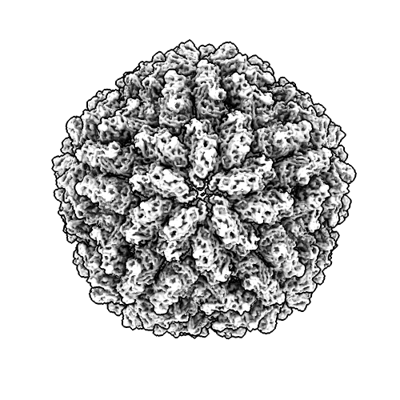 emd_15189.png emd_15189.png | 147.7 KB | ||
| Filedesc metadata | emd-15189.cif.gz | 6.8 KB | ||
| Others | emd_15189_half_map_1.map.gzemd_15189_half_map_2.map.gz | 199.7 MB 199.7 MB | ||
| Archive directory |  http://ftp.pdbj.org/pub/emdb/structures/EMD-15189ftp://ftp.pdbj.org/pub/emdb/structures/EMD-15189 http://ftp.pdbj.org/pub/emdb/structures/EMD-15189ftp://ftp.pdbj.org/pub/emdb/structures/EMD-15189 | HTTPS FTP |
-Related structure data
| Related structure data | 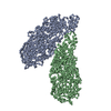 8a5tMC 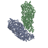 8pe4C M: atomic model generated by this map C: citing same article ( |
|---|---|
| Similar structure data |


-Links
| EMDB pages | EMDB (EBI/PDBe) / EMDataResource |
|---|
-Map
| File | Download / File: emd_15189.map.gz / Format: CCP4 / Size: 216 MB / Type: IMAGE STORED AS FLOATING POINT NUMBER (4 BYTES) | ||||||||||||||||||||||||||||||||||||
|---|---|---|---|---|---|---|---|---|---|---|---|---|---|---|---|---|---|---|---|---|---|---|---|---|---|---|---|---|---|---|---|---|---|---|---|---|---|
| Annotation | Icosahedral symmetrized map for the viral capsid of the Saccharomyces cerevisiae L-A helper virus | ||||||||||||||||||||||||||||||||||||
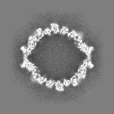
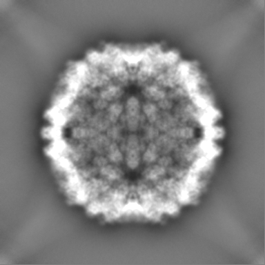
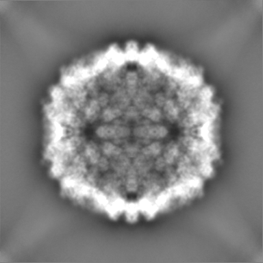
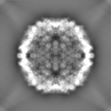
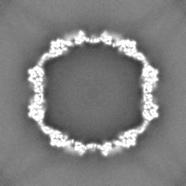
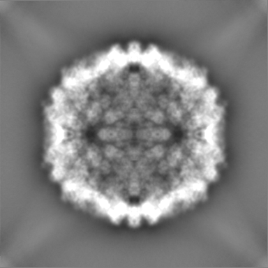
| Projections & slices | Image control
Images are generated by Spider. | ||||||||||||||||||||||||||||||||||||
| Voxel size | X=Y=Z: 1.5678 Å | ||||||||||||||||||||||||||||||||||||
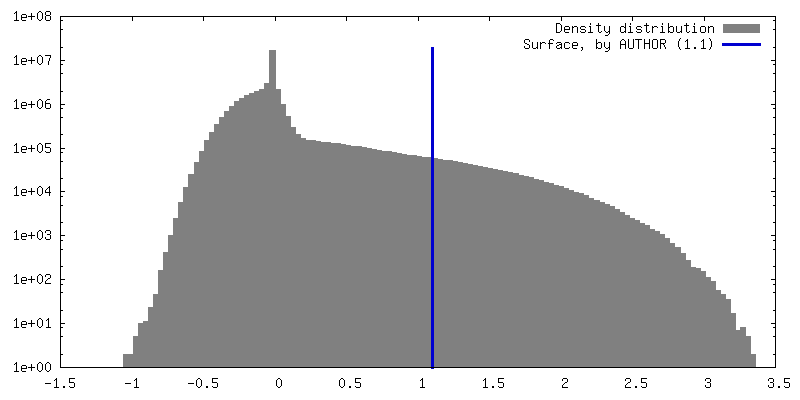
| Density |
| ||||||||||||||||||||||||||||||||||||
| Symmetry | Space group: 1 | ||||||||||||||||||||||||||||||||||||
| Details | EMDB XML:
|
 Z (Sec.)
 Y (Row.)
 X (Col.)


-Supplemental data
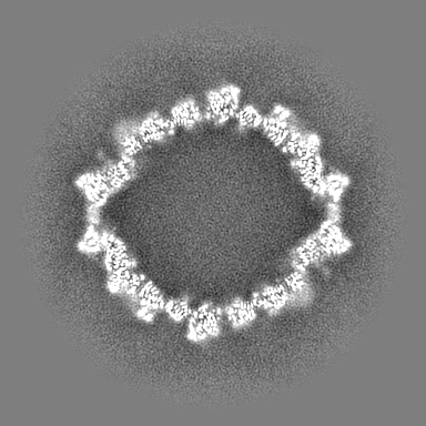
-Half map: Icosahedral symmetrized map for the viral capsid of...
| File | emd_15189_half_map_1.map | ||||||||||||
|---|---|---|---|---|---|---|---|---|---|---|---|---|---|
| Annotation | Icosahedral symmetrized map for the viral capsid of the Saccharomyces cerevisiae L-A helper virus - Half Map A | ||||||||||||
| Projections & Slices |
| ||||||||||||
| Density Histograms |


-Half map: Icosahedral symmetrized map for the viral capsid of...
| File | emd_15189_half_map_2.map | ||||||||||||
|---|---|---|---|---|---|---|---|---|---|---|---|---|---|
| Annotation | Icosahedral symmetrized map for the viral capsid of the Saccharomyces cerevisiae L-A helper virus - Half Map B | ||||||||||||
| Projections & Slices |
| ||||||||||||
| Density Histograms |


- Sample components
-Entire : Saccharomyces cerevisiae virus L-A
| Entire | Name: Saccharomyces cerevisiae virus L-A |
|---|---|
| Components |
|
-Supramolecule #1: Saccharomyces cerevisiae virus L-A
| Supramolecule | Name: Saccharomyces cerevisiae virus L-A / type: virus / ID: 1 / Parent: 0 / Macromolecule list: all / NCBI-ID: 11008 / Sci species name: Saccharomyces cerevisiae virus L-A / Virus type: VIRUS-LIKE PARTICLE / Virus isolate: SPECIES / Virus enveloped: No / Virus empty: No |
|---|---|
| Host (natural) | Organism: |
-Macromolecule #1: Major capsid protein
| Macromolecule | Name: Major capsid protein / type: protein_or_peptide / ID: 1 / Number of copies: 2 / Enantiomer: LEVO |
|---|---|
| Source (natural) | Organism: |
| Molecular weight | Theoretical: 76.070031 KDa |
| Sequence | String: MLRFVTKNSQ DKSSDLFSIC SDRGTFVAHN RVRTDFKFDN LVFNRVYGVS QKFTLVGNPT VCFNEGSSYL EGIAKKYLTL DGGLAIDNV LNELRSTCGI PGNAVASHAY NITSWRWYDN HVALLMNMLR AYHLQVLTEQ GQYSAGDIPM YHDGHVKIKL P VTIDDTAG ...String: MLRFVTKNSQ DKSSDLFSIC SDRGTFVAHN RVRTDFKFDN LVFNRVYGVS QKFTLVGNPT VCFNEGSSYL EGIAKKYLTL DGGLAIDNV LNELRSTCGI PGNAVASHAY NITSWRWYDN HVALLMNMLR AYHLQVLTEQ GQYSAGDIPM YHDGHVKIKL P VTIDDTAG PTQFAWPSDR STDSYPDWAQ FSESFPSIDV PYLDVRPLTV TEVNFVLMMM SKWHRRTNLA IDYEAPQLAD KF AYRHALT VQDADEWIEG DRTDDQFRPP SSKVMLSALR KYVNHNRLYN QFYTAAQLLA QIMMKPVPNC AEGYAWLMHD ALV NIPKFG SIRGRYPFLL SGDAALIQAT ALEDWSAIMA KPELVFTYAM QVSVALNTGL YLRRVKKTGF GTTIDDSYED GAFL QPETF VQAALACCTG QDAPLNGMSD VYVTYPDLLE FDAVTQVPIT VIEPAGYNIV DDHLVVVGVP VACSPYMIFP VAAFD TANP YCGNFVIKAA NKYLRKGAVY DKLEAWKLAW ALRVAGYDTH FKVYGDTHGL TKFYADNGDT WTHIPEFVTD GDVMEV FVT AIERRARHFV ELPRLNSPAF FRSVEVSTTI YDTHVQAGAH AVYHASRINL DYVKPVSTGI QVINAGELKN YWGSVRR TQ QGLGVVGLTM PAVMPTGEPT AGAAHEELIE QADNVLVE UniProtKB: Major capsid protein |
-Experimental details
-Structure determination
| Method | cryo EM |
|---|---|
 Processing Processing | single particle reconstruction |
| Aggregation state | particle |
-Sample preparation
| Concentration | 0.3 mg/mL |
|---|---|
| Buffer | pH: 7.4 / Component - Concentration: 200.0 mM / Component - Formula: CH3COONH4 / Component - Name: Ammoniumacetate Details: pH of the buffer was adjusted with NaOH buffer was filtered and sonicated |
| Grid | Model: Quantifoil R2/1 / Material: COPPER / Mesh: 200 / Support film - Material: CARBON / Support film - topology: HOLEY ARRAY / Pretreatment - Type: GLOW DISCHARGE / Pretreatment - Time: 25 sec. / Pretreatment - Atmosphere: AIR / Pretreatment - Pressure: 0.04 kPa |
| Vitrification | Cryogen name: ETHANE / Chamber humidity: 95 % / Chamber temperature: 277.15 K / Instrument: FEI VITROBOT MARK IV / Details: blot force 2 and blot time 6 s before plunging. |
| Details | heterogenous cell extract |
- Electron microscopy
| Microscope | TFS GLACIOS |
|---|---|
| Temperature | Min: 77.0 K / Max: 118.0 K |
| Image recording | Film or detector model: FEI FALCON III (4k x 4k) / Detector mode: INTEGRATING / Digitization - Dimensions - Width: 4096 pixel / Digitization - Dimensions - Height: 4096 pixel / Number grids imaged: 2 / Number real images: 7020 / Average exposure time: 3.61 sec. / Average electron dose: 30.0 e/Å2 |
| Electron beam | Acceleration voltage: 200 kV / Electron source:  FIELD EMISSION GUN FIELD EMISSION GUN |
| Electron optics | C2 aperture diameter: 70.0 µm / Calibrated magnification: 89297 / Illumination mode: OTHER / Imaging mode: BRIGHT FIELD / Cs: 2.7 mm / Nominal defocus max: 2.0 µm / Nominal defocus min: 1.0 µm / Nominal magnification: 92000 |
| Sample stage | Specimen holder model: FEI TITAN KRIOS AUTOGRID HOLDER / Cooling holder cryogen: NITROGEN |
+Image processing

-Atomic model buiding 1
| Initial model | PDB ID: Chain - Source name: PDB / Chain - Initial model type: experimental model |
|---|---|
| Details | The initial model was rigid-fitted by ChimeraX and refined by iterative cycles of Coot and PHENIX. |
| Refinement | Space: REAL / Protocol: RIGID BODY FIT |
| Output model | PDB-8a5t: |
