Movie
Movie Controller

[English] 日本語
 Yorodumi
- EMDB-15033: Cryo-EM structure of "BC react" conformation of Lactococcus lacti... -
+ Open data

- Basic information
| Entry |  | |||||||||
|---|---|---|---|---|---|---|---|---|---|---|
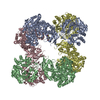
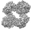
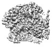
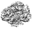
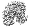
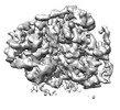
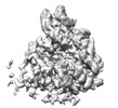
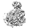
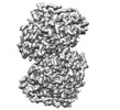
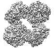
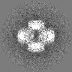
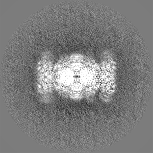
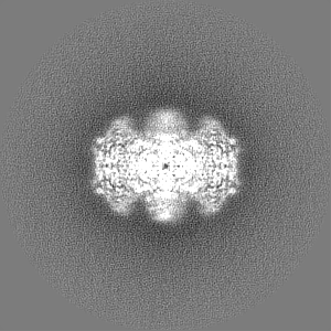
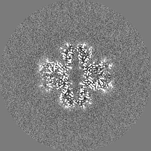
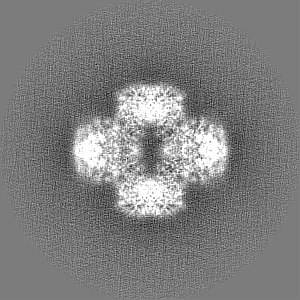
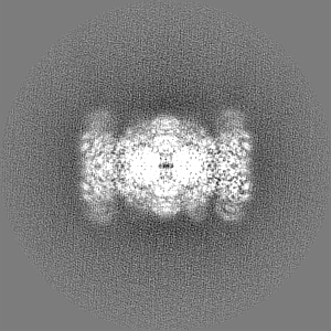
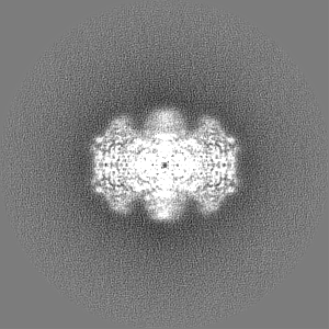
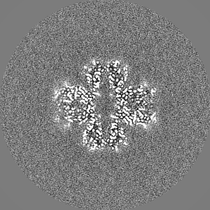
| Title | Cryo-EM structure of "BC react" conformation of Lactococcus lactis pyruvate carboxylase with acetyl-CoA | |||||||||
 Map data Map data | ||||||||||
 Sample Sample |
| |||||||||
 Keywords Keywords | Tetramer / carboxylase / biotin / LIGASE | |||||||||
| Function / homology |  Function and homology information Function and homology informationpyruvate carboxylase / pyruvate carboxylase activity / pyruvate metabolic process / gluconeogenesis / ATP binding / metal ion binding Similarity search - Function | |||||||||
| Biological species |  Lactococcus lactis (lactic acid bacteria) Lactococcus lactis (lactic acid bacteria) | |||||||||
| Method | single particle reconstruction / cryo EM / Resolution: 2.41 Å | |||||||||
 Authors Authors | Lopez-Alonso JP / Lazaro M / Gil D / Choi PH / Tong L / Valle M | |||||||||
| Funding support |  France, France,  Spain, 2 items Spain, 2 items
| |||||||||
 Citation Citation | Journal: Nat Commun / Year: 2022 Title: CryoEM structural exploration of catalytically active enzyme pyruvate carboxylase. Authors: Jorge Pedro López-Alonso / Melisa Lázaro / David Gil-Cartón / Philip H Choi / Alexandra Dodu / Liang Tong / Mikel Valle /  Abstract: Pyruvate carboxylase (PC) is a tetrameric enzyme that contains two active sites per subunit that catalyze two consecutive reactions. A mobile domain with an attached prosthetic biotin links both ...Pyruvate carboxylase (PC) is a tetrameric enzyme that contains two active sites per subunit that catalyze two consecutive reactions. A mobile domain with an attached prosthetic biotin links both reactions, an initial biotin carboxylation and the subsequent carboxyl transfer to pyruvate substrate to produce oxaloacetate. Reaction sites are at long distance, and there are several co-factors that play as allosteric regulators. Here, using cryoEM we explore the structure of active PC tetramers focusing on active sites and on the conformational space of the oligomers. The results capture the mobile domain at both active sites and expose catalytic steps of both reactions at high resolution, allowing the identification of substrates and products. The analysis of catalytically active PC tetramers reveals the role of certain motions during enzyme functioning, and the structural changes in the presence of additional cofactors expose the mechanism for allosteric regulation. | |||||||||
| History |
|
- Structure visualization
| Supplemental images |
|---|

- Downloads & links
-EMDB archive
| Map data | emd_15033.map.gz | 4.3 MB | EMDB map data format | |
|---|---|---|---|---|
| Header (meta data) | emd-15033-v30.xmlemd-15033.xml | 22.5 KB 22.5 KB | Display Display | EMDB header |
| FSC (resolution estimation) | emd_15033_fsc.xml | 10.6 KB | Display | FSC data file |
| Images | 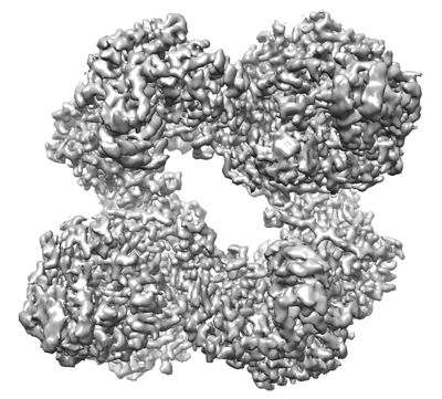 emd_15033.png emd_15033.png | 139.7 KB | ||
| Masks | emd_15033_msk_1.map | 103 MB | Mask map | |
| Filedesc metadata | emd-15033.cif.gz | 7.1 KB | ||
| Others | emd_15033_half_map_1.map.gzemd_15033_half_map_2.map.gz | 80.9 MB 81 MB | ||
| Archive directory |  http://ftp.pdbj.org/pub/emdb/structures/EMD-15033ftp://ftp.pdbj.org/pub/emdb/structures/EMD-15033 http://ftp.pdbj.org/pub/emdb/structures/EMD-15033ftp://ftp.pdbj.org/pub/emdb/structures/EMD-15033 | HTTPS FTP |
-Validation report
| Summary document | emd_15033_validation.pdf.gz | 165.7 KB | Display | EMDB validaton report |
|---|---|---|---|---|
| Full document | emd_15033_full_validation.pdf.gz | 165.3 KB | Display | |
| Data in XML | emd_15033_validation.xml.gz | 497 B | Display | |
| Data in CIF | emd_15033_validation.cif.gz | 374 B | Display | |
| Arichive directory | https://ftp.pdbj.org/pub/emdb/validation_reports/EMD-15033ftp://ftp.pdbj.org/pub/emdb/validation_reports/EMD-15033 | HTTPS FTP |
-Related structure data
| Related structure data | 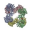 7zz3MC 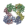 7zyyC 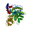 7zyzC 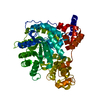 7zz0C 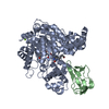 7zz1C 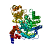 7zz2C 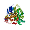 7zz4C 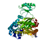 7zz5C 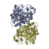 7zz6C  7zz8C C: citing same article ( M: atomic model generated by this map |
|---|---|
| Similar structure data |
-Links
| EMDB pages | EMDB (EBI/PDBe) / EMDataResource |
|---|---|
| Related items in Molecule of the Month |


-Map
| File | Download / File: emd_15033.map.gz / Format: CCP4 / Size: 103 MB / Type: IMAGE STORED AS FLOATING POINT NUMBER (4 BYTES) | ||||||||||||||||||||
|---|---|---|---|---|---|---|---|---|---|---|---|---|---|---|---|---|---|---|---|---|---|
| Voxel size | X=Y=Z: 1.06 Å | ||||||||||||||||||||
| Density |
| ||||||||||||||||||||
| Symmetry | Space group: 1 | ||||||||||||||||||||
| Details | EMDB XML:
|
-Supplemental data
-Mask #1
| File | emd_15033_msk_1.map | ||||||||||||
|---|---|---|---|---|---|---|---|---|---|---|---|---|---|
| Projections & Slices |
| ||||||||||||
| Density Histograms |
 Z
Z Y
Y X
X


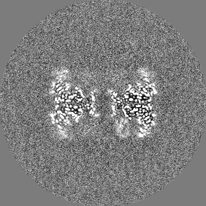
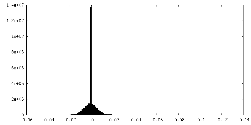
-Half map: #2
| File | emd_15033_half_map_1.map | ||||||||||||
|---|---|---|---|---|---|---|---|---|---|---|---|---|---|
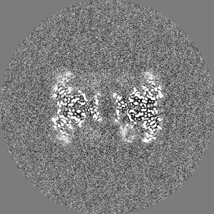
| Projections & Slices |
| ||||||||||||
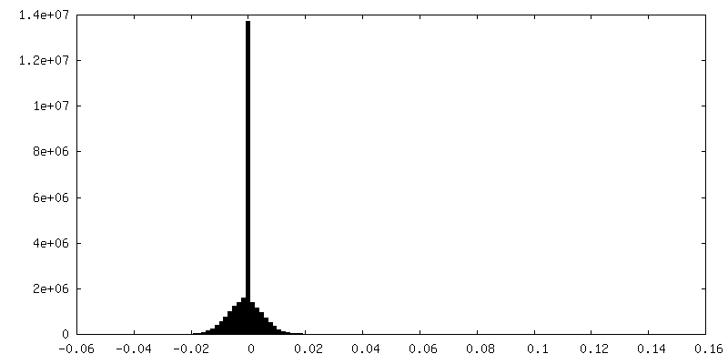
| Density Histograms |
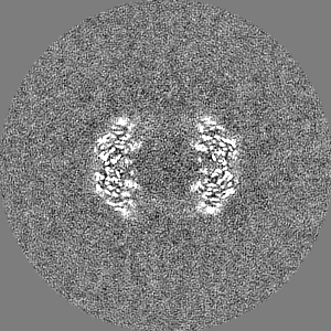
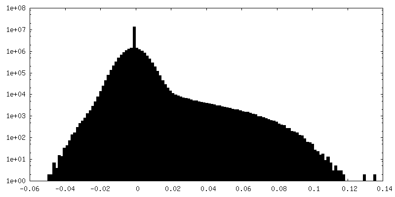
-Half map: #1
| File | emd_15033_half_map_2.map | ||||||||||||
|---|---|---|---|---|---|---|---|---|---|---|---|---|---|
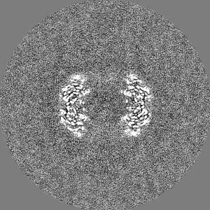
| Projections & Slices |
| ||||||||||||
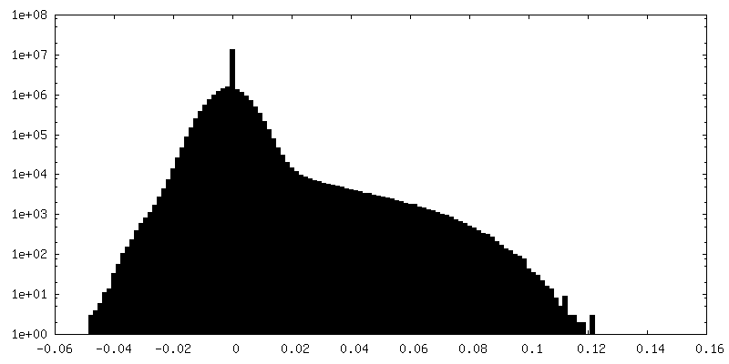
| Density Histograms |
- Sample components
+Entire : Pyruvate carboxylase with acetyl coenzyme A
+Supramolecule #1: Pyruvate carboxylase with acetyl coenzyme A
+Macromolecule #1: Pyruvate carboxylase
+Macromolecule #2: BIOTIN

+Macromolecule #3: MAGNESIUM ION
+Macromolecule #4: MANGANESE (II) ION
+Macromolecule #5: ADENOSINE-5'-TRIPHOSPHATE

+Macromolecule #6: ACETYL COENZYME *A

+Macromolecule #7: PYRUVIC ACID

+Macromolecule #8: BICARBONATE ION

+Macromolecule #9: ADENOSINE-5'-DIPHOSPHATE

+Macromolecule #10: water

-Experimental details
-Structure determination
| Method | cryo EM |
|---|---|
 Processing Processing | single particle reconstruction |
| Aggregation state | 3D array |
-Sample preparation
| Concentration | 0.05 mg/mL | |||||||||||||||||||||||||||
|---|---|---|---|---|---|---|---|---|---|---|---|---|---|---|---|---|---|---|---|---|---|---|---|---|---|---|---|---|
| Buffer | pH: 7.6 Component:
| |||||||||||||||||||||||||||
| Grid | Model: Quantifoil R2/1 / Material: COPPER / Mesh: 300 / Support film - Material: CARBON / Support film - topology: CONTINUOUS / Pretreatment - Type: GLOW DISCHARGE | |||||||||||||||||||||||||||
| Vitrification | Cryogen name: ETHANE |
- Electron microscopy
| Microscope | FEI TITAN KRIOS |
|---|---|
| Image recording | Film or detector model: GATAN K3 (6k x 4k) / Detector mode: COUNTING / Number grids imaged: 1 / Number real images: 10518 / Average exposure time: 3.99 sec. / Average electron dose: 48.0 e/Å2 |
| Electron beam | Acceleration voltage: 300 kV / Electron source:  FIELD EMISSION GUN FIELD EMISSION GUN |
| Electron optics | Illumination mode: FLOOD BEAM / Imaging mode: BRIGHT FIELD / Cs: 2.7 mm / Nominal defocus max: 2.4 µm / Nominal defocus min: 0.9 µm / Nominal magnification: 81000 |
| Experimental equipment |  Model: Titan Krios / Image courtesy: FEI Company |