ムービー
ムービー コントローラー


+ データを開く

- 基本情報
基本情報
| 登録情報 |  | |||||||||
|---|---|---|---|---|---|---|---|---|---|---|
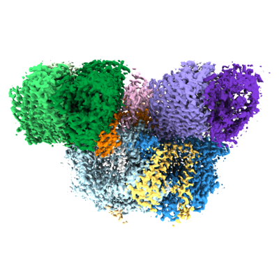
| タイトル | Cryo-EM structure of the whole photosynthetic complex from the green sulfur bacteria | |||||||||
 マップデータ マップデータ | ||||||||||
 試料 試料 |
| |||||||||
 キーワード キーワード | reaction centre / electron transport / energy transfer / green sulfur bacterium / membrane protein / light-harvesting protein complex / PHOTOSYNTHESIS | |||||||||
| 機能・相同性 |  機能・相同性情報 機能・相同性情報thylakoid / bacteriochlorophyll binding / iron-sulfur cluster binding / photosynthesis / electron transfer activity / heme binding / membrane / metal ion binding / plasma membrane 類似検索 - 分子機能 | |||||||||
| 生物種 |  Chlorobaculum tepidum TLS (バクテリア) Chlorobaculum tepidum TLS (バクテリア) | |||||||||
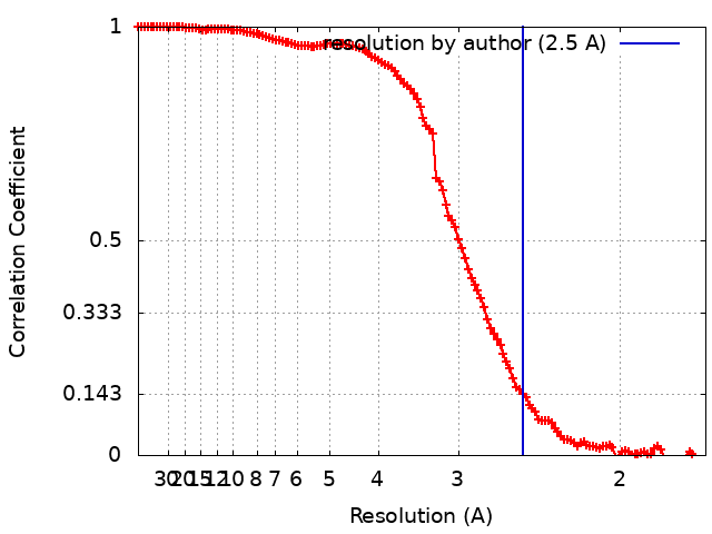
| 手法 | 単粒子再構成法 / クライオ電子顕微鏡法 / 解像度: 2.5 Å | |||||||||
 データ登録者 データ登録者 | Xie H / Tsiotis G | |||||||||
| 資金援助 |  ドイツ, 1件 ドイツ, 1件
| |||||||||
 引用 引用 | ジャーナル: Proc Natl Acad Sci U S A / 年: 2023 タイトル: Cryo-EM structure of the whole photosynthetic reaction center apparatus from the green sulfur bacterium . 著者: Hao Xie / Alexandros Lyratzakis / Radhika Khera / Myrto Koutantou / Sonja Welsch / Hartmut Michel / Georgios Tsiotis /  要旨: Light energy absorption and transfer are very important processes in photosynthesis. In green sulfur bacteria light is absorbed primarily by the chlorosomes and its energy is transferred via the ...Light energy absorption and transfer are very important processes in photosynthesis. In green sulfur bacteria light is absorbed primarily by the chlorosomes and its energy is transferred via the Fenna-Matthews-Olson (FMO) proteins to a homodimeric reaction center (RC). Here, we report the cryogenic electron microscopic structure of the intact FMO-RC apparatus from at 2.5 Å resolution. The FMO-RC apparatus presents an asymmetric architecture and contains two FMO trimers that show different interaction patterns with the RC core. Furthermore, the two permanently bound transmembrane subunits PscC, which donate electrons to the special pair, interact only with the two large PscA subunits. This structure fills an important gap in our understanding of the transfer of energy from antenna to the electron transport chain of this RC and the transfer of electrons from reduced sulfur compounds to the special pair. | |||||||||
| 履歴 |
|
- 構造の表示
構造の表示
| 添付画像 |
|---|
- ダウンロードとリンク
-EMDBアーカイブ
| マップデータ | emd_14528.map.gz | 168.1 MB | EMDBマップデータ形式 | |
|---|---|---|---|---|
| ヘッダ (付随情報) | emd-14528-v30.xmlemd-14528.xml | 28.8 KB 28.8 KB | 表示 表示 | EMDBヘッダ |
| FSC (解像度算出) | emd_14528_fsc.xml | 12.4 KB | 表示 | FSCデータファイル |
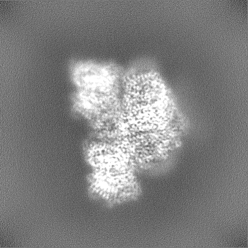
| 画像 |  emd_14528.png emd_14528.png | 143.3 KB | ||
| Filedesc metadata | emd-14528.cif.gz | 8 KB | ||
| その他 | emd_14528_half_map_1.map.gzemd_14528_half_map_2.map.gz | 165 MB 165 MB | ||
| アーカイブディレクトリ |  http://ftp.pdbj.org/pub/emdb/structures/EMD-14528ftp://ftp.pdbj.org/pub/emdb/structures/EMD-14528 http://ftp.pdbj.org/pub/emdb/structures/EMD-14528ftp://ftp.pdbj.org/pub/emdb/structures/EMD-14528 | HTTPS FTP |
-関連構造データ

-リンク
| EMDBのページ | EMDB (EBI/PDBe) / EMDataResource |
|---|---|
| 「今月の分子」の関連する項目 |


-マップ
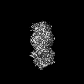
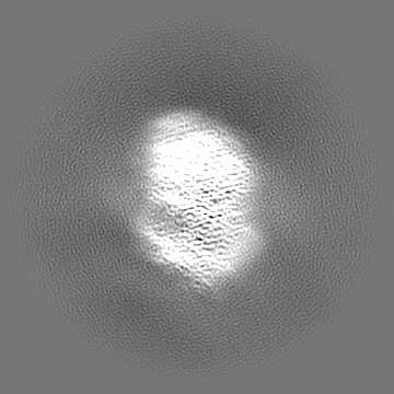
| ファイル | ダウンロード / ファイル: emd_14528.map.gz / 形式: CCP4 / 大きさ: 178 MB / タイプ: IMAGE STORED AS FLOATING POINT NUMBER (4 BYTES) | ||||||||||||||||||||||||||||||||||||
|---|---|---|---|---|---|---|---|---|---|---|---|---|---|---|---|---|---|---|---|---|---|---|---|---|---|---|---|---|---|---|---|---|---|---|---|---|---|
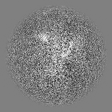
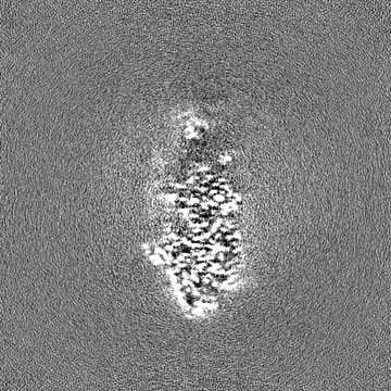
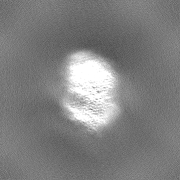
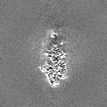
| 投影像・断面図 | 画像のコントロール
画像は Spider により作成 | ||||||||||||||||||||||||||||||||||||
| ボクセルのサイズ | X=Y=Z: 0.837 Å | ||||||||||||||||||||||||||||||||||||
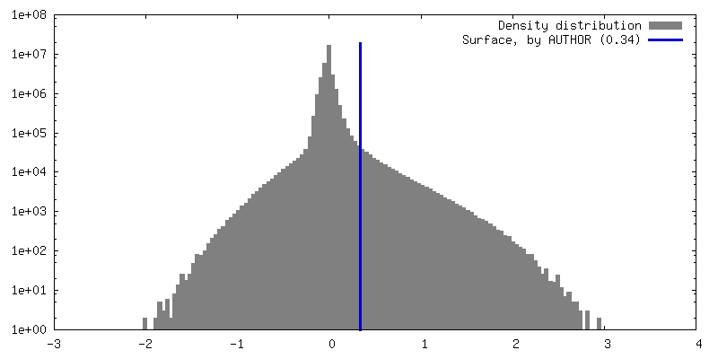
| 密度 |
| ||||||||||||||||||||||||||||||||||||
| 対称性 | 空間群: 1 | ||||||||||||||||||||||||||||||||||||
| 詳細 | EMDB XML:
|
 Z (Sec.)
 Y (Row.)
 X (Col.)


-添付データ
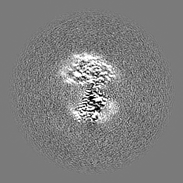
-ハーフマップ: #2
| ファイル | emd_14528_half_map_1.map | ||||||||||||
|---|---|---|---|---|---|---|---|---|---|---|---|---|---|
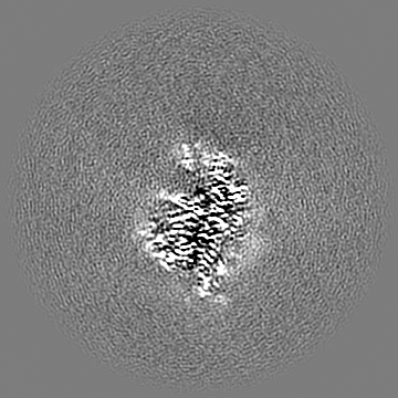
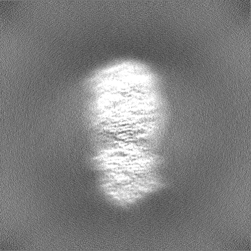
| 投影像・断面図 |
| ||||||||||||
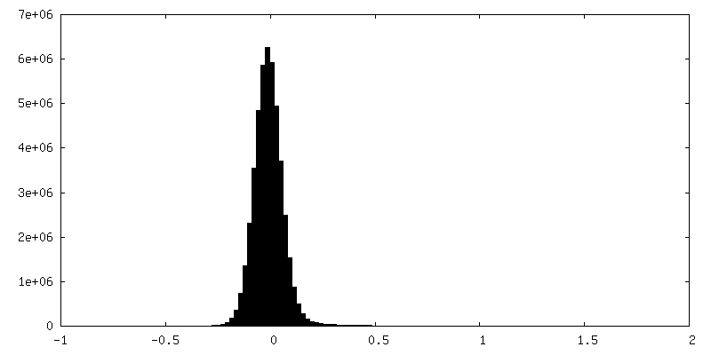
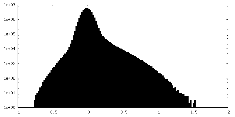
| 密度ヒストグラム |


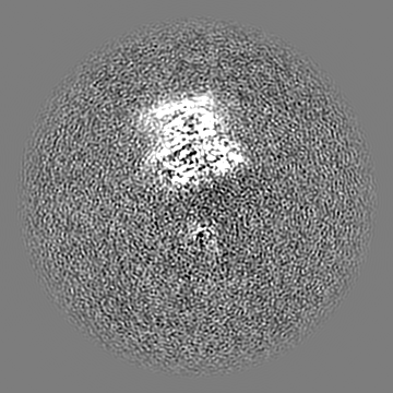
-ハーフマップ: #1
| ファイル | emd_14528_half_map_2.map | ||||||||||||
|---|---|---|---|---|---|---|---|---|---|---|---|---|---|
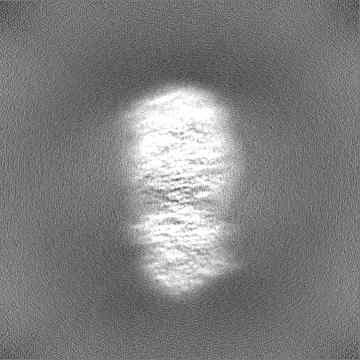
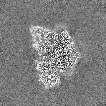
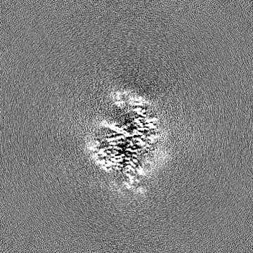
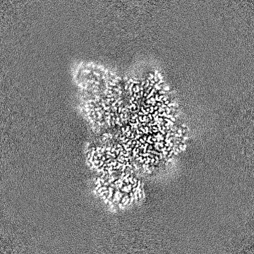
| 投影像・断面図 |
| ||||||||||||
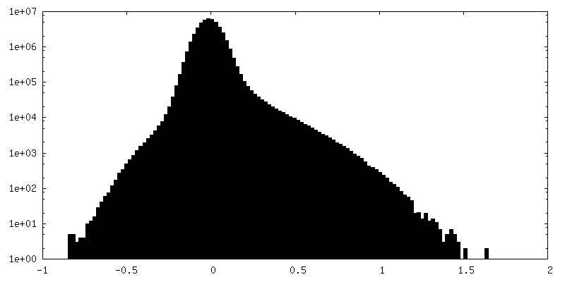
| 密度ヒストグラム |

- 試料の構成要素
+全体 : Photosystem P840 reaction center
+超分子 #1: Photosystem P840 reaction center
+分子 #1: Photosystem P840 reaction center, large subunit
+分子 #2: Photosystem P840 reaction center iron-sulfur protein
+分子 #3: Cytochrome c
+分子 #4: P840 reaction center 17 kDa protein
+分子 #5: Bacteriochlorophyll a protein
+分子 #6: Bacteriochlorophyll A isomer
+分子 #7: CHLOROPHYLL A

+分子 #8: BACTERIOCHLOROPHYLL A

+分子 #9: [(2R,3S,4S,5R,6R)-6-[(10E,12E,14E)-2,6,10,14,19,23-hexamethyl-25-...
+分子 #10: 1,2-DIPALMITOYL-PHOSPHATIDYL-GLYCEROLE

+分子 #11: [(2~{R})-2-hexadecanoyloxy-3-[(2~{S},3~{S},4~{R},5~{R},6~{S})-6-(...
+分子 #12: CALCIUM ION
+分子 #13: IRON/SULFUR CLUSTER

+分子 #14: water

-実験情報
-構造解析
| 手法 | クライオ電子顕微鏡法 |
|---|---|
 解析 解析 | 単粒子再構成法 |
| 試料の集合状態 | particle |
-試料調製
| 緩衝液 | pH: 8 |
|---|---|
| グリッド | モデル: Quantifoil / 材質: COPPER / メッシュ: 200 / 支持フィルム - 材質: CARBON / 支持フィルム - トポロジー: CONTINUOUS / 支持フィルム - Film thickness: 0.2 / 前処理 - タイプ: GLOW DISCHARGE / 前処理 - 時間: 90 sec. |
| 凍結 | 凍結剤: ETHANE / チャンバー内湿度: 100 % / チャンバー内温度: 4 K / 装置: FEI VITROBOT MARK IV |
- 電子顕微鏡法
| 顕微鏡 | FEI TITAN KRIOS |
|---|---|
| 撮影 | フィルム・検出器のモデル: GATAN K3 (6k x 4k) / デジタル化 - サイズ - 横: 4092 pixel / デジタル化 - サイズ - 縦: 5769 pixel / 平均電子線量: 45.0 e/Å2 |
| 電子線 | 加速電圧: 300 kV / 電子線源:  FIELD EMISSION GUN FIELD EMISSION GUN |
| 電子光学系 | 最大 デフォーカス(補正後): 2.5 µm / 最小 デフォーカス(補正後): 1.2 µm / 照射モード: FLOOD BEAM / 撮影モード: BRIGHT FIELD / Cs: 2.7 mm / 最大 デフォーカス(公称値): 2.5 µm / 最小 デフォーカス(公称値): 1.2 µm / 倍率(公称値): 105000 |
| 試料ステージ | 試料ホルダーモデル: FEI TITAN KRIOS AUTOGRID HOLDER ホルダー冷却材: NITROGEN |
| 実験機器 |  モデル: Titan Krios / 画像提供: FEI Company |