 Movie
Movie Controller


+ Open data

- Basic information
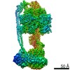
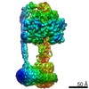
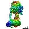
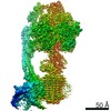
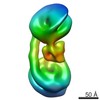
| Entry | Database: EMDB / ID: EMD-13186 | |||||||||
|---|---|---|---|---|---|---|---|---|---|---|
| Title | F1Fo-ATP synthase from Acinetobacter baumannii (state 3) | |||||||||
 Map data Map data | Cryo-em map of ATP synthase from A. baumannii (state 3). | |||||||||
 Sample Sample |
| |||||||||
 Keywords Keywords | ATP synthase / ESKAPE / Rotary ATP synthase / F1Fo / peptidisc / bioenergetics / IMP / multi-drug resistance / pathogenic / MEMBRANE PROTEIN | |||||||||
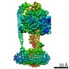
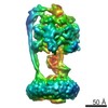
| Function / homology |  Function and homology information Function and homology informationproton motive force-driven plasma membrane ATP synthesis / proton-transporting two-sector ATPase complex, proton-transporting domain / proton-transporting ATPase activity, rotational mechanism / H+-transporting two-sector ATPase / proton-transporting ATP synthase complex / proton-transporting ATP synthase activity, rotational mechanism / ADP binding / lipid binding / ATP binding / plasma membrane Similarity search - Function | |||||||||
| Biological species |  Acinetobacter baumannii ATCC 17978 (bacteria) / Acinetobacter baumannii (strain ATCC 17978 / CIP 53.77 / LMG 1025 / NCDC KC755 / 5377) (bacteria) Acinetobacter baumannii ATCC 17978 (bacteria) / Acinetobacter baumannii (strain ATCC 17978 / CIP 53.77 / LMG 1025 / NCDC KC755 / 5377) (bacteria) | |||||||||
| Method | single particle reconstruction / cryo EM / Resolution: 4.3 Å | |||||||||
 Authors Authors | Demmer JK / Phillips BP | |||||||||
| Funding support |  United Kingdom, 1 items United Kingdom, 1 items
| |||||||||
 Citation Citation | Journal: Sci Adv / Year: 2022 Title: Structure of ATP synthase from ESKAPE pathogen . Authors: Julius K Demmer / Ben P Phillips / O Lisa Uhrig / Alain Filloux / Luke P Allsopp / Maike Bublitz / Thomas Meier / Abstract: The global spread of multidrug-resistant infections urgently calls for the identification of novel drug targets. We solved the electron cryo-microscopy structure of the FF-adenosine 5'-triphosphate ...The global spread of multidrug-resistant infections urgently calls for the identification of novel drug targets. We solved the electron cryo-microscopy structure of the FF-adenosine 5'-triphosphate (ATP) synthase from in three distinct conformational states. The nucleotide-converting F subcomplex reveals a specific self-inhibition mechanism, which supports a unidirectional ratchet mechanism to avoid wasteful ATP consumption. In the membrane-embedded F complex, the structure shows unique structural adaptations along both the entry and exit pathways of the proton-conducting a-subunit. These features, absent in mitochondrial ATP synthases, represent attractive targets for the development of next-generation therapeutics that can act directly at the culmination of bioenergetics in this clinically relevant pathogen. | |||||||||
| History |
|
- Structure visualization
| Movie |
Movie viewer |
|---|---|
| Structure viewer | EM map: SurfViewMolmilJmol/JSmol |
| Supplemental images |

- Downloads & links
-EMDB archive
| Map data | emd_13186.map.gz | 172.7 MB | EMDB map data format | |
|---|---|---|---|---|
| Header (meta data) | emd-13186-v30.xmlemd-13186.xml | 24.3 KB 24.3 KB | Display Display | EMDB header |
| Images | 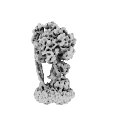 emd_13186.png emd_13186.png | 46.4 KB | ||
| Filedesc metadata | emd-13186.cif.gz | 7.6 KB | ||
| Archive directory |  http://ftp.pdbj.org/pub/emdb/structures/EMD-13186ftp://ftp.pdbj.org/pub/emdb/structures/EMD-13186 http://ftp.pdbj.org/pub/emdb/structures/EMD-13186ftp://ftp.pdbj.org/pub/emdb/structures/EMD-13186 | HTTPS FTP |
-Related structure data
| Related structure data | 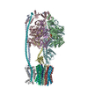 7p3wMC 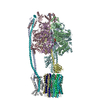 7p2yC 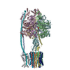 7p3nC M: atomic model generated by this map C: citing same article ( |
|---|---|
| Similar structure data |

-Links
| EMDB pages | EMDB (EBI/PDBe) / EMDataResource |
|---|---|
| Related items in Molecule of the Month |


-Map
| File | Download / File: emd_13186.map.gz / Format: CCP4 / Size: 347.6 MB / Type: IMAGE STORED AS FLOATING POINT NUMBER (4 BYTES) | ||||||||||||||||||||||||||||||||||||||||||||||||||||||||||||||||||||
|---|---|---|---|---|---|---|---|---|---|---|---|---|---|---|---|---|---|---|---|---|---|---|---|---|---|---|---|---|---|---|---|---|---|---|---|---|---|---|---|---|---|---|---|---|---|---|---|---|---|---|---|---|---|---|---|---|---|---|---|---|---|---|---|---|---|---|---|---|---|
| Annotation | Cryo-em map of ATP synthase from A. baumannii (state 3). | ||||||||||||||||||||||||||||||||||||||||||||||||||||||||||||||||||||
| Projections & slices | Image control
Images are generated by Spider. | ||||||||||||||||||||||||||||||||||||||||||||||||||||||||||||||||||||
| Voxel size | X=Y=Z: 0.85 Å | ||||||||||||||||||||||||||||||||||||||||||||||||||||||||||||||||||||
| Density |
| ||||||||||||||||||||||||||||||||||||||||||||||||||||||||||||||||||||
| Symmetry | Space group: 1 | ||||||||||||||||||||||||||||||||||||||||||||||||||||||||||||||||||||
| Details | EMDB XML:
CCP4 map header:
| ||||||||||||||||||||||||||||||||||||||||||||||||||||||||||||||||||||
 Z (Sec.)
 Y (Row.)
 X (Col.)


-Supplemental data
- Sample components
+Entire : F1Fo ATP synthase
+Supramolecule #1: F1Fo ATP synthase
+Macromolecule #1: ATP synthase subunit alpha
+Macromolecule #2: ATP synthase subunit beta
+Macromolecule #3: ATP synthase subunit c
+Macromolecule #4: ATP synthase subunit a
+Macromolecule #5: ATP synthase subunit b
+Macromolecule #6: ATP synthase subunit delta
+Macromolecule #7: ATP synthase epsilon chain
+Macromolecule #8: ATP synthase gamma chain
+Macromolecule #9: ADENOSINE-5'-TRIPHOSPHATE

+Macromolecule #10: MAGNESIUM ION
+Macromolecule #11: ADENOSINE-5'-DIPHOSPHATE

-Experimental details
-Structure determination
| Method | cryo EM |
|---|---|
 Processing Processing | single particle reconstruction |
| Aggregation state | particle |
-Sample preparation
| Concentration | 0.05 mg/mL | ||||||||||||
|---|---|---|---|---|---|---|---|---|---|---|---|---|---|
| Buffer | pH: 6.8 Component:
| ||||||||||||
| Grid | Model: PELCO Ultrathin Carbon with Lacey Carbon / Material: COPPER / Mesh: 200 / Support film - Material: CARBON / Support film - topology: LACEY / Support film - Film thickness: 300 / Pretreatment - Type: GLOW DISCHARGE / Pretreatment - Time: 30 sec. / Pretreatment - Atmosphere: OTHER | ||||||||||||
| Vitrification | Cryogen name: ETHANE / Chamber humidity: 100 % / Chamber temperature: 281 K / Instrument: FEI VITROBOT MARK IV / Details: 4ul sample, blotted for 4s at a blotforce of -4.. | ||||||||||||
| Details | Sample was applied directly from gel filtration to ultra-thin carbon in peptidiscs. |
- Electron microscopy
| Microscope | FEI TITAN KRIOS |
|---|---|
| Specialist optics | Energy filter - Slit width: 20 eV |
| Details | Data collected at Diamond Light Source (Harwell, UK) using Titan Krios G3 and automated alignments using Sherpa. Detector used was Gatan K3 including energy filter. |
| Image recording | Film or detector model: GATAN K3 (6k x 4k) / Digitization - Dimensions - Width: 5760 pixel / Digitization - Dimensions - Height: 4092 pixel / Number grids imaged: 1 / Number real images: 11490 / Average electron dose: 60.0 e/Å2 |
| Electron beam | Acceleration voltage: 300 kV / Electron source:  FIELD EMISSION GUN FIELD EMISSION GUN |
| Electron optics | C2 aperture diameter: 100.0 µm / Illumination mode: FLOOD BEAM / Imaging mode: BRIGHT FIELD / Cs: 2.7 mm / Nominal defocus max: 2.5 µm / Nominal defocus min: 1.25 µm / Nominal magnification: 85000 |
| Sample stage | Specimen holder model: FEI TITAN KRIOS AUTOGRID HOLDER / Cooling holder cryogen: NITROGEN |
| Experimental equipment |  Model: Titan Krios / Image courtesy: FEI Company |
