Movie
Movie Controller


+ Open data

- Basic information
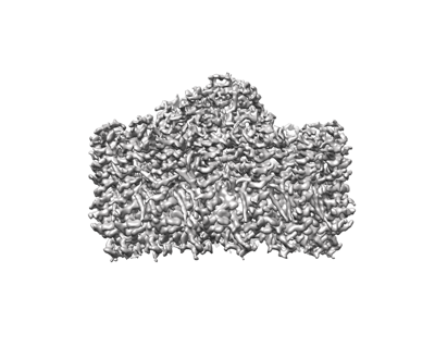
| Entry | Database: EMDB / ID: EMD-13110 | ||||||||||||
|---|---|---|---|---|---|---|---|---|---|---|---|---|---|
| Title | Cryo-EM structure of the Rhodospirillum rubrum RC-LH1 complex | ||||||||||||
 Map data Map data | 3d map | ||||||||||||
 Sample Sample |
| ||||||||||||
 Keywords Keywords | photosynthesis / light-harvesting complex / reaction centre / purple bacteria / membrane protein / STRUCTURAL PROTEIN | ||||||||||||
| Function / homology |  Function and homology information Function and homology informationorganelle inner membrane / plasma membrane-derived chromatophore membrane / plasma membrane light-harvesting complex / bacteriochlorophyll binding / photosynthetic electron transport in photosystem II / photosynthesis, light reaction / endomembrane system / metal ion binding / plasma membrane Similarity search - Function | ||||||||||||
| Biological species |  Rhodospirillum rubrum (strain ATCC 11170 / ATH 1.1.1 / DSM 467 / LMG 4362 / NCIMB 8255 / S1) (bacteria) Rhodospirillum rubrum (strain ATCC 11170 / ATH 1.1.1 / DSM 467 / LMG 4362 / NCIMB 8255 / S1) (bacteria) | ||||||||||||
| Method | single particle reconstruction / cryo EM / Resolution: 2.5 Å | ||||||||||||
 Authors Authors | Qian P / Croll TI | ||||||||||||
| Funding support |  United Kingdom, European Union, 3 items United Kingdom, European Union, 3 items
| ||||||||||||
 Citation Citation | Journal: Biochem J / Year: 2021 Title: Cryo-EM structure of the Rhodospirillum rubrum RC-LH1 complex at 2.5 Å. Authors: Pu Qian / Tristan I Croll / David J K Swainsbury / Pablo Castro-Hartmann / Nigel W Moriarty / Kasim Sader / C Neil Hunter /   Abstract: The reaction centre light-harvesting 1 (RC-LH1) complex is the core functional component of bacterial photosynthesis. We determined the cryo-electron microscopy (cryo-EM) structure of the RC-LH1 ...The reaction centre light-harvesting 1 (RC-LH1) complex is the core functional component of bacterial photosynthesis. We determined the cryo-electron microscopy (cryo-EM) structure of the RC-LH1 complex from Rhodospirillum rubrum at 2.5 Å resolution, which reveals a unique monomeric bacteriochlorophyll with a phospholipid ligand in the gap between the RC and LH1 complexes. The LH1 complex comprises a circular array of 16 αβ-polypeptide subunits that completely surrounds the RC, with a preferential binding site for a quinone, designated QP, on the inner face of the encircling LH1 complex. Quinols, initially generated at the RC QB site, are proposed to transiently occupy the QP site prior to traversing the LH1 barrier and diffusing to the cytochrome bc1 complex. Thus, the QP site, which is analogous to other such sites in recent cryo-EM structures of RC-LH1 complexes, likely reflects a general mechanism for exporting quinols from the RC-LH1 complex. | ||||||||||||
| History |
|
- Structure visualization
| Movie |
Movie viewer |
|---|---|
| Structure viewer | EM map: SurfViewMolmilJmol/JSmol |
| Supplemental images |
- Downloads & links
-EMDB archive
| Map data | emd_13110.map.gz | 2.5 MB | EMDB map data format | |
|---|---|---|---|---|
| Header (meta data) | emd-13110-v30.xmlemd-13110.xml | 25 KB 25 KB | Display Display | EMDB header |
| FSC (resolution estimation) | emd_13110_fsc.xml | 18.2 KB | Display | FSC data file |
| Images |  emd_13110.png emd_13110.png | 83.3 KB | ||
| Filedesc metadata | emd-13110.cif.gz | 7.7 KB | ||
| Archive directory |  http://ftp.pdbj.org/pub/emdb/structures/EMD-13110ftp://ftp.pdbj.org/pub/emdb/structures/EMD-13110 http://ftp.pdbj.org/pub/emdb/structures/EMD-13110ftp://ftp.pdbj.org/pub/emdb/structures/EMD-13110 | HTTPS FTP |
-Related structure data
| Related structure data |  7oy8MC M: atomic model generated by this map C: citing same article ( |
|---|---|
| Similar structure data |


-Links
| EMDB pages | EMDB (EBI/PDBe) / EMDataResource |
|---|---|
| Related items in Molecule of the Month |


-Map
| File | Download / File: emd_13110.map.gz / Format: CCP4 / Size: 64 MB / Type: IMAGE STORED AS FLOATING POINT NUMBER (4 BYTES) | ||||||||||||||||||||||||||||||||||||||||||||||||||||||||||||
|---|---|---|---|---|---|---|---|---|---|---|---|---|---|---|---|---|---|---|---|---|---|---|---|---|---|---|---|---|---|---|---|---|---|---|---|---|---|---|---|---|---|---|---|---|---|---|---|---|---|---|---|---|---|---|---|---|---|---|---|---|---|
| Annotation | 3d map | ||||||||||||||||||||||||||||||||||||||||||||||||||||||||||||
| Projections & slices | Image control
Images are generated by Spider. | ||||||||||||||||||||||||||||||||||||||||||||||||||||||||||||
| Voxel size | X=Y=Z: 1.3 Å | ||||||||||||||||||||||||||||||||||||||||||||||||||||||||||||
| Density |
| ||||||||||||||||||||||||||||||||||||||||||||||||||||||||||||
| Symmetry | Space group: 1 | ||||||||||||||||||||||||||||||||||||||||||||||||||||||||||||
| Details | EMDB XML:
CCP4 map header:
| ||||||||||||||||||||||||||||||||||||||||||||||||||||||||||||
 Z (Sec.)
 Y (Row.)
 X (Col.)


-Supplemental data
- Sample components
+Entire : RC-LH1 from rhodospirillum rubrum
+Supramolecule #1: RC-LH1 from rhodospirillum rubrum
+Macromolecule #1: Light-harvesting protein B-870 beta chain
+Macromolecule #2: Antenna complex, alpha/beta subunit
+Macromolecule #3: Photosynthetic reaction center, H-chain
+Macromolecule #4: Photosynthetic reaction center L subunit
+Macromolecule #5: Reaction center protein M chain
+Macromolecule #6: Antenna complex, alpha/beta subunit
+Macromolecule #7: Trans-Geranyl BACTERIOCHLOROPHYLL A

+Macromolecule #8: SPIRILLOXANTHIN

+Macromolecule #9: (1R)-2-{[(S)-{[(2S)-2,3-dihydroxypropyl]oxy}(hydroxy)phosphoryl]o...
+Macromolecule #10: (2R,5R,11R,14R)-5,8,11-trihydroxy-5,11-dioxido-17-oxo-2,14-bis(te...

+Macromolecule #11: BACTERIOPHEOPHYTIN A

+Macromolecule #12: 2-azanyl-5-[(2~{E},6~{E},8~{E},10~{E},12~{E},14~{E},18~{E},22~{E}...
+Macromolecule #13: UBIQUINONE-10

+Macromolecule #14: FE (III) ION
+Macromolecule #15: CHLORIDE ION
+Macromolecule #16: PHOSPHATE ION

+Macromolecule #17: DODECYL-BETA-D-MALTOSIDE

+Macromolecule #18: water

-Experimental details
-Structure determination
| Method | cryo EM |
|---|---|
 Processing Processing | single particle reconstruction |
| Aggregation state | particle |
-Sample preparation
| Concentration | 4.0 mg/mL |
|---|---|
| Buffer | pH: 8 / Component - Concentration: 20.0 mM / Component - Formula: C8H18N2O4S / Component - Name: HEPES / Details: 20 mM HEPES, 0.03% beta DDM, pH 8.0 |
| Grid | Model: Quantifoil / Material: COPPER / Mesh: 400 / Pretreatment - Type: GLOW DISCHARGE / Pretreatment - Time: 60 sec. / Pretreatment - Atmosphere: AIR |
| Vitrification | Cryogen name: ETHANE / Chamber humidity: 100 % / Chamber temperature: 277 K / Instrument: FEI VITROBOT MARK IV Details: Quantifiol R1.2/1.3 grid, glow discharged. bloting time: 2.5, bloting force 3, waiting time 0. |
| Details | The protein were solubilised using detergent beta DDM, and purified by the use of ion exchange column and size exclusion column. Monodisperse sample was used for cry-EM preparation. |
- Electron microscopy
| Microscope | FEI TITAN KRIOS |
|---|---|
| Image recording | Film or detector model: FEI FALCON IV (4k x 4k) / Number grids imaged: 1 / Number real images: 9024 / Average exposure time: 12.12 sec. / Average electron dose: 45.0 e/Å2 Details: total dose of 45 was fractionated to 42 frames within 12.12 second. |
| Electron beam | Acceleration voltage: 300 kV / Electron source:  FIELD EMISSION GUN FIELD EMISSION GUN |
| Electron optics | Illumination mode: FLOOD BEAM / Imaging mode: BRIGHT FIELD / Cs: 2.7 mm / Nominal magnification: 120000 |
| Sample stage | Specimen holder model: FEI TITAN KRIOS AUTOGRID HOLDER / Cooling holder cryogen: NITROGEN |
| Experimental equipment |  Model: Titan Krios / Image courtesy: FEI Company |