 Movie
Movie Controller


+ Open data

- Basic information
| Entry | Database: EMDB / ID: EMD-11955 | |||||||||
|---|---|---|---|---|---|---|---|---|---|---|
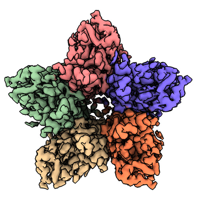
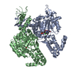
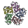
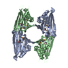
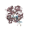
| Title | Cryo-EM structure of the green-light absorbing proteorhodopsin | |||||||||
 Map data Map data | ||||||||||
 Sample Sample |
| |||||||||
 Keywords Keywords | Light-driven proton pump / microbial rhodopsin / retinal / PROTON TRANSPORT | |||||||||
| Function / homology |  Function and homology information Function and homology informationlight-activated monoatomic ion channel activity / photoreceptor activity / phototransduction / plasma membrane Similarity search - Function | |||||||||
| Biological species |  uncultured Gammaproteobacteria bacterium (environmental samples) uncultured Gammaproteobacteria bacterium (environmental samples) | |||||||||
| Method | single particle reconstruction / cryo EM / Resolution: 2.93 Å | |||||||||
 Authors Authors | Hirschi S / Kalbermatter D | |||||||||
| Funding support |  Switzerland, 1 items Switzerland, 1 items
| |||||||||
 Citation Citation | Journal: Nat Commun / Year: 2021 Title: Cryo-EM structure and dynamics of the green-light absorbing proteorhodopsin. Authors: Stephan Hirschi / David Kalbermatter / Zöhre Ucurum / Thomas Lemmin / Dimitrios Fotiadis / Abstract: The green-light absorbing proteorhodopsin (GPR) is the archetype of bacterial light-driven proton pumps. Here, we present the 2.9 Å cryo-EM structure of pentameric GPR, resolving important ...The green-light absorbing proteorhodopsin (GPR) is the archetype of bacterial light-driven proton pumps. Here, we present the 2.9 Å cryo-EM structure of pentameric GPR, resolving important residues of the proton translocation pathway and the oligomerization interface. Superposition with the structure of a close GPR homolog and molecular dynamics simulations reveal conformational variations, which regulate the solvent access to the intra- and extracellular half channels harbouring the primary proton donor E109 and the proposed proton release group E143. We provide a mechanism for the structural rearrangements allowing hydration of the intracellular half channel, which are triggered by changing the protonation state of E109. Functional characterization of selected mutants demonstrates the importance of the molecular organization around E109 and E143 for GPR activity. Furthermore, we present evidence that helices involved in the stabilization of the protomer interfaces serve as scaffolds for facilitating the motion of the other helices. Combined with the more constrained dynamics of the pentamer compared to the monomer, these observations illustrate the previously demonstrated functional significance of GPR oligomerization. Overall, this work provides molecular insights into the structure, dynamics and function of the proteorhodopsin family that will benefit the large scientific community employing GPR as a model protein. | |||||||||
| History |
|
- Structure visualization
| Movie |
Movie viewer |
|---|---|
| Structure viewer | EM map: SurfViewMolmilJmol/JSmol |
| Supplemental images |
- Downloads & links
-EMDB archive
| Map data | emd_11955.map.gz | 1.1 MB | EMDB map data format | |
|---|---|---|---|---|
| Header (meta data) | emd-11955-v30.xmlemd-11955.xml | 10.5 KB 10.5 KB | Display Display | EMDB header |
| Images |  emd_11955.png emd_11955.png | 198.3 KB | ||
| Filedesc metadata | emd-11955.cif.gz | 5.1 KB | ||
| Archive directory |  http://ftp.pdbj.org/pub/emdb/structures/EMD-11955ftp://ftp.pdbj.org/pub/emdb/structures/EMD-11955 http://ftp.pdbj.org/pub/emdb/structures/EMD-11955ftp://ftp.pdbj.org/pub/emdb/structures/EMD-11955 | HTTPS FTP |
-Related structure data
| Related structure data |  7b03MC M: atomic model generated by this map C: citing same article ( |
|---|---|
| Similar structure data |
-Links
| EMDB pages | EMDB (EBI/PDBe) / EMDataResource |
|---|---|
| Related items in Molecule of the Month |


-Map
| File | Download / File: emd_11955.map.gz / Format: CCP4 / Size: 1.2 MB / Type: IMAGE STORED AS FLOATING POINT NUMBER (4 BYTES) | ||||||||||||||||||||||||||||||||||||||||||||||||||||||||||||||||||||
|---|---|---|---|---|---|---|---|---|---|---|---|---|---|---|---|---|---|---|---|---|---|---|---|---|---|---|---|---|---|---|---|---|---|---|---|---|---|---|---|---|---|---|---|---|---|---|---|---|---|---|---|---|---|---|---|---|---|---|---|---|---|---|---|---|---|---|---|---|---|
| Projections & slices | Image control
Images are generated by Spider. generated in cubic-lattice coordinate | ||||||||||||||||||||||||||||||||||||||||||||||||||||||||||||||||||||
| Voxel size | X=Y=Z: 1.29 Å | ||||||||||||||||||||||||||||||||||||||||||||||||||||||||||||||||||||
| Density |
| ||||||||||||||||||||||||||||||||||||||||||||||||||||||||||||||||||||
| Symmetry | Space group: 1 | ||||||||||||||||||||||||||||||||||||||||||||||||||||||||||||||||||||
| Details | EMDB XML:
CCP4 map header:
| ||||||||||||||||||||||||||||||||||||||||||||||||||||||||||||||||||||
 X (Sec.)
 Y (Row.)
 Z (Col.)


-Supplemental data
- Sample components
-Entire : Pentamer of the green-light absorbing proteorhodopsin
| Entire | Name: Pentamer of the green-light absorbing proteorhodopsin |
|---|---|
| Components |
|
-Supramolecule #1: Pentamer of the green-light absorbing proteorhodopsin
| Supramolecule | Name: Pentamer of the green-light absorbing proteorhodopsin / type: complex / ID: 1 / Parent: 0 / Macromolecule list: #1 |
|---|---|
| Source (natural) | Organism: uncultured Gammaproteobacteria bacterium (environmental samples) |
-Macromolecule #1: Proteorhodopsin
| Macromolecule | Name: Proteorhodopsin / type: protein_or_peptide / ID: 1 / Details: Expressed without signal sequence. / Number of copies: 5 / Enantiomer: LEVO |
|---|---|
| Source (natural) | Organism: uncultured Gammaproteobacteria bacterium (environmental samples) |
| Molecular weight | Theoretical: 25.611842 KDa |
| Recombinant expression | Organism: |
| Sequence | String: MAGGGDLDAS DYTGVSFWLV TAALLASTVF FFVERDRVSA KWKTSLTVSG LVTGIAFWHY MYMRGVWIET GDSPTVFRYI DWLLTVPLL ICEFYLILAA ATNVAGSLFK KLLVGSLVML VFGYMGEAGI MAAWPAFIIG CLAWVYMIYE LWAGEGKSAC N TASPAVQS ...String: MAGGGDLDAS DYTGVSFWLV TAALLASTVF FFVERDRVSA KWKTSLTVSG LVTGIAFWHY MYMRGVWIET GDSPTVFRYI DWLLTVPLL ICEFYLILAA ATNVAGSLFK KLLVGSLVML VFGYMGEAGI MAAWPAFIIG CLAWVYMIYE LWAGEGKSAC N TASPAVQS AYNTMMYIII FGWAIYPVGY FTGYLMGDGG SALNLNLIYN LADFVNKILF GLIIWNVAVK ESSNA UniProtKB: Green-light absorbing proteorhodopsin |
-Macromolecule #2: RETINAL
| Macromolecule | Name: RETINAL / type: ligand / ID: 2 / Number of copies: 5 / Formula: RET |
|---|---|
| Molecular weight | Theoretical: 284.436 Da |
| Chemical component information |  ChemComp-RET: |
-Experimental details
-Structure determination
| Method | cryo EM |
|---|---|
 Processing Processing | single particle reconstruction |
| Aggregation state | particle |
-Sample preparation
| Concentration | 3.5 mg/mL |
|---|---|
| Buffer | pH: 7.5 |
| Grid | Model: Quantifoil R1.2/1.3 / Material: COPPER / Pretreatment - Type: GLOW DISCHARGE / Pretreatment - Time: 30 sec. / Pretreatment - Atmosphere: AIR / Pretreatment - Pressure: 0.025 kPa |
| Vitrification | Cryogen name: ETHANE / Chamber humidity: 100 % / Chamber temperature: 277 K / Instrument: FEI VITROBOT MARK IV |
- Electron microscopy
| Microscope | FEI TITAN KRIOS |
|---|---|
| Image recording | Film or detector model: GATAN K3 (6k x 4k) / Average electron dose: 1.36 e/Å2 |
| Electron beam | Acceleration voltage: 300 kV / Electron source:  FIELD EMISSION GUN FIELD EMISSION GUN |
| Electron optics | Illumination mode: SPOT SCAN / Imaging mode: BRIGHT FIELD |
| Experimental equipment |  Model: Titan Krios / Image courtesy: FEI Company |
-Image processing
| Startup model | Type of model: NONE |
|---|---|
| Final reconstruction | Applied symmetry - Point group: C5 (5 fold cyclic) / Resolution.type: BY AUTHOR / Resolution: 2.93 Å / Resolution method: FSC 0.143 CUT-OFF / Software - Name: cryoSPARC Details: Final map was obtained after applying density modification using Resolve Cryo-EM in Phenix. Number images used: 717107 |
| Initial angle assignment | Type: ANGULAR RECONSTITUTION / Software - Name: RELION (ver. 3.1) |
| Final angle assignment | Type: ANGULAR RECONSTITUTION / Software - Name: cryoSPARC |
