ムービー
ムービー コントローラー


+ データを開く

- 基本情報
基本情報
| 登録情報 | データベース: EMDB / ID: EMD-11437 | |||||||||
|---|---|---|---|---|---|---|---|---|---|---|
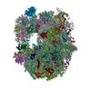
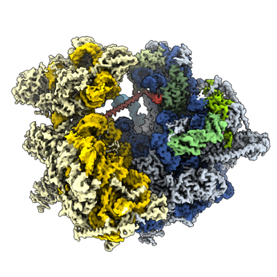
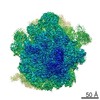
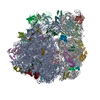
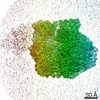
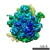
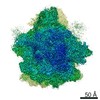
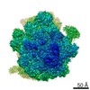
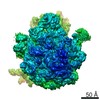
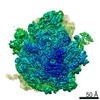
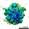
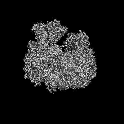
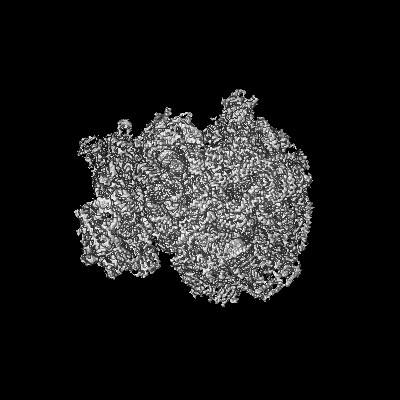
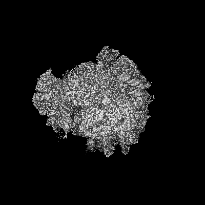
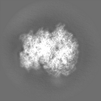
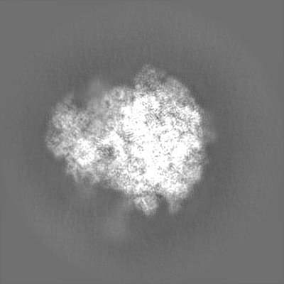
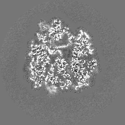
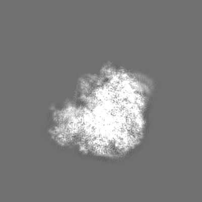
| タイトル | Structure of the Paranosema locustae ribosome in complex with Lso2 | |||||||||
 マップデータ マップデータ | Combined focused maps, state 2 | |||||||||
 試料 試料 |
| |||||||||
 キーワード キーワード | Microsporidia / Pathogen / Ribosome / Hibernation / Genome Compaction | |||||||||
| 生物種 |  Paranosema locustae (菌類) Paranosema locustae (菌類) | |||||||||
| 手法 | 単粒子再構成法 / クライオ電子顕微鏡法 / 解像度: 2.9 Å | |||||||||
 データ登録者 データ登録者 | Ehrenbolger K / Jespersen N / Sharma H / Sokolova YY / Tokarev YS / Vossbrinck CR / Barandun J | |||||||||
| 資金援助 |  スウェーデン, 1件 スウェーデン, 1件
| |||||||||
 引用 引用 | ジャーナル: PLoS Biol / 年: 2020 タイトル: Differences in structure and hibernation mechanism highlight diversification of the microsporidian ribosome. 著者: Kai Ehrenbolger / Nathan Jespersen / Himanshu Sharma / Yuliya Y Sokolova / Yuri S Tokarev / Charles R Vossbrinck / Jonas Barandun /   要旨: Assembling and powering ribosomes are energy-intensive processes requiring fine-tuned cellular control mechanisms. In organisms operating under strict nutrient limitations, such as pathogenic ...Assembling and powering ribosomes are energy-intensive processes requiring fine-tuned cellular control mechanisms. In organisms operating under strict nutrient limitations, such as pathogenic microsporidia, conservation of energy via ribosomal hibernation and recycling is critical. The mechanisms by which hibernation is achieved in microsporidia, however, remain poorly understood. Here, we present the cryo-electron microscopy structure of the ribosome from Paranosema locustae spores, bound by the conserved eukaryotic hibernation and recycling factor Lso2. The microsporidian Lso2 homolog adopts a V-shaped conformation to bridge the mRNA decoding site and the large subunit tRNA binding sites, providing a reversible ribosome inactivation mechanism. Although microsporidian ribosomes are highly compacted, the P. locustae ribosome retains several rRNA segments absent in other microsporidia, and represents an intermediate state of rRNA reduction. In one case, the near complete reduction of an expansion segment has resulted in a single bound nucleotide, which may act as an architectural co-factor to stabilize a protein-protein interface. The presented structure highlights the reductive evolution in these emerging pathogens and sheds light on a conserved mechanism for eukaryotic ribosome hibernation. | |||||||||
| 履歴 |
|
- 構造の表示
構造の表示
| ムービー |
 ムービービューア ムービービューア |
|---|---|
| 構造ビューア | EMマップ: SurfViewMolmilJmol/JSmol |
| 添付画像 |
- ダウンロードとリンク
-EMDBアーカイブ
| マップデータ | emd_11437.map.gz | 218 MB | EMDBマップデータ形式 | |
|---|---|---|---|---|
| ヘッダ (付随情報) | emd-11437-v30.xmlemd-11437.xml | 91.9 KB 91.9 KB | 表示 表示 | EMDBヘッダ |
| FSC (解像度算出) | emd_11437_fsc.xml | 18.2 KB | 表示 | FSCデータファイル |
| 画像 |  emd_11437.png emd_11437.png | 190 KB | ||
| Filedesc metadata | emd-11437.cif.gz | 17.5 KB | ||
| その他 | emd_11437_additional_1.map.gzemd_11437_additional_2.map.gzemd_11437_additional_3.map.gzemd_11437_half_map_1.map.gzemd_11437_half_map_2.map.gz | 4.7 MB 16.7 MB 8.2 MB 217.8 MB 217.8 MB | ||
| アーカイブディレクトリ |  http://ftp.pdbj.org/pub/emdb/structures/EMD-11437ftp://ftp.pdbj.org/pub/emdb/structures/EMD-11437 http://ftp.pdbj.org/pub/emdb/structures/EMD-11437ftp://ftp.pdbj.org/pub/emdb/structures/EMD-11437 | HTTPS FTP |
-関連構造データ
| 関連構造データ |  6zu5MC M: このマップから作成された原子モデル C: 同じ文献を引用 ( |
|---|---|
| 類似構造データ | |
| 電子顕微鏡画像生データ | EMPIAR-11076 (タイトル: Single particle cryo-EM dataset of the Paranosema locustae ribosome bound to Lso2 Data size: 2.0 TB Data #1: Unaligned multi-frame micrographs of the Paranosema locustae Lso2-ribosome complex - 20 frames [micrographs - multiframe] Data #2: Unaligned multi-frame micrographs of the Paranosema locustae Lso2-ribosome complex - 40 frames [micrographs - multiframe]) |

-リンク
| EMDBのページ | EMDB (EBI/PDBe) / EMDataResource |
|---|---|
| 「今月の分子」の関連する項目 |

-マップ
| ファイル | ダウンロード / ファイル: emd_11437.map.gz / 形式: CCP4 / 大きさ: 244.1 MB / タイプ: IMAGE STORED AS FLOATING POINT NUMBER (4 BYTES) | ||||||||||||||||||||||||||||||||||||||||||||||||||||||||||||||||||||
|---|---|---|---|---|---|---|---|---|---|---|---|---|---|---|---|---|---|---|---|---|---|---|---|---|---|---|---|---|---|---|---|---|---|---|---|---|---|---|---|---|---|---|---|---|---|---|---|---|---|---|---|---|---|---|---|---|---|---|---|---|---|---|---|---|---|---|---|---|---|
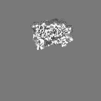
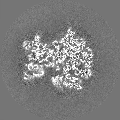
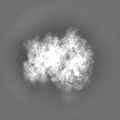
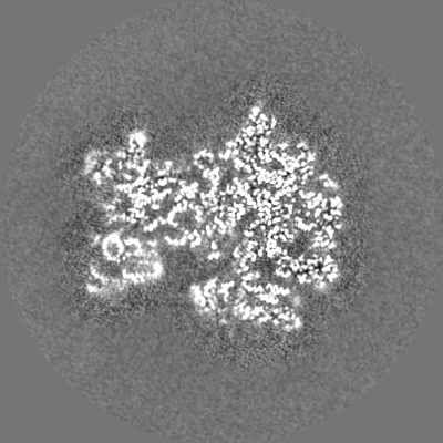
| 注釈 | Combined focused maps, state 2 | ||||||||||||||||||||||||||||||||||||||||||||||||||||||||||||||||||||
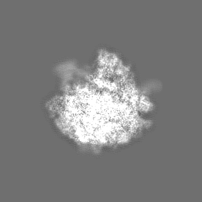
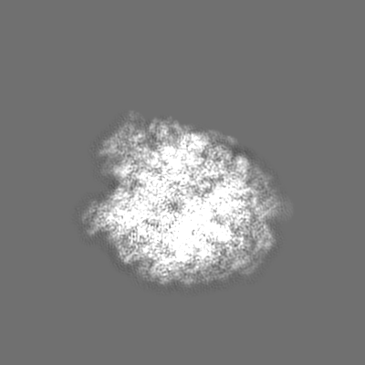
| 投影像・断面図 | 画像のコントロール
画像は Spider により作成 | ||||||||||||||||||||||||||||||||||||||||||||||||||||||||||||||||||||
| ボクセルのサイズ | X=Y=Z: 1.04 Å | ||||||||||||||||||||||||||||||||||||||||||||||||||||||||||||||||||||
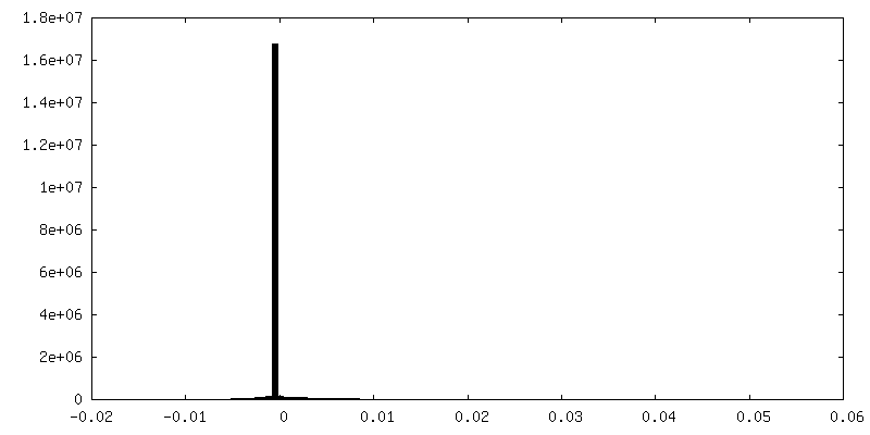
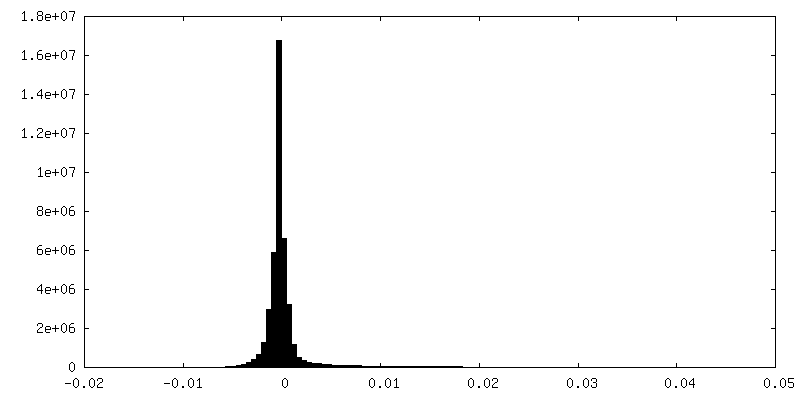
| 密度 |
| ||||||||||||||||||||||||||||||||||||||||||||||||||||||||||||||||||||
| 対称性 | 空間群: 1 | ||||||||||||||||||||||||||||||||||||||||||||||||||||||||||||||||||||
| 詳細 | EMDB XML:
CCP4マップ ヘッダ情報:
| ||||||||||||||||||||||||||||||||||||||||||||||||||||||||||||||||||||
 X (Sec.)
 Y (Row.)
 Z (Col.)


-添付データ
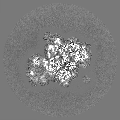
-追加マップ: Additional map 3 (SSU head focused)
| ファイル | emd_11437_additional_1.map | ||||||||||||
|---|---|---|---|---|---|---|---|---|---|---|---|---|---|
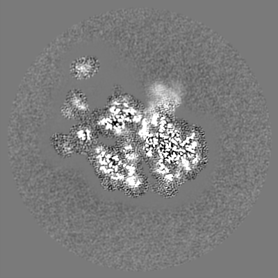
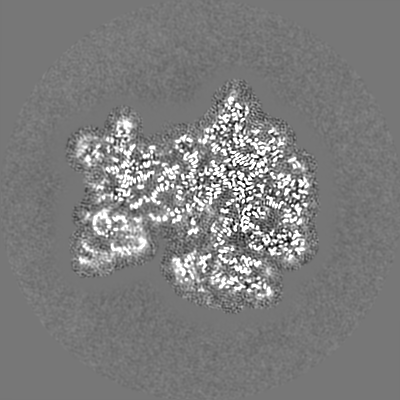
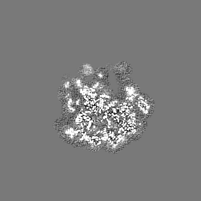
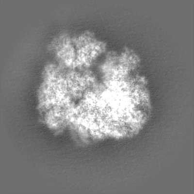
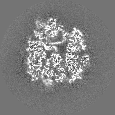
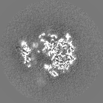
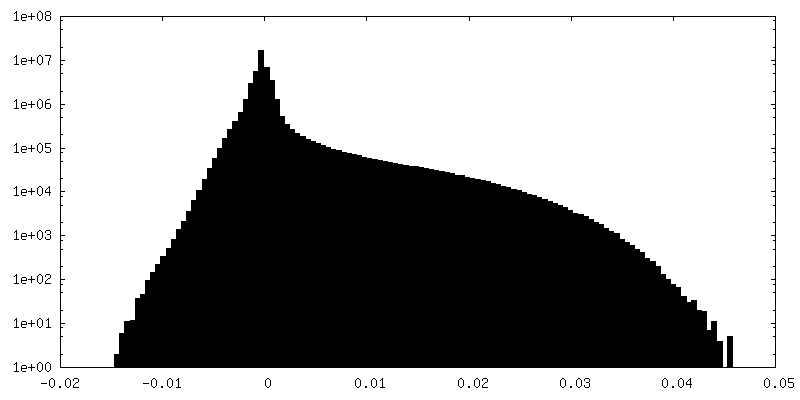
| 注釈 | Additional map 3 (SSU head focused) | ||||||||||||
| 投影像・断面図 |
| ||||||||||||
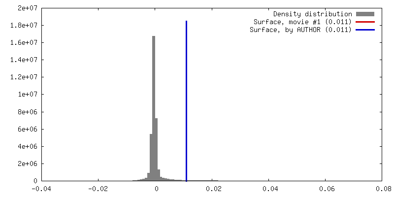
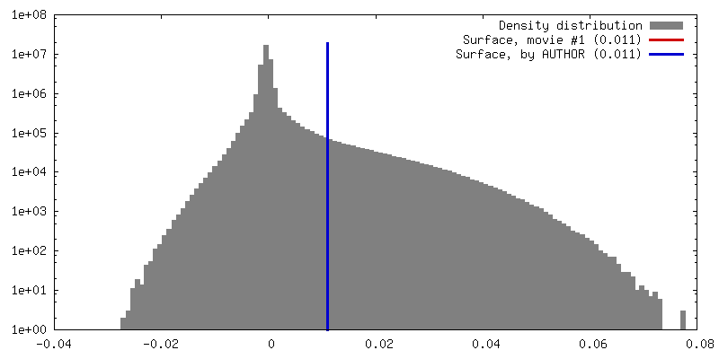
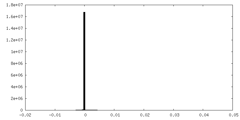
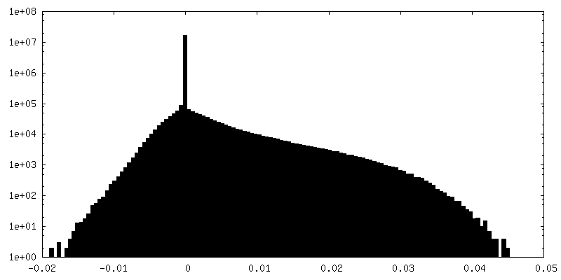
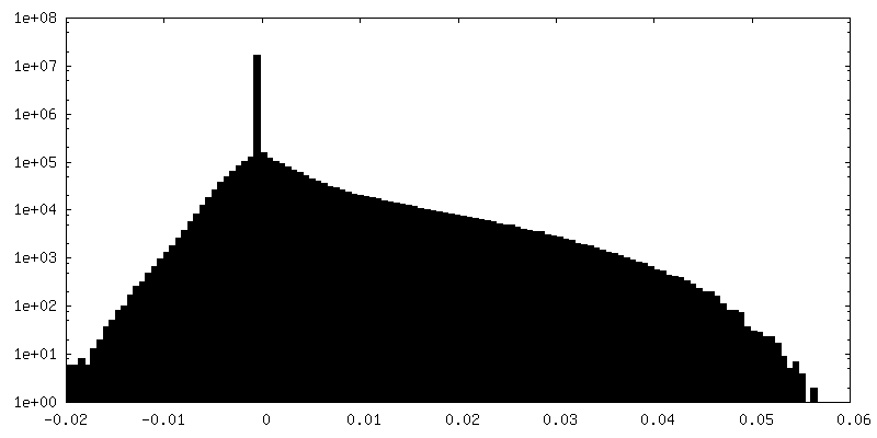
| 密度ヒストグラム |


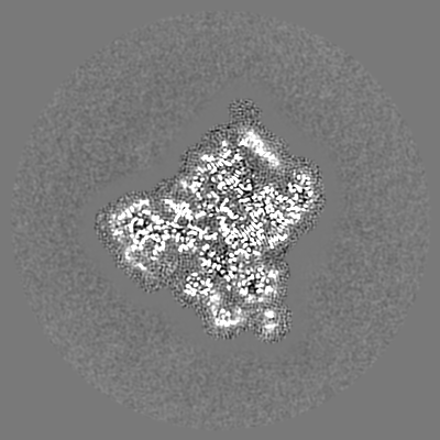
-追加マップ: Additional map 1 (LSU focused)
| ファイル | emd_11437_additional_2.map | ||||||||||||
|---|---|---|---|---|---|---|---|---|---|---|---|---|---|
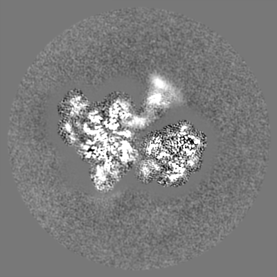
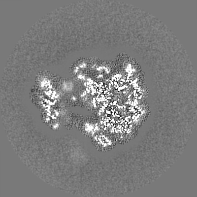
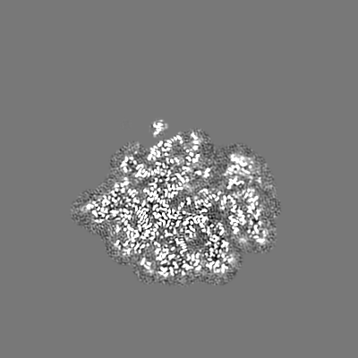
| 注釈 | Additional map 1 (LSU focused) | ||||||||||||
| 投影像・断面図 |
| ||||||||||||
| 密度ヒストグラム |


-追加マップ: Additional map 2 (SSU body focused)
| ファイル | emd_11437_additional_3.map | ||||||||||||
|---|---|---|---|---|---|---|---|---|---|---|---|---|---|
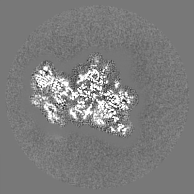
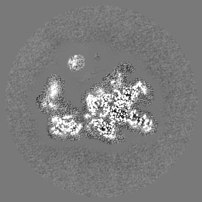
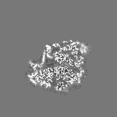
| 注釈 | Additional map 2 (SSU body focused) | ||||||||||||
| 投影像・断面図 |
| ||||||||||||
| 密度ヒストグラム |

-ハーフマップ: Combined focused half map 1, state 2
| ファイル | emd_11437_half_map_1.map | ||||||||||||
|---|---|---|---|---|---|---|---|---|---|---|---|---|---|
| 注釈 | Combined focused half map 1, state 2 | ||||||||||||
| 投影像・断面図 |
| ||||||||||||
| 密度ヒストグラム |


-ハーフマップ: Combined focused half map 2, state 2
| ファイル | emd_11437_half_map_2.map | ||||||||||||
|---|---|---|---|---|---|---|---|---|---|---|---|---|---|
| 注釈 | Combined focused half map 2, state 2 | ||||||||||||
| 投影像・断面図 |
| ||||||||||||
| 密度ヒストグラム |


- 試料の構成要素
+全体 : Lso2 bound to the ribosome of Paranosema locustae
+超分子 #1: Lso2 bound to the ribosome of Paranosema locustae
+分子 #1: 25S rRNA
+分子 #2: 5S rRNA
+分子 #43: 18S rRNA
+分子 #3: uL2
+分子 #4: uL15
+分子 #5: uL3
+分子 #6: eL29
+分子 #7: uL4
+分子 #8: eL30
+分子 #9: uL18
+分子 #10: eL31
+分子 #11: eL6
+分子 #12: eL32
+分子 #13: uL30
+分子 #14: eL33
+分子 #15: eL8
+分子 #16: eL34
+分子 #17: uL6
+分子 #18: uL29
+分子 #19: uL16
+分子 #20: eL36
+分子 #21: uL5
+分子 #22: eL37
+分子 #23: eL13
+分子 #24: eL39
+分子 #25: eL14
+分子 #26: eL40
+分子 #27: eL15
+分子 #28: Lso2
+分子 #29: uL13
+分子 #30: eL42
+分子 #31: uL22
+分子 #32: eL43
+分子 #33: eL18
+分子 #34: eL19
+分子 #35: eL20
+分子 #36: eL21
+分子 #37: eL22
+分子 #38: uL14
+分子 #39: eL24
+分子 #40: uL23
+分子 #41: uL24
+分子 #42: eL27
+分子 #44: uS2
+分子 #45: eS26
+分子 #46: eS1
+分子 #47: eS27
+分子 #48: uS5
+分子 #49: eS28
+分子 #50: uS3
+分子 #51: uS14
+分子 #52: eS4
+分子 #53: eS30
+分子 #54: uS7
+分子 #55: eS6
+分子 #56: RACK1
+分子 #57: eS7
+分子 #58: eS8
+分子 #59: uS4
+分子 #60: eS10
+分子 #61: uS17
+分子 #62: uS15
+分子 #63: uS11
+分子 #64: uS19
+分子 #65: uS9
+分子 #66: eS17
+分子 #67: uS13
+分子 #68: eS19
+分子 #69: uS10
+分子 #70: eS21
+分子 #71: uS8
+分子 #72: uS12
+分子 #73: eS24
+分子 #74: eS25
+分子 #75: MAGNESIUM ION
+分子 #76: ZINC ION
+分子 #77: ADENOSINE MONOPHOSPHATE

-実験情報
-構造解析
| 手法 | クライオ電子顕微鏡法 |
|---|---|
 解析 解析 | 単粒子再構成法 |
| 試料の集合状態 | particle |
-試料調製
| 緩衝液 | pH: 7.5 詳細: 30 mM Tris-HCl, pH 7.5, 25 mM KCl, 5 mM magnesium acetate, 1 mM DTT, 1 mM EDTA |
|---|---|
| グリッド | モデル: Quantifoil R1.2/1.3 / 材質: COPPER / メッシュ: 300 / 前処理 - タイプ: GLOW DISCHARGE / 前処理 - 時間: 30 sec. |
| 凍結 | 凍結剤: ETHANE / チャンバー内湿度: 100 % / チャンバー内温度: 298 K / 装置: FEI VITROBOT MARK I |
- 電子顕微鏡法
| 顕微鏡 | FEI TITAN KRIOS |
|---|---|
| 撮影 | フィルム・検出器のモデル: GATAN K2 QUANTUM (4k x 4k) 撮影したグリッド数: 1 / 平均電子線量: 28.6 e/Å2 |
| 電子線 | 加速電圧: 300 kV / 電子線源:  FIELD EMISSION GUN FIELD EMISSION GUN |
| 電子光学系 | 照射モード: OTHER / 撮影モード: BRIGHT FIELD / Cs: 2.7 mm |
| 試料ステージ | 試料ホルダーモデル: FEI TITAN KRIOS AUTOGRID HOLDER ホルダー冷却材: NITROGEN |
| 実験機器 |  モデル: Titan Krios / 画像提供: FEI Company |