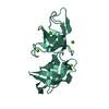
Protein or peptide: Rod shape-determining protein MreC
Keywords
bacterial cell wall elongation / STRUCTURAL PROTEIN
Function / homology
Cell/Rod shape-determining protein MreC, domain 1 / Rod shape-determining protein MreC / Cell/Rod shape-determining protein MreC, domain 2 / rod shape-determining protein MreC / regulation of cell shape / Cell shape-determining protein MreC
Function and homology information
Biological species
Pseudomonas aeruginosa (bacteria)
Method
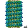
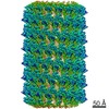
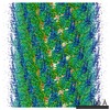
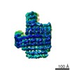
helical reconstruction / cryo EM / Resolution: 3.5 Å
Journal: Nat Commun / Year: 2021 Title: Self-association of MreC as a regulatory signal in bacterial cell wall elongation. Authors: Alexandre Martins / Carlos Contreras-Martel / Manon Janet-Maitre / Mayara M Miyachiro / Leandro F Estrozi / Daniel Maragno Trindade / Caíque C Malospirito / Fernanda Rodrigues-Costa / ...Authors: Alexandre Martins / Carlos Contreras-Martel / Manon Janet-Maitre / Mayara M Miyachiro / Leandro F Estrozi / Daniel Maragno Trindade / Caíque C Malospirito / Fernanda Rodrigues-Costa / Lionel Imbert / Viviana Job / Guy Schoehn / Ina Attrée / Andréa Dessen / Abstract: The elongasome, or Rod system, is a protein complex that controls cell wall formation in rod-shaped bacteria. MreC is a membrane-associated elongasome component that co-localizes with the ...The elongasome, or Rod system, is a protein complex that controls cell wall formation in rod-shaped bacteria. MreC is a membrane-associated elongasome component that co-localizes with the cytoskeletal element MreB and regulates the activity of cell wall biosynthesis enzymes, in a process that may be dependent on MreC self-association. Here, we use electron cryo-microscopy and X-ray crystallography to determine the structure of a self-associated form of MreC from Pseudomonas aeruginosa in atomic detail. MreC monomers interact in head-to-tail fashion. Longitudinal and lateral interfaces are essential for oligomerization in vitro, and a phylogenetic analysis of proteobacterial MreC sequences indicates the prevalence of the identified interfaces. Our results are consistent with a model where MreC's ability to alternate between self-association and interaction with the cell wall biosynthesis machinery plays a key role in the regulation of elongasome activity.
History
Deposition
Jul 1, 2020
-
Header (metadata) release
Mar 17, 2021
-
Map release
Mar 17, 2021
-
Update
Jul 10, 2024
-
Current status
Jul 10, 2024
Processing site: PDBe / Status: Released
-
Structure visualization
Movie
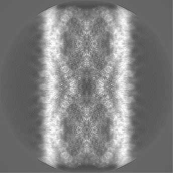
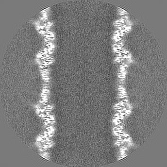
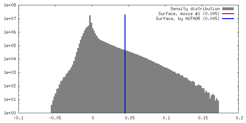
Surface view with section colored by density value
Model: Quantifoil / Material: COPPER / Mesh: 300 / Support film - Material: CARBON / Support film - topology: HOLEY / Pretreatment - Type: GLOW DISCHARGE / Pretreatment - Time: 45 sec. / Details: 30 mA
Vitrification
Cryogen name: ETHANE / Chamber humidity: 100 % / Chamber temperature: 293 K / Instrument: FEI VITROBOT MARK IV
-
Electron microscopy
Microscope
TFS GLACIOS
Image recording
Film or detector model: FEI FALCON II (4k x 4k) / Detector mode: COUNTING / Digitization - Frames/image: 2-20 / Number real images: 1200 / Average electron dose: 43.0 e/Å2
Electron beam
Acceleration voltage: 200 kV / Electron source: FIELD EMISSION GUN
In the structure databanks used in Yorodumi, some data are registered as the other names, "COVID-19 virus" and "2019-nCoV". Here are the details of the virus and the list of structure data.
Jan 31, 2019. EMDB accession codes are about to change! (news from PDBe EMDB page)
EMDB accession codes are about to change! (news from PDBe EMDB page)
The allocation of 4 digits for EMDB accession codes will soon come to an end. Whilst these codes will remain in use, new EMDB accession codes will include an additional digit and will expand incrementally as the available range of codes is exhausted. The current 4-digit format prefixed with “EMD-” (i.e. EMD-XXXX) will advance to a 5-digit format (i.e. EMD-XXXXX), and so on. It is currently estimated that the 4-digit codes will be depleted around Spring 2019, at which point the 5-digit format will come into force.
The EM Navigator/Yorodumi systems omit the EMD- prefix.
Related info.:Q: What is EMD? / ID/Accession-code notation in Yorodumi/EM Navigator
Yorodumi is a browser for structure data from EMDB, PDB, SASBDB, etc.
This page is also the successor to EM Navigator detail page, and also detail information page/front-end page for Omokage search.
The word "yorodu" (or yorozu) is an old Japanese word meaning "ten thousand". "mi" (miru) is to see.
Related info.:EMDB / PDB / SASBDB / Comparison of 3 databanks / Yorodumi Search / Aug 31, 2016. New EM Navigator & Yorodumi / Yorodumi Papers / Jmol/JSmol / Function and homology information / Changes in new EM Navigator and Yorodumi
 Movie
Movie Controller


 Open data
 Basic information
 Map data
 Sample
 Keywords
 Function and homology information
 Pseudomonas aeruginosa (bacteria)
 Authors
 Brazil,
  France, 7 items
  Citation
 Structure visualization
 Downloads & links
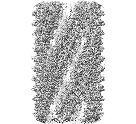 emd_11275.png
 http://ftp.pdbj.org/pub/emdb/structures/EMD-11275
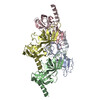
 Z (Sec.)
 Y (Row.)
 X (Col.)


 Sample components
 Processing
 Electron microscopy
 FIELD EMISSION GUN
