 Movie
Movie Controller

[English] 日本語
 Yorodumi
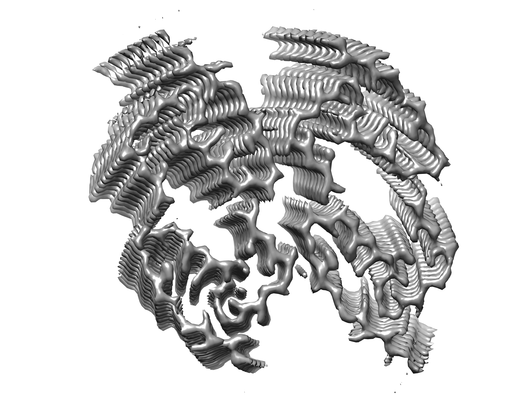
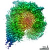
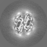
- EMDB-11163: Amyloid fibril morphology ii (in vitro) from murine SAA1.1 protein -
+ Open data

- Basic information
| Entry | Database: EMDB / ID: EMD-11163 | |||||||||||||||||||||
|---|---|---|---|---|---|---|---|---|---|---|---|---|---|---|---|---|---|---|---|---|---|---|
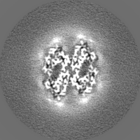
| Title | Amyloid fibril morphology ii (in vitro) from murine SAA1.1 protein | |||||||||||||||||||||
 Map data Map data | in vitro mSAA amyloid fibril morphology ii | |||||||||||||||||||||
 Sample Sample |
| |||||||||||||||||||||
 Keywords Keywords | systemic amyloidosis / misfolding disease / inflammation / prion / PROTEIN FIBRIL | |||||||||||||||||||||
| Function / homology | Serum amyloid A protein / Serum amyloid A protein / Serum amyloid A proteins signature. / Serum amyloid A proteins / response to stilbenoid / high-density lipoprotein particle / acute-phase response / Serum amyloid A-2 protein Function and homology information Function and homology information | |||||||||||||||||||||
| Biological species |  | |||||||||||||||||||||
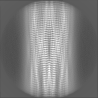
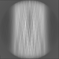
| Method | helical reconstruction / cryo EM / Resolution: 2.95 Å | |||||||||||||||||||||
 Authors Authors | Bansal A / Schmidt M | |||||||||||||||||||||
| Funding support |  Germany, 6 items Germany, 6 items
| |||||||||||||||||||||
 Citation Citation | Journal: Nat Commun / Year: 2021 Title: AA amyloid fibrils from diseased tissue are structurally different from in vitro formed SAA fibrils. Authors: Akanksha Bansal / Matthias Schmidt / Matthies Rennegarbe / Christian Haupt / Falk Liberta / Sabrina Stecher / Ioana Puscalau-Girtu / Alexander Biedermann / Marcus Fändrich / Abstract: Systemic AA amyloidosis is a world-wide occurring protein misfolding disease of humans and animals. It arises from the formation of amyloid fibrils from serum amyloid A (SAA) protein. Using cryo ...Systemic AA amyloidosis is a world-wide occurring protein misfolding disease of humans and animals. It arises from the formation of amyloid fibrils from serum amyloid A (SAA) protein. Using cryo electron microscopy we here show that amyloid fibrils which were purified from AA amyloidotic mice are structurally different from fibrils formed from recombinant SAA protein in vitro. Ex vivo amyloid fibrils consist of fibril proteins that contain more residues within their ordered parts and possess a higher β-sheet content than in vitro fibril proteins. They are also more resistant to proteolysis than their in vitro formed counterparts. These data suggest that pathogenic amyloid fibrils may originate from proteolytic selection, allowing specific fibril morphologies to proliferate and to cause damage to the surrounding tissue. | |||||||||||||||||||||
| History |
|
- Structure visualization
| Movie |
Movie viewer |
|---|---|
| Structure viewer | EM map: SurfViewMolmilJmol/JSmol |
| Supplemental images |
- Downloads & links
-EMDB archive
| Map data | emd_11163.map.gz | 2.4 MB | EMDB map data format | |
|---|---|---|---|---|
| Header (meta data) | emd-11163-v30.xmlemd-11163.xml | 16 KB 16 KB | Display Display | EMDB header |
| Images |  emd_11163.png emd_11163.png | 83.8 KB | ||
| Filedesc metadata | emd-11163.cif.gz | 5.6 KB | ||
| Others | emd_11163_half_map_1.map.gzemd_11163_half_map_2.map.gz | 22.4 MB 22.2 MB | ||
| Archive directory |  http://ftp.pdbj.org/pub/emdb/structures/EMD-11163ftp://ftp.pdbj.org/pub/emdb/structures/EMD-11163 http://ftp.pdbj.org/pub/emdb/structures/EMD-11163ftp://ftp.pdbj.org/pub/emdb/structures/EMD-11163 | HTTPS FTP |
-Validation report
| Summary document | emd_11163_validation.pdf.gz | 797.3 KB | Display | EMDB validaton report |
|---|---|---|---|---|
| Full document | emd_11163_full_validation.pdf.gz | 796.9 KB | Display | |
| Data in XML | emd_11163_validation.xml.gz | 11 KB | Display | |
| Data in CIF | emd_11163_validation.cif.gz | 12.5 KB | Display | |
| Arichive directory | https://ftp.pdbj.org/pub/emdb/validation_reports/EMD-11163ftp://ftp.pdbj.org/pub/emdb/validation_reports/EMD-11163 | HTTPS FTP |
-Related structure data
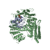
| Related structure data |  6zcgMC  6zcfC  6zchC M: atomic model generated by this map C: citing same article ( |
|---|---|
| Similar structure data | |
| EM raw data | EMPIAR-11027 (Title: Cryo electron microscopy of in vitro recombinant SAA1.1 amyloid fibrils Data size: 525.4 Data #1: Unaligned multiframe micrographs of ex-vivo murine SAA1 [micrographs - multiframe] Data #2: Thumbnails for micrographs of ex-vivo murine SAA1 [micrographs - single frame]) |


-Links
| EMDB pages | EMDB (EBI/PDBe) / EMDataResource |
|---|---|
| Related items in Molecule of the Month |

-Map
| File | Download / File: emd_11163.map.gz / Format: CCP4 / Size: 12.9 MB / Type: IMAGE STORED AS FLOATING POINT NUMBER (4 BYTES) | ||||||||||||||||||||||||||||||||||||||||||||||||||||||||||||||||||||
|---|---|---|---|---|---|---|---|---|---|---|---|---|---|---|---|---|---|---|---|---|---|---|---|---|---|---|---|---|---|---|---|---|---|---|---|---|---|---|---|---|---|---|---|---|---|---|---|---|---|---|---|---|---|---|---|---|---|---|---|---|---|---|---|---|---|---|---|---|---|
| Annotation | in vitro mSAA amyloid fibril morphology ii | ||||||||||||||||||||||||||||||||||||||||||||||||||||||||||||||||||||
| Voxel size | X=Y=Z: 1.04 Å | ||||||||||||||||||||||||||||||||||||||||||||||||||||||||||||||||||||
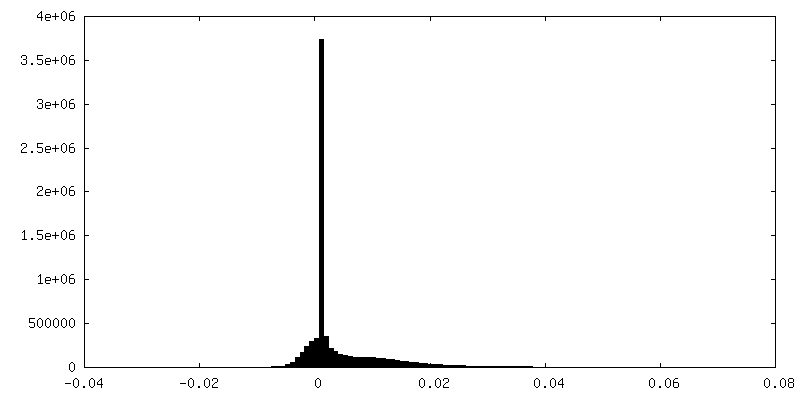
| Density |
| ||||||||||||||||||||||||||||||||||||||||||||||||||||||||||||||||||||
| Symmetry | Space group: 1 | ||||||||||||||||||||||||||||||||||||||||||||||||||||||||||||||||||||
| Details | EMDB XML:
CCP4 map header:
| ||||||||||||||||||||||||||||||||||||||||||||||||||||||||||||||||||||
-Supplemental data
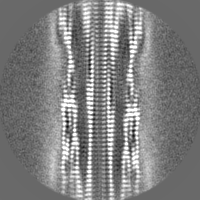
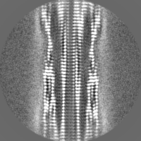
-Half map: in vitro mSAA amyloid fibril morphology ii half map 2
| File | emd_11163_half_map_1.map | ||||||||||||
|---|---|---|---|---|---|---|---|---|---|---|---|---|---|
| Annotation | in vitro mSAA amyloid fibril morphology ii half map 2 | ||||||||||||
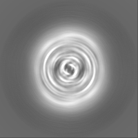
| Projections & Slices |
| ||||||||||||
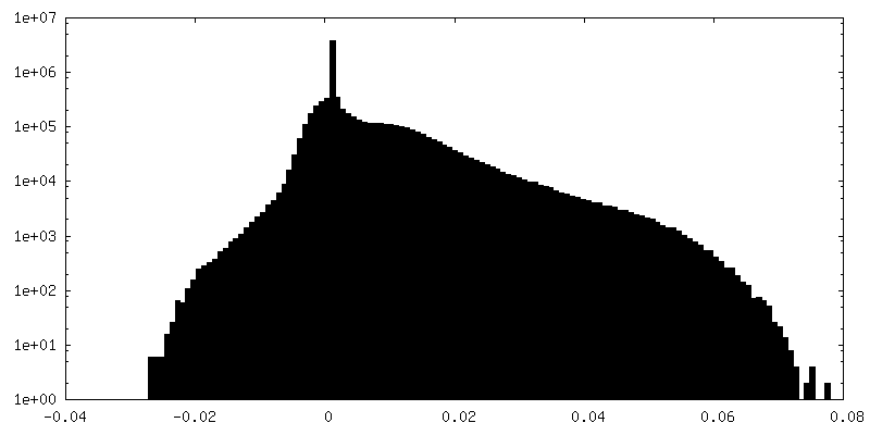
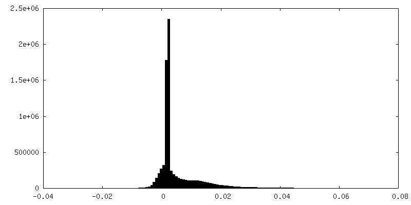
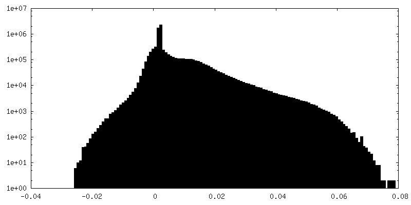
| Density Histograms |
 Z
Z Y
Y X
X


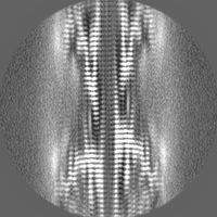
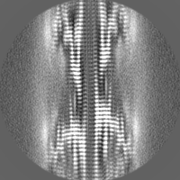
-Half map: in vitro mSAA amyloid fibril morphology ii half map 1
| File | emd_11163_half_map_2.map | ||||||||||||
|---|---|---|---|---|---|---|---|---|---|---|---|---|---|
| Annotation | in vitro mSAA amyloid fibril morphology ii half map 1 | ||||||||||||
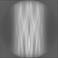
| Projections & Slices |
| ||||||||||||
| Density Histograms |
- Sample components
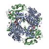
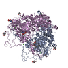
-Entire : Serum amyloid A1 (SAA1) amyloid fibril
| Entire | Name: Serum amyloid A1 (SAA1) amyloid fibril |
|---|---|
| Components |
|
-Supramolecule #1: Serum amyloid A1 (SAA1) amyloid fibril
| Supramolecule | Name: Serum amyloid A1 (SAA1) amyloid fibril / type: complex / ID: 1 / Parent: 0 / Macromolecule list: all / Details: in vitro murine SAA amyloid fibril morphology ii |
|---|---|
| Source (natural) | Organism: |
-Macromolecule #1: Serum amyloid A-2 protein
| Macromolecule | Name: Serum amyloid A-2 protein / type: protein_or_peptide / ID: 1 / Number of copies: 24 / Enantiomer: LEVO |
|---|---|
| Source (natural) | Organism: |
| Molecular weight | Theoretical: 11.622629 KDa |
| Recombinant expression | Organism:  |
| Sequence | String: GFFSFIGEAF QGAGDMWRAY TDMKEAGWKD GDKYFHARGN YDAAQRGPGG VWAAEKISDA RESFQEFFGR GHEDTMADQE ANRHGRSGK DPNYYRPPGL PAKY UniProtKB: Serum amyloid A-2 protein |
-Experimental details
-Structure determination
| Method | cryo EM |
|---|---|
 Processing Processing | helical reconstruction |
| Aggregation state | helical array |
-Sample preparation
| Concentration | 0.2 mg/mL |
|---|---|
| Buffer | pH: 8.5 / Component - Concentration: 10.0 mM / Component - Formula: (HOCH2)3CNH2 / Component - Name: Tris(hydroxymethyl)aminomethane / Details: 10mM Tris(hydroxymethyl)aminomethane (Tris) |
| Vitrification | Cryogen name: ETHANE / Chamber humidity: 96 % / Instrument: FEI VITROBOT MARK III |
- Electron microscopy
| Microscope | FEI TITAN KRIOS |
|---|---|
| Image recording | Film or detector model: GATAN K2 SUMMIT (4k x 4k) / Detector mode: COUNTING / Average exposure time: 12.0 sec. / Average electron dose: 40.0 e/Å2 |
| Electron beam | Acceleration voltage: 300 kV / Electron source:  FIELD EMISSION GUN FIELD EMISSION GUN |
| Electron optics | Illumination mode: FLOOD BEAM / Imaging mode: BRIGHT FIELD / Cs: 2.7 mm |
| Sample stage | Cooling holder cryogen: NITROGEN |
| Experimental equipment |  Model: Titan Krios / Image courtesy: FEI Company |
-Image processing
| Final reconstruction | Applied symmetry - Helical parameters - Δz: 4.74874 Å Applied symmetry - Helical parameters - Δ&Phi: -1.6 ° Applied symmetry - Helical parameters - Axial symmetry: C2 (2 fold cyclic) Resolution.type: BY AUTHOR / Resolution: 2.95 Å / Resolution method: FSC 0.143 CUT-OFF / Software - Name: RELION (ver. 3.0) / Number images used: 21355 |
|---|---|
| Segment selection | Number selected: 80724 |
| Startup model | Type of model: NONE |
| Final angle assignment | Type: NOT APPLICABLE |
-Atomic model buiding 1
| Refinement | Space: REAL / Protocol: BACKBONE TRACE / Target criteria: Correlation coefficient |
|---|---|
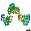
| Output model | PDB-6zcg: |
