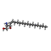
登録情報 データベース : EMDB / ID : EMD-10263タイトル Structure of Coxsackievirus A10 complexed with its receptor KREMEN1 複合体 : Coxsackievirus A10複合体 : Coxsackievirus A10タンパク質・ペプチド : Capsid protein VP1タンパク質・ペプチド : Coxsackievirus VP2タンパク質・ペプチド : Capsid protein VP3タンパク質・ペプチド : Coxsackievirus VP4複合体 : KREMEN1タンパク質・ペプチド : Kremen protein 1リガンド : SPHINGOSINEリガンド : 2-acetamido-2-deoxy-beta-D-glucopyranose / / / / / / 機能・相同性 分子機能 ドメイン・相同性 構成要素
/ / / / / / / / / / / / / / / / / / / / / / / / / / / / / / / / / / / / / / / / / / / / / / / / / / / / / / / / / / / / / / / / / / / / / / / / / / / / / / / / / / / / / / / / / / / / / / / / / / / / / / / / / / / / / / 生物種 / Homo sapiens (ヒト)手法 / / 解像度 : 3.9 Å Zhao Y / Zhou D 資金援助 Organization Grant number 国 Medical Research Council (United Kingdom) MR/N00065X/1 Wellcome Trust 101122/Z/13/Z Cancer Research UK C375/A17721
ジャーナル : Nat Commun / 年 : 2020タイトル : Hand-foot-and-mouth disease virus receptor KREMEN1 binds the canyon of Coxsackie Virus A10.著者 : Yuguang Zhao / Daming Zhou / Tao Ni / Dimple Karia / Abhay Kotecha / Xiangxi Wang / Zihe Rao / E Yvonne Jones / Elizabeth E Fry / Jingshan Ren / David I Stuart / 要旨 : Coxsackievirus A10 (CV-A10) is responsible for an escalating number of severe infections in children, but no prophylactics or therapeutics are currently available. KREMEN1 (KRM1) is the entry ... Coxsackievirus A10 (CV-A10) is responsible for an escalating number of severe infections in children, but no prophylactics or therapeutics are currently available. KREMEN1 (KRM1) is the entry receptor for the largest receptor-group of hand-foot-and-mouth disease causing viruses, which includes CV-A10. We report here structures of CV-A10 mature virus alone and in complex with KRM1 as well as of the CV-A10 A-particle. The receptor spans the viral canyon with a large footprint on the virus surface. The footprint has some overlap with that seen for the neonatal Fc receptor complexed with enterovirus E6 but is larger and distinct from that of another enterovirus receptor SCARB2. Reduced occupancy of a particle-stabilising pocket factor in the complexed virus and the presence of both unbound and expanded virus particles suggests receptor binding initiates a cascade of conformational changes that produces expanded particles primed for viral uncoating. 履歴 登録 2019年8月27日 - ヘッダ(付随情報) 公開 2019年9月4日 - マップ公開 2020年1月15日 - 更新 2024年11月6日 - 現状 2024年11月6日 処理サイト : PDBe / 状態 : 公開
すべて表示 表示を減らす
 データを開く
 基本情報
基本情報 マップデータ
 試料
試料 キーワード
キーワード 機能・相同性情報
 Coxsackievirus A10 (コクサッキーウイルス) /
  Homo sapiens (ヒト)
 データ登録者
 英国, 3件
  引用
引用 ジャーナル: Nat Commun / 年: 2020


 構造の表示
構造の表示 ムービービューア
 SurfView
 Molmil
 Jmol/JSmol
 ダウンロードとリンク
 emd_10263.map.gz
 EMDBマップデータ形式
 emd-10263-v30.xml
 emd-10263.xml
 EMDBヘッダ
 emd_10263.png
 emd-10263.cif.gz
 http://ftp.pdbj.org/pub/emdb/structures/EMD-10263
 ftp://ftp.pdbj.org/pub/emdb/structures/EMD-10263
 emd_10263_validation.pdf.gz
 EMDB検証レポート
 emd_10263_full_validation.pdf.gz
 emd_10263_validation.xml.gz
 emd_10263_validation.cif.gz
 https://ftp.pdbj.org/pub/emdb/validation_reports/EMD-10263
 ftp://ftp.pdbj.org/pub/emdb/validation_reports/EMD-10263
 リンク
リンク EMDB (EBI/PDBe) /
  EMDataResource
 マップ
マップ ダウンロード / ファイル: emd_10263.map.gz / 形式: CCP4 / 大きさ: 244.1 MB / タイプ: IMAGE STORED AS FLOATING POINT NUMBER (4 BYTES)
 試料の構成要素
 解析
解析 試料調製
試料調製 電子顕微鏡法
 FIELD EMISSION GUN
 画像解析
画像解析
 ムービー
ムービー コントローラー


 Z (Sec.)
 Y (Row.)
 X (Col.)


