 Movie
Movie Controller


+ Open data

- Basic information
| Entry | Database: EMDB / ID: EMD-0731 | |||||||||
|---|---|---|---|---|---|---|---|---|---|---|
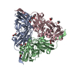
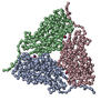
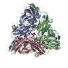
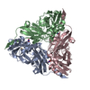
| Title | CryoEM map and model of Nitrite Reductase at pH 8.1 | |||||||||
 Map data Map data | ||||||||||
 Sample Sample |
| |||||||||
 Keywords Keywords | Cu containing nitrite reductase / OXIDOREDUCTASE | |||||||||
| Function / homology |  Function and homology information Function and homology informationdenitrification pathway / nitrite reductase (NO-forming) / nitrite reductase (NO-forming) activity / nitrate assimilation / periplasmic space / copper ion binding Similarity search - Function | |||||||||
| Biological species |  Achromobacter cycloclastes (bacteria) Achromobacter cycloclastes (bacteria) | |||||||||
| Method | single particle reconstruction / cryo EM / Resolution: 2.85 Å | |||||||||
 Authors Authors | Adachi N / Yamaguchi T / Moriya T / Kawasaki M / Koiwai K / Shinoda A / Yamada Y / Yumoto F / Kohzuma T / Senda T | |||||||||
| Funding support |  Japan, 1 items Japan, 1 items
| |||||||||
 Citation Citation | Journal: J Struct Biol / Year: 2021 Title: 2.85 and 2.99 Å resolution structures of 110 kDa nitrite reductase determined by 200 kV cryogenic electron microscopy. Authors: Naruhiko Adachi / Takahide Yamaguchi / Toshio Moriya / Masato Kawasaki / Kotaro Koiwai / Akira Shinoda / Yusuke Yamada / Fumiaki Yumoto / Takamitsu Kohzuma / Toshiya Senda / Abstract: Cu-containing nitrite reductases (NiRs) are 110 kDa enzymes that play central roles in denitrification. Although the NiRs have been well studied, with over 100 Protein Data Bank entries, such issues ...Cu-containing nitrite reductases (NiRs) are 110 kDa enzymes that play central roles in denitrification. Although the NiRs have been well studied, with over 100 Protein Data Bank entries, such issues as crystal packing, photoreduction, and lack of high pH cases have impeded structural analysis of their catalytic mechanisms. Here we show the cryogenic electron microscopy (cryo-EM) structures of Achromobacter cycloclastes NiR (AcNiR) at pH 6.2 and 8.1. The optimization of 3D-reconstruction parameters achieved 2.99 and 2.85 Å resolution. Comprehensive comparisons with cryo-EM and 56 AcNiR crystal structures suggested crystallographic artifacts in residues 185-215 and His255' due to packing and photoreduction, respectively. We used a newly developed map comparison method to detect structural change around the type 2 Cu site. While the theoretical estimation of coordinate errors of cryo-EM structures remains difficult, combined analysis using X-ray and cryo-EM structures will allow deeper insight into the local structural changes of proteins. | |||||||||
| History |
|
- Structure visualization
| Movie |
Movie viewer |
|---|---|
| Structure viewer | EM map: SurfViewMolmilJmol/JSmol |
| Supplemental images |

- Downloads & links
-EMDB archive
| Map data | emd_0731.map.gz | 355.2 MB | EMDB map data format | |
|---|---|---|---|---|
| Header (meta data) | emd-0731-v30.xmlemd-0731.xml | 13.1 KB 13.1 KB | Display Display | EMDB header |
| Images | 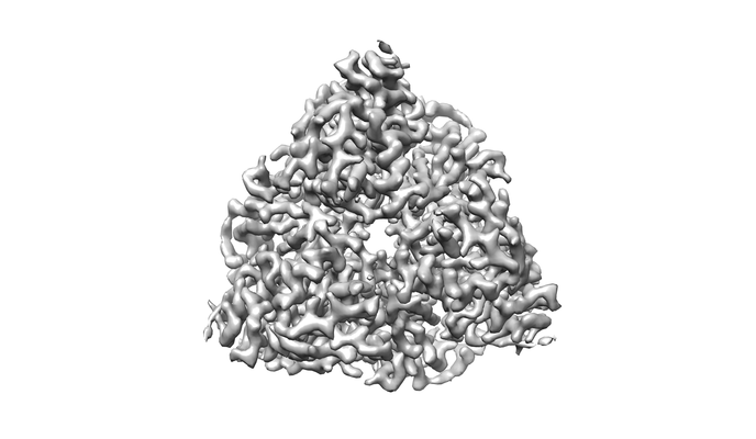 emd_0731.png emd_0731.png | 65.2 KB | ||
| Filedesc metadata | emd-0731.cif.gz | 5.5 KB | ||
| Archive directory |  http://ftp.pdbj.org/pub/emdb/structures/EMD-0731ftp://ftp.pdbj.org/pub/emdb/structures/EMD-0731 http://ftp.pdbj.org/pub/emdb/structures/EMD-0731ftp://ftp.pdbj.org/pub/emdb/structures/EMD-0731 | HTTPS FTP |
-Related structure data
| Related structure data |  6kngMC 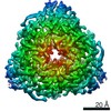 0730C 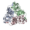 6knfC M: atomic model generated by this map C: citing same article ( |
|---|---|
| Similar structure data | |
| EM raw data | EMPIAR-10581 (Title: CryoEM map and model of Nitrite Reductase at pH 8.1 / Data size: 1.0 TB Data #1: CryoEM map and model of Nitrite Reductase at pH 8.1 [micrographs - multiframe]) |


-Links
| EMDB pages | EMDB (EBI/PDBe) / EMDataResource |
|---|---|
| Related items in Molecule of the Month |


-Map
| File | Download / File: emd_0731.map.gz / Format: CCP4 / Size: 437.9 MB / Type: IMAGE STORED AS FLOATING POINT NUMBER (4 BYTES) | ||||||||||||||||||||||||||||||||||||||||||||||||||||||||||||
|---|---|---|---|---|---|---|---|---|---|---|---|---|---|---|---|---|---|---|---|---|---|---|---|---|---|---|---|---|---|---|---|---|---|---|---|---|---|---|---|---|---|---|---|---|---|---|---|---|---|---|---|---|---|---|---|---|---|---|---|---|---|
| Projections & slices | Image control
Images are generated by Spider. | ||||||||||||||||||||||||||||||||||||||||||||||||||||||||||||
| Voxel size | X=Y=Z: 0.88 Å | ||||||||||||||||||||||||||||||||||||||||||||||||||||||||||||
| Density |
| ||||||||||||||||||||||||||||||||||||||||||||||||||||||||||||
| Symmetry | Space group: 1 | ||||||||||||||||||||||||||||||||||||||||||||||||||||||||||||
| Details | EMDB XML:
CCP4 map header:
| ||||||||||||||||||||||||||||||||||||||||||||||||||||||||||||
 Z (Sec.)
 Y (Row.)
 X (Col.)


-Supplemental data
- Sample components
-Entire : Nitrite reductase
| Entire | Name: Nitrite reductase |
|---|---|
| Components |
|
-Supramolecule #1: Nitrite reductase
| Supramolecule | Name: Nitrite reductase / type: organelle_or_cellular_component / ID: 1 / Parent: 0 / Macromolecule list: #1 |
|---|---|
| Source (natural) | Organism: Achromobacter cycloclastes (bacteria) |
| Molecular weight | Theoretical: 110 KDa |
-Macromolecule #1: Copper-containing nitrite reductase
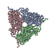
| Macromolecule | Name: Copper-containing nitrite reductase / type: protein_or_peptide / ID: 1 / Number of copies: 3 / Enantiomer: LEVO / EC number: nitrite reductase (NO-forming) |
|---|---|
| Source (natural) | Organism: Achromobacter cycloclastes (bacteria) |
| Molecular weight | Theoretical: 36.621332 KDa |
| Sequence | String: VDISTLPRVK VDLVKPPFVH AHDQVAKTGP RVVEFTMTIE EKKLVIDREG TEIHAMTFNG SVPGPLMVVH ENDYVELRLI NPDTNTLLH NIDFHAATGA LGGGALTQVN PGEETTLRFK ATKPGVFVYH CAPEGMVPWH VTSGMNGAIM VLPRDGLKDE K GQPLTYDK ...String: VDISTLPRVK VDLVKPPFVH AHDQVAKTGP RVVEFTMTIE EKKLVIDREG TEIHAMTFNG SVPGPLMVVH ENDYVELRLI NPDTNTLLH NIDFHAATGA LGGGALTQVN PGEETTLRFK ATKPGVFVYH CAPEGMVPWH VTSGMNGAIM VLPRDGLKDE K GQPLTYDK IYYVGEQDFY VPKDEAGNYK KYETPGEAYE DAVKAMRTLT PTHIVFNGAV GALTGDHALT AAVGERVLVV HS QANRDTR PHLIGGHGDY VWATGKFRNP PDLDQETWLI PGGTAGAAFY TFRQPGVYAY VNHNLIEAFE LGAAGHFKVT GEW NDDLMT SVVKPASM UniProtKB: Copper-containing nitrite reductase |
-Macromolecule #2: COPPER (II) ION
| Macromolecule | Name: COPPER (II) ION / type: ligand / ID: 2 / Number of copies: 6 / Formula: CU |
|---|---|
| Molecular weight | Theoretical: 63.546 Da |
| Chemical component information |  ChemComp-CU: |
-Experimental details
-Structure determination
| Method | cryo EM |
|---|---|
 Processing Processing | single particle reconstruction |
| Aggregation state | particle |
-Sample preparation
| Concentration | 1.1 mg/mL |
|---|---|
| Buffer | pH: 8.1 / Component - Concentration: 100.0 mM / Component - Name: pottasium phosphate |
| Grid | Model: Quantifoil R1.2/1.3 / Material: COPPER / Mesh: 300 / Support film - Material: CARBON / Support film - topology: HOLEY / Pretreatment - Type: GLOW DISCHARGE / Pretreatment - Time: 30 sec. / Pretreatment - Atmosphere: AIR / Details: The grid was washed by acetone prior to use. |
| Vitrification | Cryogen name: ETHANE / Chamber humidity: 100 % / Chamber temperature: 291 K / Instrument: FEI VITROBOT MARK IV / Details: Blotting time was 15 seconds.. |
| Details | This sample was mono-disperse. |
- Electron microscopy
| Microscope | FEI TALOS ARCTICA |
|---|---|
| Image recording | Film or detector model: FEI FALCON III (4k x 4k) / Detector mode: COUNTING / Number grids imaged: 1 / Number real images: 694 / Average exposure time: 56.37 sec. / Average electron dose: 50.0 e/Å2 |
| Electron beam | Acceleration voltage: 200 kV / Electron source:  FIELD EMISSION GUN FIELD EMISSION GUN |
| Electron optics | C2 aperture diameter: 50.0 µm / Illumination mode: FLOOD BEAM / Imaging mode: BRIGHT FIELD / Cs: 2.7 mm / Nominal defocus max: 3.0 µm / Nominal defocus min: 1.0 µm / Nominal magnification: 120000 |
| Sample stage | Cooling holder cryogen: NITROGEN |
| Experimental equipment |  Model: Talos Arctica / Image courtesy: FEI Company |
-Image processing
| Startup model | Type of model: OTHER Details: An ab initio model was generated using RELION3's own implementation of Stochastic Gradient Descent (SGD) algorithm and low-pass filtered to 25 A for use as an initial model for 3D refinement. |
|---|---|
| Final reconstruction | Applied symmetry - Point group: C3 (3 fold cyclic) / Resolution.type: BY AUTHOR / Resolution: 2.85 Å / Resolution method: FSC 0.143 CUT-OFF / Software - Name: RELION (ver. 3) / Number images used: 89513 |
| Initial angle assignment | Type: MAXIMUM LIKELIHOOD / Software - Name: RELION (ver. 3) |
| Final angle assignment | Type: MAXIMUM LIKELIHOOD / Software - Name: RELION (ver. 3) |
