 ムービー
ムービー コントローラー


+ データを開く

- 基本情報
基本情報
| 登録情報 | データベース: EMDB / ID: EMD-8838 | |||||||||
|---|---|---|---|---|---|---|---|---|---|---|
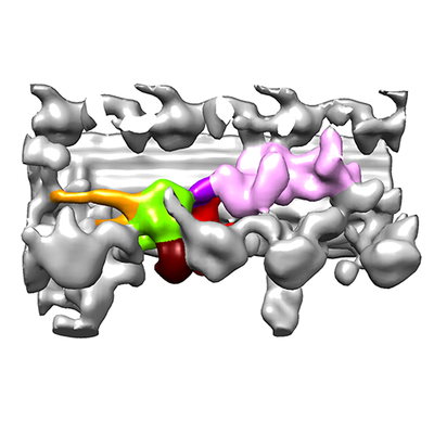
| タイトル | Sub-tomogram average of I1 dynein Pre class | |||||||||
 マップデータ マップデータ | Sub-tomogram average of I1 dynein Pre class: the two dynein heavy chains of sea urchin sperm flagellar I1 dynein complex are in the pre-powerstroke state. | |||||||||
 試料 試料 |
| |||||||||
| 生物種 |  | |||||||||
| 手法 | サブトモグラム平均法 / クライオ電子顕微鏡法 / 解像度: 31.0 Å | |||||||||
 データ登録者 データ登録者 | Lin J / Nicastro D | |||||||||
| 資金援助 |  米国, 2件 米国, 2件
| |||||||||
 引用 引用 | ジャーナル: Science / 年: 2018 タイトル: Asymmetric distribution and spatial switching of dynein activity generates ciliary motility. 著者: Jianfeng Lin / Daniela Nicastro / 要旨: Motile cilia and flagella are essential, highly conserved organelles, and their motility is driven by the coordinated activities of multiple dynein isoforms. The prevailing "switch-point" hypothesis ...Motile cilia and flagella are essential, highly conserved organelles, and their motility is driven by the coordinated activities of multiple dynein isoforms. The prevailing "switch-point" hypothesis posits that dyneins are asymmetrically activated to drive flagellar bending. To test this model, we applied cryo-electron tomography to visualize activity states of individual dyneins relative to their locations along beating flagella of sea urchin sperm cells. As predicted, bending was generated by the asymmetric distribution of dynein activity on opposite sides of the flagellum. However, contrary to predictions, most dyneins were in their active state, and the smaller population of conformationally inactive dyneins switched flagellar sides relative to the bending direction. Thus, our data suggest a "switch-inhibition" mechanism in which force imbalance is generated by inhibiting, rather than activating, dyneins on alternating sides of the flagellum. | |||||||||
| 履歴 |
|
- 構造の表示
構造の表示
| ムービー |
 ムービービューア ムービービューア |
|---|---|
| 構造ビューア | EMマップ: SurfViewMolmilJmol/JSmol |
| 添付画像 |
- ダウンロードとリンク
-EMDBアーカイブ
| マップデータ | emd_8838.map.gz | 587.5 KB | EMDBマップデータ形式 | |
|---|---|---|---|---|
| ヘッダ (付随情報) | emd-8838-v30.xmlemd-8838.xml | 11.2 KB 11.2 KB | 表示 表示 | EMDBヘッダ |
| 画像 |  emd_8838.png emd_8838.png | 93.7 KB | ||
| アーカイブディレクトリ |  http://ftp.pdbj.org/pub/emdb/structures/EMD-8838ftp://ftp.pdbj.org/pub/emdb/structures/EMD-8838 http://ftp.pdbj.org/pub/emdb/structures/EMD-8838ftp://ftp.pdbj.org/pub/emdb/structures/EMD-8838 | HTTPS FTP |
-検証レポート
| 文書・要旨 | emd_8838_validation.pdf.gz | 77.8 KB | 表示 | EMDB検証レポート |
|---|---|---|---|---|
| 文書・詳細版 | emd_8838_full_validation.pdf.gz | 77 KB | 表示 | |
| XML形式データ | emd_8838_validation.xml.gz | 494 B | 表示 | |
| アーカイブディレクトリ | https://ftp.pdbj.org/pub/emdb/validation_reports/EMD-8838ftp://ftp.pdbj.org/pub/emdb/validation_reports/EMD-8838 | HTTPS FTP |
-関連構造データ
| 関連構造データ |  8835C  8836C 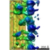 8837C C: 同じ文献を引用 ( |
|---|---|
| 類似構造データ | |
| 電子顕微鏡画像生データ | EMPIAR-10157 (タイトル: Cryo electron tomography of sea urchin sperm flagella Data size: 15.8 Data #1: Tilt series of sea urchin sperm flagella that were frozen while they were actively beating. [tilt series]) |
-リンク
| EMDBのページ | EMDB (EBI/PDBe) / EMDataResource |
|---|
-マップ
| ファイル | ダウンロード / ファイル: emd_8838.map.gz / 形式: CCP4 / 大きさ: 704.1 KB / タイプ: IMAGE STORED AS FLOATING POINT NUMBER (4 BYTES) | ||||||||||||||||||||||||||||||||||||||||||||||||||||||||||||||||||||
|---|---|---|---|---|---|---|---|---|---|---|---|---|---|---|---|---|---|---|---|---|---|---|---|---|---|---|---|---|---|---|---|---|---|---|---|---|---|---|---|---|---|---|---|---|---|---|---|---|---|---|---|---|---|---|---|---|---|---|---|---|---|---|---|---|---|---|---|---|---|
| 注釈 | Sub-tomogram average of I1 dynein Pre class: the two dynein heavy chains of sea urchin sperm flagellar I1 dynein complex are in the pre-powerstroke state. | ||||||||||||||||||||||||||||||||||||||||||||||||||||||||||||||||||||
| 投影像・断面図 | 画像のコントロール
画像は Spider により作成 これらの図は立方格子座標系で作成されたものです | ||||||||||||||||||||||||||||||||||||||||||||||||||||||||||||||||||||
| ボクセルのサイズ | X=Y=Z: 9.856 Å | ||||||||||||||||||||||||||||||||||||||||||||||||||||||||||||||||||||
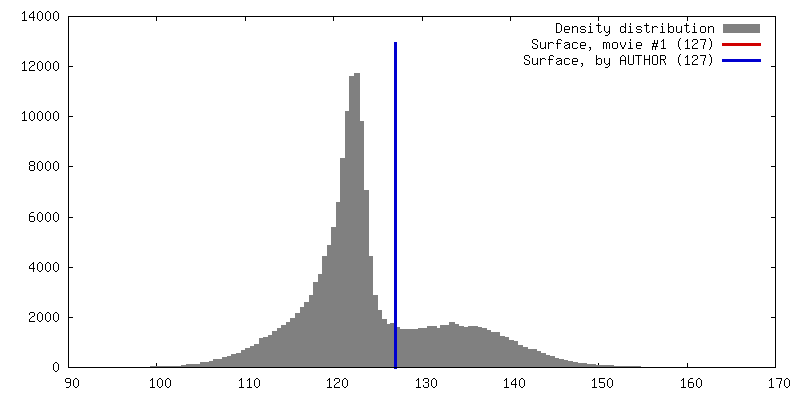
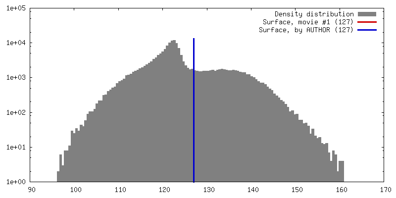
| 密度 |
| ||||||||||||||||||||||||||||||||||||||||||||||||||||||||||||||||||||
| 対称性 | 空間群: 1 | ||||||||||||||||||||||||||||||||||||||||||||||||||||||||||||||||||||
| 詳細 | EMDB XML:
CCP4マップ ヘッダ情報:
| ||||||||||||||||||||||||||||||||||||||||||||||||||||||||||||||||||||
 Z (Sec.)
 Y (Row.)
 X (Col.)


-添付データ
- 試料の構成要素
-全体 : Sea urchin sperm
| 全体 | 名称: Sea urchin sperm |
|---|---|
| 要素 |
|
-超分子 #1: Sea urchin sperm
| 超分子 | 名称: Sea urchin sperm / タイプ: cell / ID: 1 / 親要素: 0 / 含まれる分子: #1 詳細: Sperm cells were actively swimming in the artificial sea water. |
|---|---|
| 由来(天然) | 生物種: |
-実験情報
-構造解析
| 手法 | クライオ電子顕微鏡法 |
|---|---|
 解析 解析 | サブトモグラム平均法 |
| 試料の集合状態 | cell |
-試料調製
| 緩衝液 | pH: 8 構成要素:
| ||||||||||||||||||
|---|---|---|---|---|---|---|---|---|---|---|---|---|---|---|---|---|---|---|---|
| グリッド | モデル: Quantifoil R2/2 / 材質: COPPER / メッシュ: 200 / 支持フィルム - 材質: CARBON / 支持フィルム - トポロジー: HOLEY / 前処理 - タイプ: GLOW DISCHARGE | ||||||||||||||||||
| 凍結 | 凍結剤: ETHANE / 装置: HOMEMADE PLUNGER |
- 電子顕微鏡法
| 顕微鏡 | FEI TECNAI F30 |
|---|---|
| 特殊光学系 | エネルギーフィルター - 名称: GIF |
| 撮影 | フィルム・検出器のモデル: GATAN MULTISCAN / 平均電子線量: 1.5 e/Å2 |
| 電子線 | 加速電圧: 300 kV / 電子線源:  FIELD EMISSION GUN FIELD EMISSION GUN |
| 電子光学系 | C2レンズ絞り径: 100.0 µm / 照射モード: FLOOD BEAM / 撮影モード: BRIGHT FIELD / 最大 デフォーカス(公称値): 8.0 µm / 最小 デフォーカス(公称値): 6.0 µm / 倍率(公称値): 13500 |
| 試料ステージ | ホルダー冷却材: NITROGEN |
| 実験機器 |  モデル: Tecnai F30 / 画像提供: FEI Company |
-画像解析
| 最終 再構成 | 解像度のタイプ: BY AUTHOR / 解像度: 31.0 Å / 解像度の算出法: FSC 0.5 CUT-OFF / ソフトウェア - 名称: PEET (ver. 1.9.0) / 使用したサブトモグラム数: 2700 |
|---|---|
| 抽出 | トモグラム数: 41 / 使用した粒子像数: 6481 / ソフトウェア - 名称: PEET (ver. 1.9.0) |
| 最終 3次元分類 | ソフトウェア - 名称: PEET (ver. 1.9.0) |
| 最終 角度割当 | タイプ: OTHER |
