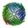
Movie
Movie Controller

[English] 日本語
 Yorodumi
- EMDB-68251: Cryo-EM structure of mouse heavy-chain apoferritin at 1.24 A on C... -
+ Open data

- Basic information
| Entry |  | |||||||||
|---|---|---|---|---|---|---|---|---|---|---|
| Title | Cryo-EM structure of mouse heavy-chain apoferritin at 1.24 A on CRYO ARM 200 II | |||||||||
 Map data Map data | Sharpened map | |||||||||
 Sample Sample |
| |||||||||
 Keywords Keywords | apoferritin / METAL BINDING PROTEIN | |||||||||
| Function / homology |  Function and homology information Function and homology informationIron uptake and transport / Golgi Associated Vesicle Biogenesis / ferroxidase / negative regulation of ferroptosis / ferroxidase activity / protein complex oligomerization / autolysosome / negative regulation of fibroblast proliferation / endocytic vesicle lumen / Neutrophil degranulation ...Iron uptake and transport / Golgi Associated Vesicle Biogenesis / ferroxidase / negative regulation of ferroptosis / ferroxidase activity / protein complex oligomerization / autolysosome / negative regulation of fibroblast proliferation / endocytic vesicle lumen / Neutrophil degranulation / ferric iron binding / autophagosome / iron ion transport / ferrous iron binding / intracellular iron ion homeostasis / immune response / iron ion binding / negative regulation of cell population proliferation / mitochondrion / extracellular region / membrane / identical protein binding / cytosol / cytoplasm Similarity search - Function | |||||||||
| Biological species |   Mus musculus (house mouse) Mus musculus (house mouse) | |||||||||
| Method | single particle reconstruction / cryo EM / Resolution: 1.24 Å | |||||||||
 Authors Authors | Danev R / Yanagisawa H / Yamashita K / Eisenstein F / Kikkawa M | |||||||||
| Funding support |  Japan, 2 items Japan, 2 items
| |||||||||
 Citation Citation | Journal: IUCrJ / Year: 2026 Title: Atomic resolution cryo-EM at 200 keV. Authors: Radostin Danev / Haruaki Yanagisawa / Keitaro Yamashita / Fabian Eisenstein / Masahide Kikkawa /  Abstract: Atomic resolution in cryo-electron microscopy was first demonstrated six years ago. This was accomplished using 300 kV electron microscopes equipped with new hardware that provided narrower energy ...Atomic resolution in cryo-electron microscopy was first demonstrated six years ago. This was accomplished using 300 kV electron microscopes equipped with new hardware that provided narrower energy spread, aberration correction and energy filtering. Here, we report the achievement of 1.24 Å atomic resolution on an upgraded 200 kV electron microscope featuring a cold field emission gun, a high-resolution objective lens polepiece and an energy filter. These components transform the instrument into a cost-effective single-particle cryo-EM platform with performance comparable to that of significantly more expensive 300 kV systems. The microscope can also be operated at 100 kV and by using a high-speed hybrid-pixel detector we were able to reach sub-2 Å resolution. | |||||||||
| History |
|
- Structure visualization
| Supplemental images |
|---|

- Downloads & links
-EMDB archive
| Map data | emd_68251.map.gz | 306.9 MB | EMDB map data format | |
|---|---|---|---|---|
| Header (meta data) | emd-68251-v30.xmlemd-68251.xml | 28.3 KB 28.3 KB | Display Display | EMDB header |
| FSC (resolution estimation) | emd_68251_fsc.xml | 21.2 KB | Display | FSC data file |
| Images |  emd_68251.png emd_68251.png | 251.9 KB | ||
| Masks | emd_68251_msk_1.map | 325 MB | Mask map | |
| Filedesc metadata | emd-68251.cif.gz | 7.2 KB | ||
| Others | emd_68251_additional_1.map.gzemd_68251_half_map_1.map.gzemd_68251_half_map_2.map.gz | 68.8 MB 300.7 MB 300.7 MB | ||
| Archive directory |  https://data.pdbj.org/pub/emdb/structures/EMD-68251ftp://data.pdbj.org/pub/emdb/structures/EMD-68251 https://data.pdbj.org/pub/emdb/structures/EMD-68251ftp://data.pdbj.org/pub/emdb/structures/EMD-68251 | HTTPS FTP |
-Related structure data
| Related structure data |  22fxMC M: atomic model generated by this map C: citing same article ( |
|---|---|
| Similar structure data |

-Links
| EMDB pages | EMDB (EBI/PDBe) / EMDataResource |
|---|---|
| Related items in Molecule of the Month |

-Map
| File | Download / File: emd_68251.map.gz / Format: CCP4 / Size: 325 MB / Type: IMAGE STORED AS FLOATING POINT NUMBER (4 BYTES) | ||||||||||||||||||||||||||||||||||||
|---|---|---|---|---|---|---|---|---|---|---|---|---|---|---|---|---|---|---|---|---|---|---|---|---|---|---|---|---|---|---|---|---|---|---|---|---|---|
| Annotation | Sharpened map | ||||||||||||||||||||||||||||||||||||
| Projections & slices | Image control
Images are generated by Spider. | ||||||||||||||||||||||||||||||||||||
| Voxel size | X=Y=Z: 0.56953 Å | ||||||||||||||||||||||||||||||||||||
| Density |
| ||||||||||||||||||||||||||||||||||||
| Symmetry | Space group: 1 | ||||||||||||||||||||||||||||||||||||
| Details | EMDB XML:
|
 Z (Sec.)
 Y (Row.)
 X (Col.)


-Supplemental data
-Mask #1
| File | emd_68251_msk_1.map | ||||||||||||
|---|---|---|---|---|---|---|---|---|---|---|---|---|---|
| Projections & Slices |
| ||||||||||||
| Density Histograms |


-Additional map: Fo-Fc difference density map
| File | emd_68251_additional_1.map | ||||||||||||
|---|---|---|---|---|---|---|---|---|---|---|---|---|---|
| Annotation | Fo-Fc difference density map | ||||||||||||
| Projections & Slices |
| ||||||||||||
| Density Histograms |


-Half map: #2
| File | emd_68251_half_map_1.map | ||||||||||||
|---|---|---|---|---|---|---|---|---|---|---|---|---|---|
| Projections & Slices |
| ||||||||||||
| Density Histograms |


-Half map: #1
| File | emd_68251_half_map_2.map | ||||||||||||
|---|---|---|---|---|---|---|---|---|---|---|---|---|---|
| Projections & Slices |
| ||||||||||||
| Density Histograms |


- Sample components
-Entire : homo 24-mer mouse heavy-chain apoferritin
| Entire | Name: homo 24-mer mouse heavy-chain apoferritin |
|---|---|
| Components |
|
-Supramolecule #1: homo 24-mer mouse heavy-chain apoferritin
| Supramolecule | Name: homo 24-mer mouse heavy-chain apoferritin / type: complex / ID: 1 / Parent: 0 / Macromolecule list: #1 |
|---|---|
| Source (natural) | Organism: Mus musculus (house mouse) |
| Molecular weight | Theoretical: 500 KDa |
-Macromolecule #1: Ferritin heavy chain
| Macromolecule | Name: Ferritin heavy chain / type: protein_or_peptide / ID: 1 / Number of copies: 1 / Enantiomer: LEVO / EC number: ferroxidase |
|---|---|
| Source (natural) | Organism: Mus musculus (house mouse) |
| Molecular weight | Theoretical: 21.097631 KDa |
| Recombinant expression | Organism:   Escherichia coli 'BL21-Gold(DE3)pLysS AG' (bacteria) Escherichia coli 'BL21-Gold(DE3)pLysS AG' (bacteria) |
| Sequence | String: MTTASPSQVR QNYHQDAEAA INRQINLELY ASYVYLSMSC YFDRDDVALK NFAKYFLHQS HEEREHAEKL MKLQNQRGGR IFLQDIKKP DRDDWESGLN AMECALHLEK SVNQSLLELH KLATDKNDPH LCDFIETYYL SEQVKSIKEL GDHVTNLRKM G APEAGMAE YLFDKHTLGH GDES UniProtKB: Ferritin heavy chain |
-Macromolecule #2: FE (III) ION
| Macromolecule | Name: FE (III) ION / type: ligand / ID: 2 / Number of copies: 1 / Formula: FE |
|---|---|
| Molecular weight | Theoretical: 55.845 Da |
-Macromolecule #3: ZINC ION
| Macromolecule | Name: ZINC ION / type: ligand / ID: 3 / Number of copies: 1 / Formula: ZN |
|---|---|
| Molecular weight | Theoretical: 65.409 Da |
-Macromolecule #4: water
| Macromolecule | Name: water / type: ligand / ID: 4 / Number of copies: 201 / Formula: HOH |
|---|---|
| Molecular weight | Theoretical: 18.015 Da |
| Chemical component information |  ChemComp-HOH: |
-Experimental details
-Structure determination
| Method | cryo EM |
|---|---|
 Processing Processing | single particle reconstruction |
| Aggregation state | particle |
-Sample preparation
| Concentration | 1.9 mg/mL |
|---|---|
| Buffer | pH: 7.5 Details: 20 mM HEPES-NaOH pH 7.5, 300 mM NaCl, 1 mM dithiothreitol (DTT) |
| Grid | Model: UltrAuFoil / Material: GOLD / Mesh: 300 / Support film - Material: GOLD / Support film - topology: HOLEY ARRAY / Pretreatment - Type: GLOW DISCHARGE / Pretreatment - Time: 45 sec. / Pretreatment - Atmosphere: AIR / Pretreatment - Pressure: 0.04 kPa / Details: R0.6/1 |
| Vitrification | Cryogen name: ETHANE / Chamber humidity: 100 % / Chamber temperature: 278 K / Instrument: FEI VITROBOT MARK IV / Details: 3 ul sample, 20 s blot time. |
- Electron microscopy
| Microscope | JEOL CRYO ARM 200 |
|---|---|
| Temperature | Min: 87.0 K / Max: 87.0 K |
| Alignment procedure | Coma free - Residual tilt: 0.5 mrad |
| Specialist optics | Energy filter - Name: In-column Omega Filter / Energy filter - Slit width: 20 eV |
| Image recording | Film or detector model: GATAN K3 (6k x 4k) / Number grids imaged: 1 / Number real images: 13654 / Average exposure time: 1.51 sec. / Average electron dose: 53.4 e/Å2 |
| Electron beam | Acceleration voltage: 200 kV / Electron source:  FIELD EMISSION GUN FIELD EMISSION GUN |
| Electron optics | C2 aperture diameter: 100.0 µm / Calibrated defocus max: 0.8 µm / Calibrated defocus min: 0.2 µm / Calibrated magnification: 163612 / Illumination mode: FLOOD BEAM / Imaging mode: BRIGHT FIELD / Cs: 1.5 mm / Nominal defocus max: 0.8 µm / Nominal defocus min: 0.2 µm / Nominal magnification: 150000 |
| Sample stage | Specimen holder model: JEOL CRYOSPECPORTER / Cooling holder cryogen: NITROGEN |