 ムービー
ムービー コントローラー


+ データを開く

- 基本情報
基本情報
| 登録情報 | データベース: EMDB / ID: EMD-6737 | |||||||||
|---|---|---|---|---|---|---|---|---|---|---|
| タイトル | Natural chromatin is heterogeneous and self associates in vitro | |||||||||
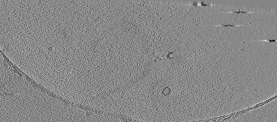
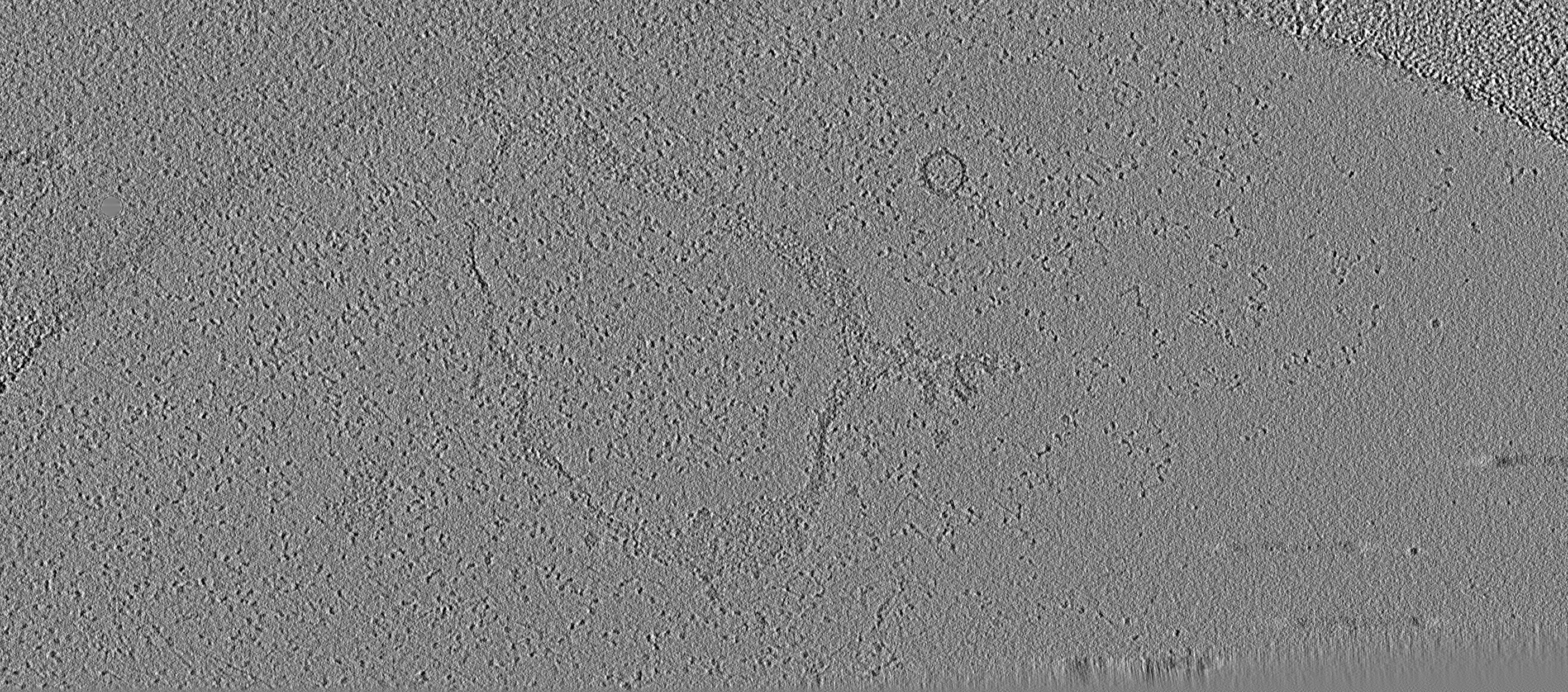
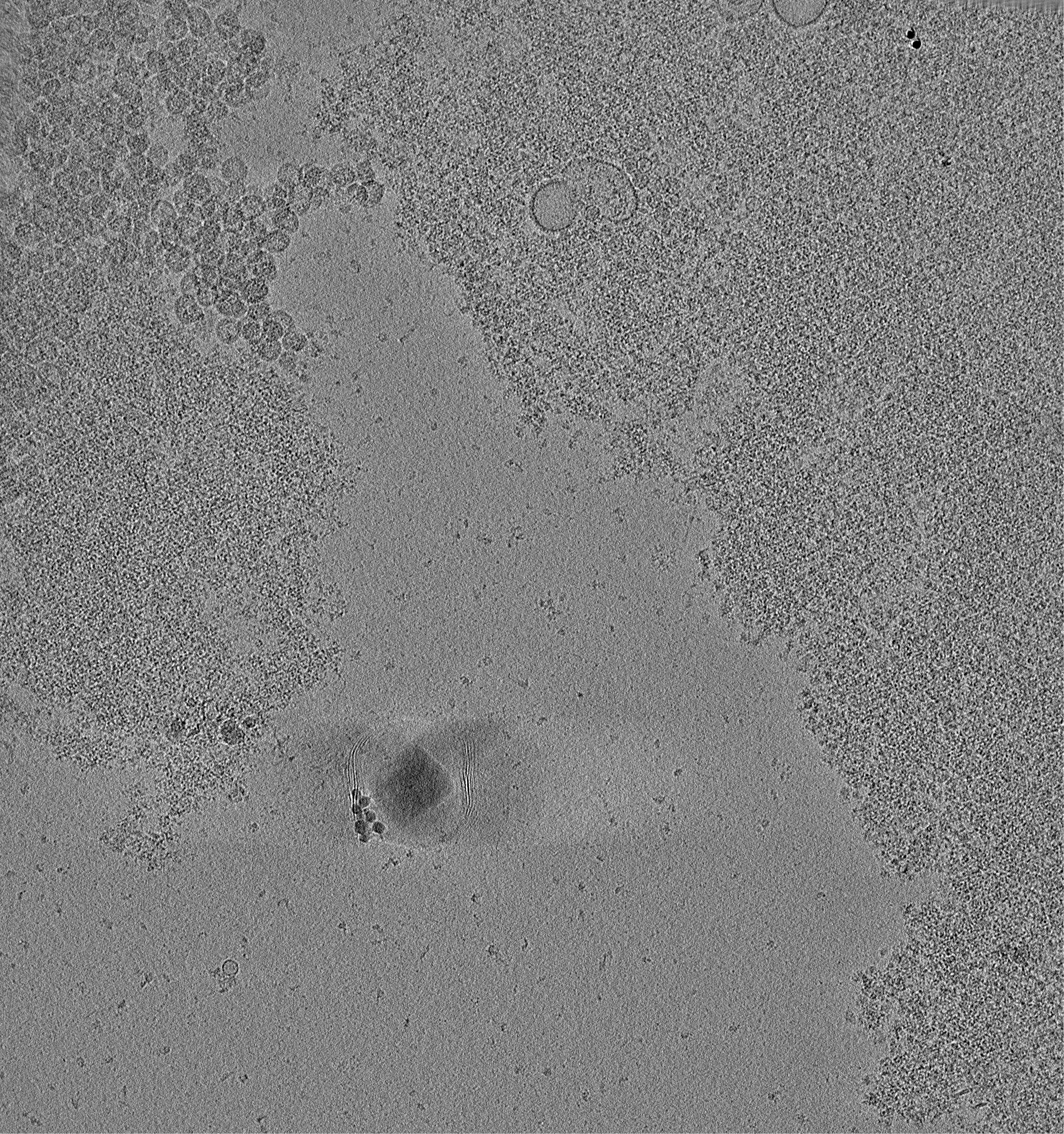
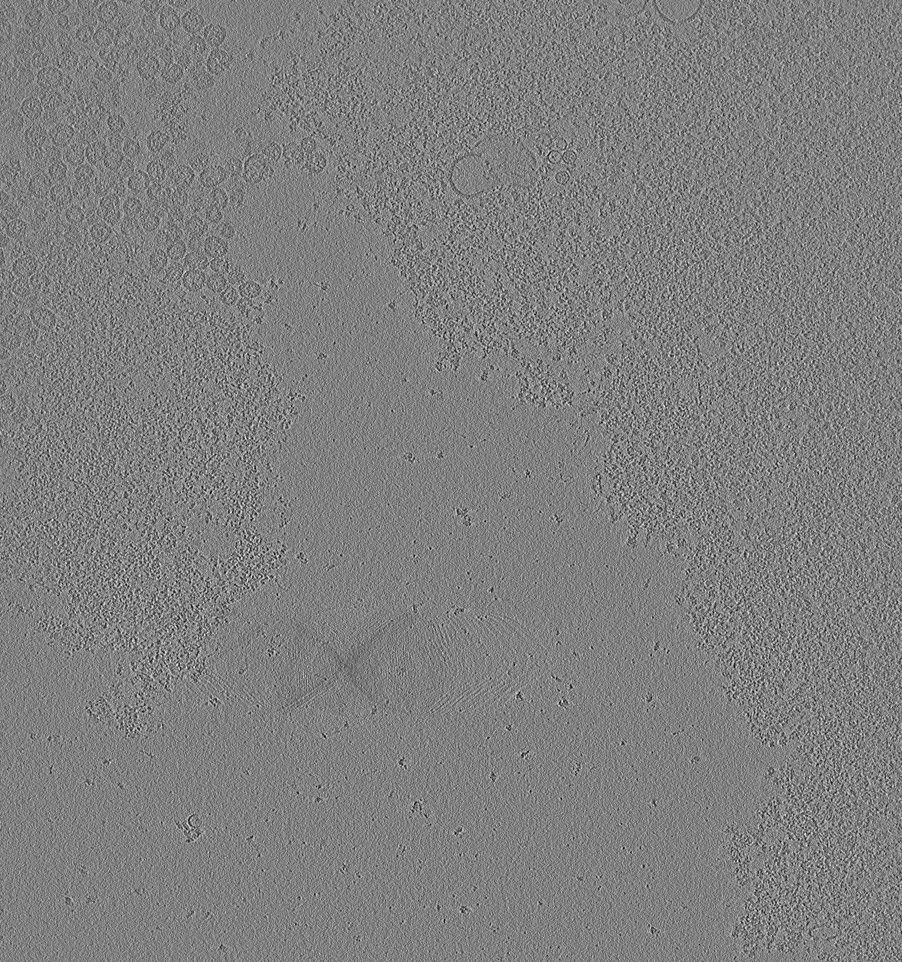
 マップデータ マップデータ | Cryotomogram of picoplankton chromatin in 5mM EDTA. | |||||||||
 試料 試料 |
| |||||||||
| 生物種 |  Ostreococcus tauri (植物) Ostreococcus tauri (植物) | |||||||||
| 手法 | 電子線トモグラフィー法 / クライオ電子顕微鏡法 | |||||||||
 データ登録者 データ登録者 | Cai S / Song Y / Chen C / Shi J / Gan L | |||||||||
 引用 引用 | ジャーナル: Mol Biol Cell / 年: 2018 タイトル: Natural chromatin is heterogeneous and self-associates in vitro. 著者: Shujun Cai / Yajiao Song / Chen Chen / Jian Shi / Lu Gan /  要旨: The 30-nm fiber is commonly formed by oligonucleosome arrays in vitro but rarely found inside cells. To determine how chromatin higher-order structure is controlled, we used electron cryotomography ...The 30-nm fiber is commonly formed by oligonucleosome arrays in vitro but rarely found inside cells. To determine how chromatin higher-order structure is controlled, we used electron cryotomography (cryo-ET) to study the undigested natural chromatin released from two single-celled organisms in which 30-nm fibers have not been observed in vivo: picoplankton and yeast. In the presence of divalent cations, most of the chromatin from both organisms is condensed into a large mass in vitro. Rare irregular 30-nm fibers, some of which include face-to-face nucleosome interactions, do form at the periphery of this mass. In the absence of divalent cations, picoplankton chromatin decondenses into open zigzags. By contrast, yeast chromatin mostly remains condensed, with very few open motifs. Yeast chromatin packing is largely unchanged in the absence of linker histone and mildly decondensed when histones are more acetylated. Natural chromatin is therefore generally nonpermissive of regular motifs, even at the level of oligonucleosomes. | |||||||||
| 履歴 |
|
- 構造の表示
構造の表示
| ムービー |
 ムービービューア ムービービューア |
|---|---|
| 添付画像 |
- ダウンロードとリンク
-EMDBアーカイブ
| マップデータ | emd_6737.map.gz | 836.8 MB | EMDBマップデータ形式 | |
|---|---|---|---|---|
| ヘッダ (付随情報) | emd-6737-v30.xmlemd-6737.xml | 12.8 KB 12.8 KB | 表示 表示 | EMDBヘッダ |
| 画像 |  emd_6737.png emd_6737.png | 700.4 KB | ||
| その他 | emd_6737_additional.map.gz | 3.6 GB | ||
| アーカイブディレクトリ |  http://ftp.pdbj.org/pub/emdb/structures/EMD-6737ftp://ftp.pdbj.org/pub/emdb/structures/EMD-6737 http://ftp.pdbj.org/pub/emdb/structures/EMD-6737ftp://ftp.pdbj.org/pub/emdb/structures/EMD-6737 | HTTPS FTP |
-検証レポート
| 文書・要旨 | emd_6737_validation.pdf.gz | 78.2 KB | 表示 | EMDB検証レポート |
|---|---|---|---|---|
| 文書・詳細版 | emd_6737_full_validation.pdf.gz | 77.3 KB | 表示 | |
| XML形式データ | emd_6737_validation.xml.gz | 499 B | 表示 | |
| アーカイブディレクトリ | https://ftp.pdbj.org/pub/emdb/validation_reports/EMD-6737ftp://ftp.pdbj.org/pub/emdb/validation_reports/EMD-6737 | HTTPS FTP |
-関連構造データ
| 電子顕微鏡画像生データ | EMPIAR-10098 (タイトル: Cryo-ET of natural chromatin from Ostreococcus tauri and Saccharyomyces cerevisiae Data size: 40.1 Data #1: Tilt series of chromatin release from picoplankton and budding yeast [class averages]) |
|---|
-リンク
| EMDBのページ | EMDB (EBI/PDBe) / EMDataResource |
|---|
-マップ
| ファイル | ダウンロード / ファイル: emd_6737.map.gz / 形式: CCP4 / 大きさ: 1.1 GB / タイプ: IMAGE STORED AS SIGNED INTEGER (2 BYTES) | ||||||||||||||||||||||||||||||||||||||||||||||||||||||||||||
|---|---|---|---|---|---|---|---|---|---|---|---|---|---|---|---|---|---|---|---|---|---|---|---|---|---|---|---|---|---|---|---|---|---|---|---|---|---|---|---|---|---|---|---|---|---|---|---|---|---|---|---|---|---|---|---|---|---|---|---|---|---|
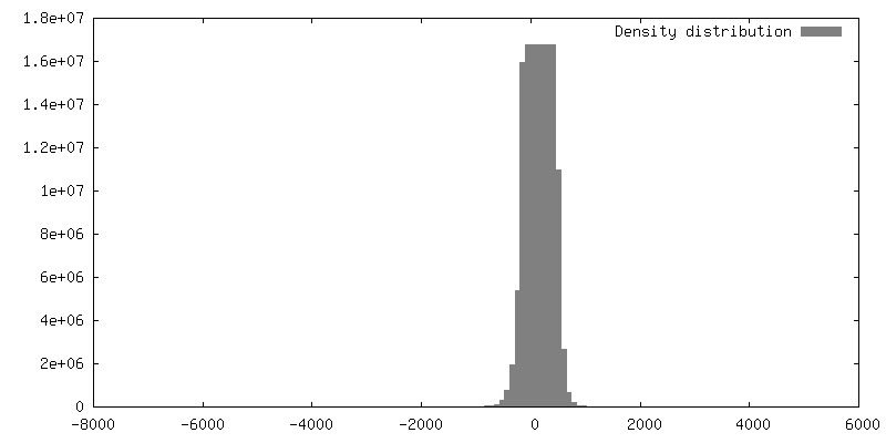
| 注釈 | Cryotomogram of picoplankton chromatin in 5mM EDTA. | ||||||||||||||||||||||||||||||||||||||||||||||||||||||||||||
| 投影像・断面図 | 画像のコントロール
画像は Spider により作成 これらの図は立方格子座標系で作成されたものです | ||||||||||||||||||||||||||||||||||||||||||||||||||||||||||||
| ボクセルのサイズ | X=Y=Z: 9.12 Å | ||||||||||||||||||||||||||||||||||||||||||||||||||||||||||||
| 密度 |
| ||||||||||||||||||||||||||||||||||||||||||||||||||||||||||||
| 対称性 | 空間群: 1 | ||||||||||||||||||||||||||||||||||||||||||||||||||||||||||||
| 詳細 | EMDB XML:
CCP4マップ ヘッダ情報:
| ||||||||||||||||||||||||||||||||||||||||||||||||||||||||||||
 Z (Sec.)
 Y (Row.)
 X (Col.)


-添付データ
-追加マップ: Cryotomogram of yeast chromatin in 50mM EDTA.
| ファイル | emd_6737_additional.map | ||||||||||||
|---|---|---|---|---|---|---|---|---|---|---|---|---|---|
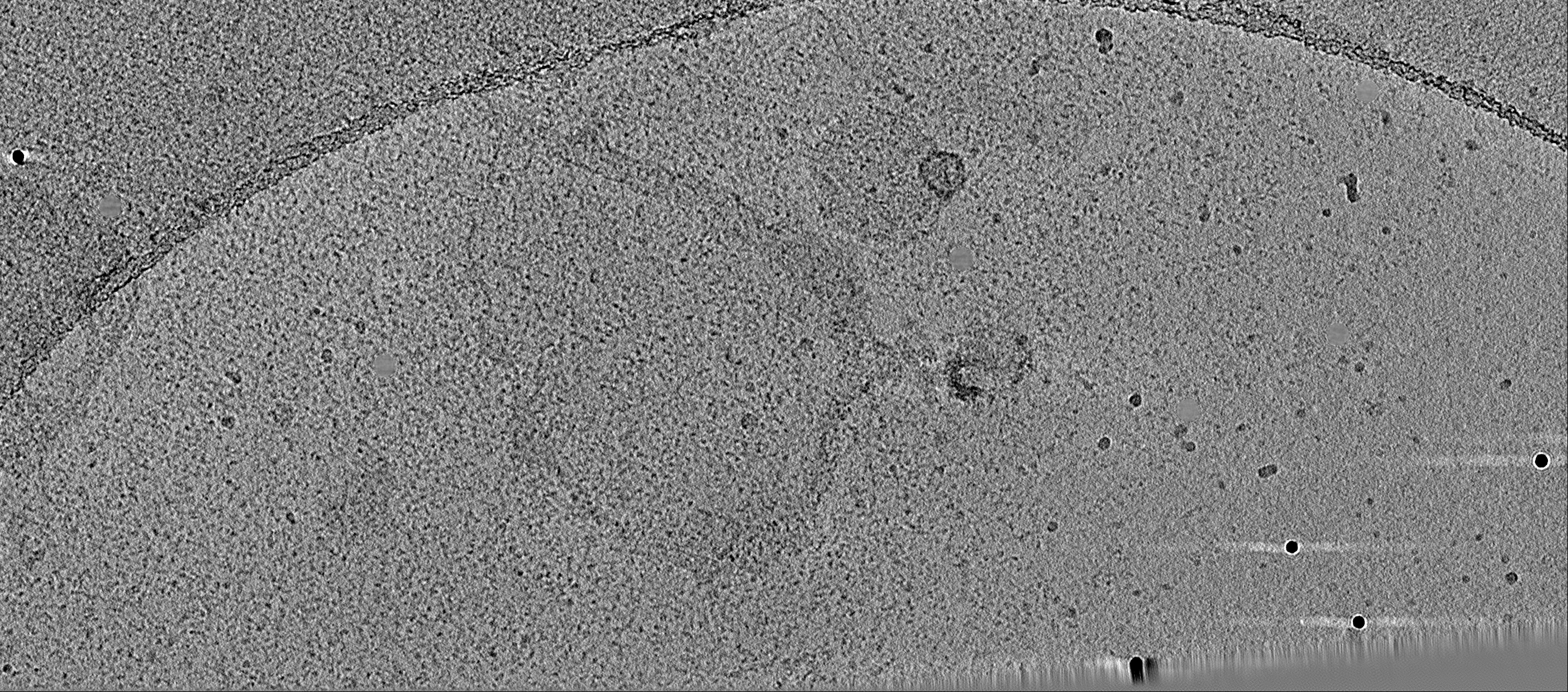
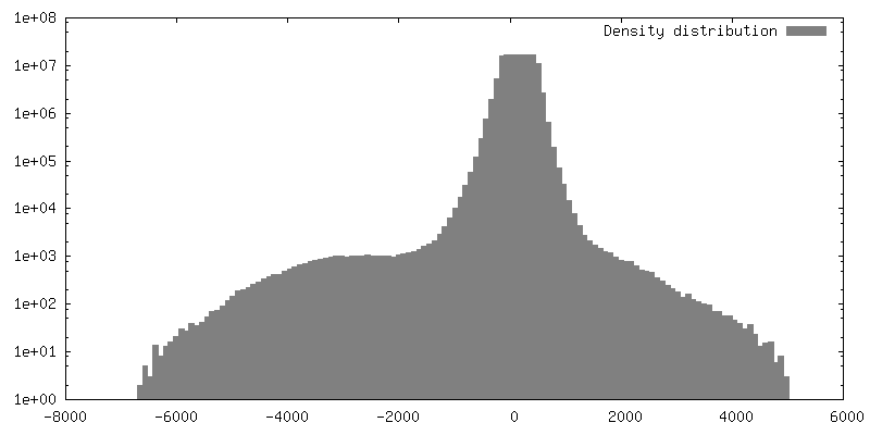
| 注釈 | Cryotomogram of yeast chromatin in 50mM EDTA. | ||||||||||||
| 投影像・断面図 |
| ||||||||||||
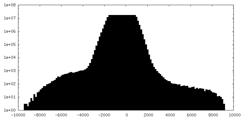
| 密度ヒストグラム |


- 試料の構成要素
-全体 : Chromatin released from the picoplankton Ostreococcus tauri, in 5...
| 全体 | 名称: Chromatin released from the picoplankton Ostreococcus tauri, in 5mM EDTA |
|---|---|
| 要素 |
|
-超分子 #1: Chromatin released from the picoplankton Ostreococcus tauri, in 5...
| 超分子 | 名称: Chromatin released from the picoplankton Ostreococcus tauri, in 5mM EDTA タイプ: organelle_or_cellular_component / ID: 1 / 親要素: 0 |
|---|---|
| 由来(天然) | 生物種: Ostreococcus tauri (植物) / 株: RCC 745 |
-実験情報
-構造解析
| 手法 | クライオ電子顕微鏡法 |
|---|---|
 解析 解析 | 電子線トモグラフィー法 |
| 試料の集合状態 | filament |
-試料調製
| 緩衝液 | pH: 7 構成要素:
| ||||||||||||
|---|---|---|---|---|---|---|---|---|---|---|---|---|---|
| グリッド | モデル: Protochips CF-4/2-2C-T / 材質: COPPER / メッシュ: 200 / 支持フィルム - 材質: CARBON / 支持フィルム - トポロジー: HOLEY / 前処理 - タイプ: PLASMA CLEANING / 詳細: 15mA, Emitech K100X | ||||||||||||
| 凍結 | 凍結剤: NITROGEN / チャンバー内湿度: 100 % / チャンバー内温度: 298 K / 装置: FEI VITROBOT MARK IV | ||||||||||||
| 切片作成 | その他: NO SECTIONING | ||||||||||||
| 位置合わせマーカー | Manufacturer: Sigma / 直径: 10 nm |
- 電子顕微鏡法
| 顕微鏡 | FEI TITAN KRIOS |
|---|---|
| 撮影 | フィルム・検出器のモデル: FEI FALCON II (4k x 4k) 検出モード: INTEGRATING / デジタル化 - サイズ - 横: 4096 pixel / デジタル化 - サイズ - 縦: 4096 pixel / 撮影したグリッド数: 1 / 実像数: 62 / 平均電子線量: 2.4 e/Å2 |
| 電子線 | 加速電圧: 300 kV / 電子線源:  FIELD EMISSION GUN FIELD EMISSION GUN |
| 電子光学系 | 最小 デフォーカス(補正後): 11.0 µm / 倍率(補正後): 15678 / 照射モード: FLOOD BEAM / 撮影モード: BRIGHT FIELD / Cs: 2.7 mm / 最小 デフォーカス(公称値): 8.0 µm / 倍率(公称値): 8700 |
| 試料ステージ | 試料ホルダーモデル: FEI TITAN KRIOS AUTOGRID HOLDER ホルダー冷却材: NITROGEN |
| 実験機器 |  モデル: Titan Krios / 画像提供: FEI Company |
-画像解析
| 最終 再構成 | アルゴリズム: BACK PROJECTION / ソフトウェア - 名称: IMOD (ver. 4.9) / 使用した粒子像数: 59 |
|---|---|
| CTF補正 | ソフトウェア - 名称: IMOD (ver. 4.8) |
