 Movie
Movie Controller

[English] 日本語
 Yorodumi
- EMDB-60563: Cryo-EM structure of uropathogenic Escherichia coli 2:1 CysK:CdiA... -
+ Open data

- Basic information
| Entry |  | |||||||||
|---|---|---|---|---|---|---|---|---|---|---|
| Title | Cryo-EM structure of uropathogenic Escherichia coli 2:1 CysK:CdiA complex | |||||||||
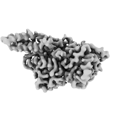
 Map data Map data | ||||||||||
 Sample Sample |
| |||||||||
 Keywords Keywords | RNase / Complex / Contact-dependent growth inhibition / Toxin | |||||||||
| Biological species |  | |||||||||
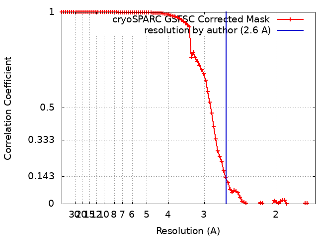
| Method | single particle reconstruction / cryo EM / Resolution: 2.6 Å | |||||||||
 Authors Authors | Feng Z / Yashiro Y / Tomita K | |||||||||
| Funding support |  Japan, 2 items Japan, 2 items
| |||||||||
 Citation Citation | Journal: Nucleic Acids Res / Year: 2025 Title: Mechanism of activation of contact-dependent growth inhibition tRNase toxin by the amino acid biogenesis factor CysK in the bacterial competition system. Authors: Zhaohang Feng / Yuka Yashiro / Kozo Tomita / Abstract: Contact-dependent growth inhibition (CDI) is a bacterial competition mechanism, wherein the C-terminal toxin domain of CdiA protein (CdiA-CT) is transferred from one bacterium to another, impeding ...Contact-dependent growth inhibition (CDI) is a bacterial competition mechanism, wherein the C-terminal toxin domain of CdiA protein (CdiA-CT) is transferred from one bacterium to another, impeding the growth of the toxin recipient. In uropathogenic Escherichia coli 536, CdiA-CT (CdiA-CTEC536) is a tRNA anticodon endonuclease that requires a cysteine biogenesis factor, CysK, for its activity. However, the mechanism underlying tRNA recognition and cleavage by CdiA-CTEC536 remains unresolved. Here, we present the cryo-EM structure of the CysK:CdiA-CTEC536:tRNA ternary complex. The interaction between CdiA-CTEC536 and CysK stabilizes the CdiA-CTEC536 structure and facilitates tRNA binding and the formation of the CdiA-CTEC536 catalytic core structure. The bottom-half of the tRNA interacts exclusively with CdiA-CTEC536 and the α-helices of CdiA-CTEC536 engage with the minor and major grooves of the bottom-half of tRNA, positioning the tRNA anticodon loop at the CdiA-CTEC536 catalytic site for tRNA cleavage. Thus, CysK serves as a platform facilitating the recognition and cleavage of substrate tRNAs by CdiA-CTEC536. | |||||||||
| History |
|
- Structure visualization
| Supplemental images |
|---|
- Downloads & links
-EMDB archive
| Map data | emd_60563.map.gz | 31.9 MB |  EMDB map data format EMDB map data format | |
|---|---|---|---|---|
| Header (meta data) | emd-60563-v30.xmlemd-60563.xml | 20.8 KB 20.8 KB | Display Display | EMDB header |
| FSC (resolution estimation) | emd_60563_fsc.xml | 8.4 KB | Display | FSC data file |
| Images |  emd_60563.png emd_60563.png | 56.9 KB | ||
| Masks | emd_60563_msk_1.map | 64 MB | Mask map | |
| Filedesc metadata | emd-60563.cif.gz | 5.9 KB | ||
| Others | emd_60563_half_map_1.map.gzemd_60563_half_map_2.map.gz | 59.4 MB 59.4 MB | ||
| Archive directory |  http://ftp.pdbj.org/pub/emdb/structures/EMD-60563ftp://ftp.pdbj.org/pub/emdb/structures/EMD-60563 http://ftp.pdbj.org/pub/emdb/structures/EMD-60563ftp://ftp.pdbj.org/pub/emdb/structures/EMD-60563 | HTTPS FTP |
-Related structure data


-Links
| EMDB pages | EMDB (EBI/PDBe) / EMDataResource |
|---|
-Map
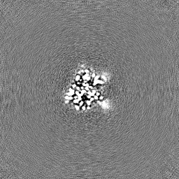
| File | Download / File: emd_60563.map.gz / Format: CCP4 / Size: 64 MB / Type: IMAGE STORED AS FLOATING POINT NUMBER (4 BYTES) | ||||||||||||||||||||||||||||||||||||
|---|---|---|---|---|---|---|---|---|---|---|---|---|---|---|---|---|---|---|---|---|---|---|---|---|---|---|---|---|---|---|---|---|---|---|---|---|---|
| Projections & slices | Image control
Images are generated by Spider. | ||||||||||||||||||||||||||||||||||||
| Voxel size | X=Y=Z: 0.83 Å | ||||||||||||||||||||||||||||||||||||
| Density |
| ||||||||||||||||||||||||||||||||||||
| Symmetry | Space group: 1 | ||||||||||||||||||||||||||||||||||||
| Details | EMDB XML:
|
 Z (Sec.)
 Y (Row.)
 X (Col.)


-Supplemental data
-Mask #1
| File | emd_60563_msk_1.map | ||||||||||||
|---|---|---|---|---|---|---|---|---|---|---|---|---|---|
| Projections & Slices |
| ||||||||||||
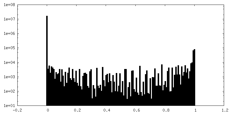
| Density Histograms |


-Half map: #1
| File | emd_60563_half_map_1.map | ||||||||||||
|---|---|---|---|---|---|---|---|---|---|---|---|---|---|
| Projections & Slices |
| ||||||||||||
| Density Histograms |


-Half map: #2
| File | emd_60563_half_map_2.map | ||||||||||||
|---|---|---|---|---|---|---|---|---|---|---|---|---|---|
| Projections & Slices |
| ||||||||||||
| Density Histograms |


- Sample components
-Entire : 2 CysK in complex with 1 CdiA-CT.
| Entire | Name: 2 CysK in complex with 1 CdiA-CT. |
|---|---|
| Components |
|
-Supramolecule #1: 2 CysK in complex with 1 CdiA-CT.
| Supramolecule | Name: 2 CysK in complex with 1 CdiA-CT. / type: complex / ID: 1 / Parent: 0 / Macromolecule list: all |
|---|---|
| Source (natural) | Organism: |
| Molecular weight | Theoretical: 90 KDa |
-Macromolecule #1: CysK
| Macromolecule | Name: CysK / type: protein_or_peptide / ID: 1 / Enantiomer: LEVO |
|---|---|
| Source (natural) | Organism: |
| Recombinant expression | Organism: |
| Sequence | String: MSKIFEDNSL TIGHTPLVRL NRIGNGRILA KVESRNPSFS V(LLP)CRIGANMI WDAEKRGVLK PGVELVEPTS GNTGIA LAY VAAARGYKLT LTMPETMSIE RRKLLKALGA NLVLTEGAKG MKGAIQKAEE IVASNPEKYL LLQQFSNPAN PEIHEKT TG PEIWEDTDGQ ...String: MSKIFEDNSL TIGHTPLVRL NRIGNGRILA KVESRNPSFS V(LLP)CRIGANMI WDAEKRGVLK PGVELVEPTS GNTGIA LAY VAAARGYKLT LTMPETMSIE RRKLLKALGA NLVLTEGAKG MKGAIQKAEE IVASNPEKYL LLQQFSNPAN PEIHEKT TG PEIWEDTDGQ VDVFIAGVGT GGTLTGVSRY IKGTKGKTDL ISVAVEPTDS PVIAQALAGE EIKPGPHKIQ GIGAGFIP A NLDLKLVDKV IGITNEEAIS TARRLMEEEG ILAGISSGAA VAAALKLQED ESFTNKNIVV ILPSSGERYL STALFADLF TEKELQQ |
-Macromolecule #2: CdiA-CT
| Macromolecule | Name: CdiA-CT / type: protein_or_peptide / ID: 2 / Enantiomer: LEVO |
|---|---|
| Source (natural) | Organism: |
| Recombinant expression | Organism: |
| Sequence | String: MHHHHHHVEN NALSLVARGC AVAAPCRTKV AEQLLEIGAK AGMAGLAGAA VKDMADRMTS DELEHLITLQ MMGNDEITTK YLSSLHDKY GSGAASNPNI GKDLTDAEKV ELGGSGSGTG TPPPSENDPK QQNEKTVDKL NQKQESAIKK IDNTIKNALK D HDIIGTLK ...String: MHHHHHHVEN NALSLVARGC AVAAPCRTKV AEQLLEIGAK AGMAGLAGAA VKDMADRMTS DELEHLITLQ MMGNDEITTK YLSSLHDKY GSGAASNPNI GKDLTDAEKV ELGGSGSGTG TPPPSENDPK QQNEKTVDKL NQKQESAIKK IDNTIKNALK D HDIIGTLK DMDGKPVPKE NGGYWDAMQE MQNTLRGLRN HADTLKNVNN PEAQAAYGRA TDAINKIESA LKGYGI |
-Experimental details
-Structure determination
| Method | cryo EM |
|---|---|
 Processing Processing | single particle reconstruction |
| Aggregation state | particle |
-Sample preparation
| Concentration | 1.6 mg/mL | |||||||||||||||
|---|---|---|---|---|---|---|---|---|---|---|---|---|---|---|---|---|
| Buffer | pH: 7 Component:
Details: 25mM Tris-HCl,50mM NaCl,2mM MgCl2, 10mM 2-mercaptoethanol | |||||||||||||||
| Vitrification | Cryogen name: ETHANE / Chamber humidity: 100 % / Chamber temperature: 277.15 K / Instrument: FEI VITROBOT MARK IV |
- Electron microscopy
| Microscope | FEI TITAN KRIOS |
|---|---|
| Image recording | Film or detector model: GATAN K3 BIOQUANTUM (6k x 4k) / Digitization - Dimensions - Width: 4092 pixel / Digitization - Dimensions - Height: 5760 pixel / Number grids imaged: 1 / Number real images: 7044 / Average exposure time: 1.0 sec. / Average electron dose: 50.0 e/Å2 |
| Electron beam | Acceleration voltage: 300 kV / Electron source:  FIELD EMISSION GUN FIELD EMISSION GUN |
| Electron optics | Illumination mode: FLOOD BEAM / Imaging mode: BRIGHT FIELD / Cs: 2.7 mm / Nominal defocus max: 1.8 µm / Nominal defocus min: 0.8 µm / Nominal magnification: 105000 |
| Sample stage | Cooling holder cryogen: NITROGEN |
| Experimental equipment |  Model: Titan Krios / Image courtesy: FEI Company |
+Image processing
-Atomic model buiding 1
| Initial model | PDB ID: Chain - Source name: PDB / Chain - Initial model type: experimental model |
|---|

