 Movie
Movie Controller

[English] 日本語
 Yorodumi
- EMDB-52140: In situ human nuclear ring subunit of nuclear pore complex (FIB-l... -
+ Open data

- Basic information
| Entry | 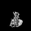 | |||||||||
|---|---|---|---|---|---|---|---|---|---|---|
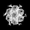
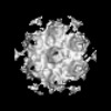
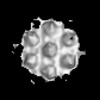
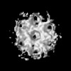
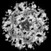
| Title | In situ human nuclear ring subunit of nuclear pore complex (FIB-lamella data of mock infected macrophages) | |||||||||
 Map data Map data | Nuclear ring of NPC from mock infected macrophages - masked | |||||||||
 Sample Sample |
| |||||||||
 Keywords Keywords | Nuclear envelope / Nuclear pore / Nucleocytoplasmic transport / NPC / STRUCTURAL PROTEIN / MEMBRANE PROTEIN | |||||||||
| Biological species |  Homo sapiens (human) Homo sapiens (human) | |||||||||
| Method | subtomogram averaging / cryo EM / Resolution: 30.4 Å | |||||||||
 Authors Authors | Kreysing JP / Welsch S / Turonova B / Beck M | |||||||||
| Funding support |  Germany, 2 items Germany, 2 items
| |||||||||
 Citation Citation | Journal: Cell / Year: 2025 Title: Passage of the HIV capsid cracks the nuclear pore. Authors: Jan Philipp Kreysing / Maziar Heidari / Vojtech Zila / Sergio Cruz-León / Agnieszka Obarska-Kosinska / Vibor Laketa / Lara Rohleder / Sonja Welsch / Jürgen Köfinger / Beata Turoňová / ...Authors: Jan Philipp Kreysing / Maziar Heidari / Vojtech Zila / Sergio Cruz-León / Agnieszka Obarska-Kosinska / Vibor Laketa / Lara Rohleder / Sonja Welsch / Jürgen Köfinger / Beata Turoňová / Gerhard Hummer / Hans-Georg Kräusslich / Martin Beck / Abstract: Upon infection, human immunodeficiency virus type 1 (HIV-1) releases its cone-shaped capsid into the cytoplasm of infected T cells and macrophages. The capsid enters the nuclear pore complex (NPC), ...Upon infection, human immunodeficiency virus type 1 (HIV-1) releases its cone-shaped capsid into the cytoplasm of infected T cells and macrophages. The capsid enters the nuclear pore complex (NPC), driven by interactions with numerous phenylalanine-glycine (FG)-repeat nucleoporins (FG-Nups). Whether NPCs structurally adapt to capsid passage and whether capsids are modified during passage remains unknown, however. Here, we combined super-resolution and correlative microscopy with cryoelectron tomography and molecular simulations to study the nuclear entry of HIV-1 capsids in primary human macrophages. Our data indicate that cytosolically bound cyclophilin A is stripped off capsids entering the NPC, and the capsid hexagonal lattice remains largely intact inside and beyond the central channel. Strikingly, the NPC scaffold rings frequently crack during capsid passage, consistent with computer simulations indicating the need for NPC widening. The unique cone shape of the HIV-1 capsid facilitates its entry into NPCs and helps to crack their rings. | |||||||||
| History |
|
- Structure visualization
| Supplemental images |
|---|

- Downloads & links
-EMDB archive
| Map data | emd_52140.map.gz | 482.5 KB |  EMDB map data format EMDB map data format | |
|---|---|---|---|---|
| Header (meta data) | emd-52140-v30.xmlemd-52140.xml | 19 KB 19 KB | Display Display | EMDB header |
| FSC (resolution estimation) | emd_52140_fsc.xml | 4.1 KB | Display | FSC data file |
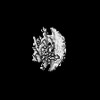
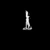
| Images |  emd_52140.png emd_52140.png | 47.1 KB | ||
| Filedesc metadata | emd-52140.cif.gz | 4.7 KB | ||
| Others | emd_52140_additional_1.map.gzemd_52140_half_map_1.map.gzemd_52140_half_map_2.map.gz | 5 MB 5 MB 5 MB | ||
| Archive directory |  http://ftp.pdbj.org/pub/emdb/structures/EMD-52140ftp://ftp.pdbj.org/pub/emdb/structures/EMD-52140 http://ftp.pdbj.org/pub/emdb/structures/EMD-52140ftp://ftp.pdbj.org/pub/emdb/structures/EMD-52140 | HTTPS FTP |
-Related structure data
| Related structure data | C: citing same article ( |
|---|


-Links
| EMDB pages | EMDB (EBI/PDBe) / EMDataResource |
|---|
-Map
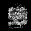
| File | Download / File: emd_52140.map.gz / Format: CCP4 / Size: 5.4 MB / Type: IMAGE STORED AS FLOATING POINT NUMBER (4 BYTES) | ||||||||||||||||||||||||||||||||||||
|---|---|---|---|---|---|---|---|---|---|---|---|---|---|---|---|---|---|---|---|---|---|---|---|---|---|---|---|---|---|---|---|---|---|---|---|---|---|
| Annotation | Nuclear ring of NPC from mock infected macrophages - masked | ||||||||||||||||||||||||||||||||||||
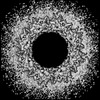
| Projections & slices | Image control
Images are generated by Spider. | ||||||||||||||||||||||||||||||||||||
| Voxel size | X=Y=Z: 9.656 Å | ||||||||||||||||||||||||||||||||||||
| Density |
| ||||||||||||||||||||||||||||||||||||
| Symmetry | Space group: 1 | ||||||||||||||||||||||||||||||||||||
| Details | EMDB XML:
|
 Z (Sec.)
 Y (Row.)
 X (Col.)


-Supplemental data
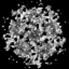
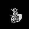
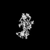
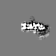
-Additional map: Nuclear ring of NPC from mock infected macrophages - unmasked
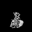
| File | emd_52140_additional_1.map | ||||||||||||
|---|---|---|---|---|---|---|---|---|---|---|---|---|---|
| Annotation | Nuclear ring of NPC from mock infected macrophages - unmasked | ||||||||||||
| Projections & Slices |
| ||||||||||||
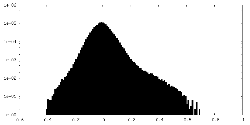
| Density Histograms |


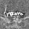


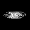
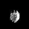
-Half map: Nuclear ring of NPC from mock infected macrophages - half2
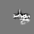
| File | emd_52140_half_map_1.map | ||||||||||||
|---|---|---|---|---|---|---|---|---|---|---|---|---|---|
| Annotation | Nuclear ring of NPC from mock infected macrophages - half2 | ||||||||||||
| Projections & Slices |
| ||||||||||||
| Density Histograms |
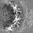


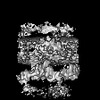
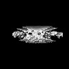
-Half map: Nuclear ring of NPC from mock infected macrophages - half1
| File | emd_52140_half_map_2.map | ||||||||||||
|---|---|---|---|---|---|---|---|---|---|---|---|---|---|
| Annotation | Nuclear ring of NPC from mock infected macrophages - half1 | ||||||||||||
| Projections & Slices |
| ||||||||||||
| Density Histograms |


- Sample components
-Entire : In situ human nuclear ring subunit of nuclear pore complex (FIB-l...
| Entire | Name: In situ human nuclear ring subunit of nuclear pore complex (FIB-lamella data of mock infected macrophages) |
|---|---|
| Components |
|
-Supramolecule #1: In situ human nuclear ring subunit of nuclear pore complex (FIB-l...
| Supramolecule | Name: In situ human nuclear ring subunit of nuclear pore complex (FIB-lamella data of mock infected macrophages) type: cell / ID: 1 / Parent: 0 |
|---|---|
| Source (natural) | Organism: Homo sapiens (human) |
-Experimental details
-Structure determination
| Method | cryo EM |
|---|---|
 Processing Processing | subtomogram averaging |
| Aggregation state | cell |
-Sample preparation
| Buffer | pH: 7.4 |
|---|---|
| Grid | Model: Quantifoil R2/2 / Material: GOLD / Mesh: 200 / Pretreatment - Type: GLOW DISCHARGE |
| Vitrification | Cryogen name: ETHANE / Chamber temperature: 310 K / Instrument: LEICA EM GP |
- Electron microscopy
| Microscope | TFS KRIOS |
|---|---|
| Specialist optics | Energy filter - Name: TFS Selectris X / Energy filter - Slit width: 10 eV |
| Image recording | Film or detector model: FEI FALCON IV (4k x 4k) / Average electron dose: 2.2 e/Å2 |
| Electron beam | Acceleration voltage: 300 kV / Electron source:  FIELD EMISSION GUN FIELD EMISSION GUN |
| Electron optics | Illumination mode: FLOOD BEAM / Imaging mode: BRIGHT FIELD / Nominal defocus max: 4.0 µm / Nominal defocus min: 2.0 µm / Nominal magnification: 53000 |
| Experimental equipment |  Model: Titan Krios / Image courtesy: FEI Company |
-Image processing
| Final reconstruction | Resolution.type: BY AUTHOR / Resolution: 30.4 Å / Resolution method: FSC 0.143 CUT-OFF / Software - Name: novaSTA / Number subtomograms used: 571 |
|---|---|
| Extraction | Number tomograms: 52 / Number images used: 940 / Software - Name: novaSTA |
| Final angle assignment | Type: OTHER / Software - Name: novaSTA / Details: Cross correlations |
| FSC plot (resolution estimation) |  |
