ribonucleotide reductase / class Ia / mechanistic inhibition / OXIDOREDUCTASE
Function / homology
Function and homology information
ribonucleoside diphosphate metabolic process / 2'-deoxyribonucleotide biosynthetic process / nucleobase-containing small molecule interconversion / ribonucleoside-diphosphate reductase complex / ribonucleoside-diphosphate reductase / ribonucleoside-diphosphate reductase activity, thioredoxin disulfide as acceptor / deoxyribonucleotide biosynthetic process / protein folding chaperone / iron ion binding / ATP binding ...ribonucleoside diphosphate metabolic process / 2'-deoxyribonucleotide biosynthetic process / nucleobase-containing small molecule interconversion / ribonucleoside-diphosphate reductase complex / ribonucleoside-diphosphate reductase / ribonucleoside-diphosphate reductase activity, thioredoxin disulfide as acceptor / deoxyribonucleotide biosynthetic process / protein folding chaperone / iron ion binding / ATP binding / identical protein binding / cytoplasm / cytosol Similarity search - Function
ATP-cone domain / ATP cone domain / ATP-cone domain profile. / Ribonucleotide reductase, class I , alpha subunit / Ribonucleotide reductase large subunit signature. / Ribonucleoside-diphosphate reductase large subunit / Ribonucleotide reductase R1 subunit, N-terminal / Ribonucleotide reductase large subunit, N-terminal / Ribonucleotide reductase, all-alpha domain / Ribonucleotide reductase large subunit, C-terminal ...ATP-cone domain / ATP cone domain / ATP-cone domain profile. / Ribonucleotide reductase, class I , alpha subunit / Ribonucleotide reductase large subunit signature. / Ribonucleoside-diphosphate reductase large subunit / Ribonucleotide reductase R1 subunit, N-terminal / Ribonucleotide reductase large subunit, N-terminal / Ribonucleotide reductase, all-alpha domain / Ribonucleotide reductase large subunit, C-terminal / Ribonucleotide reductase, barrel domain / Ribonucleotide reductase small subunit, acitve site / Ribonucleotide reductase small subunit signature. / Ribonucleotide reductase small subunit / Ribonucleotide reductase small subunit family / Ribonucleotide reductase, small chain / Ribonucleotide reductase-like / Ferritin-like superfamily Similarity search - Domain/homology
National Institutes of Health/National Institute of General Medical Sciences (NIH/NIGMS)
1R35GM126982-01
United States
National Institutes of Health/National Institute of General Medical Sciences (NIH/NIGMS)
GM047274
United States
National Institutes of Health/National Institute of General Medical Sciences (NIH/NIGMS)
GM29595
United States
National Institutes of Health/National Institute of General Medical Sciences (NIH/NIGMS)
1F32GM145072-01
United States
Citation
Journal: Proc Natl Acad Sci U S A / Year: 2024 Title: 2.6-Å resolution cryo-EM structure of a class Ia ribonucleotide reductase trapped with mechanism-based inhibitor NCDP. Authors: Dana E Westmoreland / Patricia R Feliciano / Gyunghoon Kang / Chang Cui / Albert Kim / JoAnne Stubbe / Daniel G Nocera / Catherine L Drennan / Abstract: Ribonucleotide reductases (RNRs) reduce ribonucleotides to deoxyribonucleotides using radical-based chemistry. For class Ia RNRs, the radical species is stored in a separate subunit (β2) from the ...Ribonucleotide reductases (RNRs) reduce ribonucleotides to deoxyribonucleotides using radical-based chemistry. For class Ia RNRs, the radical species is stored in a separate subunit (β2) from the subunit housing the active site (α2), requiring the formation of a short-lived α2β2 complex and long-range radical transfer (RT). RT occurs via proton-coupled electron transfer (PCET) over a long distance (~32-Å) and involves the formation and decay of multiple amino acid radical species. Here, we use cryogenic electron microscopy and a mechanism-based inhibitor 2'-azido-2'-deoxycytidine-5'-diphosphate (NCDP) to trap a wild-type α2β2 complex of class Ia RNR. We find that one α subunit has turned over and that the other is trapped, bound to β in a midturnover state. Instead of NCDP in the active site, forward RT has resulted in N loss, migration of the third nitrogen from the ribose C2' to C3' positions, and attachment of this nitrogen to the sulfur of cysteine-225. In this study, an inhibitor has been visualized as an adduct to an RNR. Additionally, this structure reveals the positions of PCET residues following forward RT, complementing the previous structure that depicted a preturnover PCET pathway and suggesting how PCET is gated at the α-β interface. This NCDP-trapped structure is also of sufficient resolution (2.6 Å) to visualize water molecules, allowing us to evaluate the proposal that water molecules are proton acceptors and donors as part of the PCET process.
Supramolecule #1: Active state of class Ia ribonucleotide reductase trapped with me...
Supramolecule
Name: Active state of class Ia ribonucleotide reductase trapped with mechanism-based inhibitor N3CDP type: complex / ID: 1 / Parent: 0 / Macromolecule list: #1-#2
In the structure databanks used in Yorodumi, some data are registered as the other names, "COVID-19 virus" and "2019-nCoV". Here are the details of the virus and the list of structure data.
Jan 31, 2019. EMDB accession codes are about to change! (news from PDBe EMDB page)
EMDB accession codes are about to change! (news from PDBe EMDB page)
The allocation of 4 digits for EMDB accession codes will soon come to an end. Whilst these codes will remain in use, new EMDB accession codes will include an additional digit and will expand incrementally as the available range of codes is exhausted. The current 4-digit format prefixed with “EMD-” (i.e. EMD-XXXX) will advance to a 5-digit format (i.e. EMD-XXXXX), and so on. It is currently estimated that the 4-digit codes will be depleted around Spring 2019, at which point the 5-digit format will come into force.
The EM Navigator/Yorodumi systems omit the EMD- prefix.
Related info.:Q: What is EMD? / ID/Accession-code notation in Yorodumi/EM Navigator
Yorodumi is a browser for structure data from EMDB, PDB, SASBDB, etc.
This page is also the successor to EM Navigator detail page, and also detail information page/front-end page for Omokage search.
The word "yorodu" (or yorozu) is an old Japanese word meaning "ten thousand". "mi" (miru) is to see.
Related info.:EMDB / PDB / SASBDB / Comparison of 3 databanks / Yorodumi Search / Aug 31, 2016. New EM Navigator & Yorodumi / Yorodumi Papers / Jmol/JSmol / Function and homology information / Changes in new EM Navigator and Yorodumi
 Movie
Movie Controller

 Yorodumi
 Open data
 Basic information
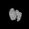
 Map data
 Sample
 Keywords
 Function and homology information
 Authors
 United States, 4 items
  Citation
 Structure visualization
 Downloads & links
 emd_46711.png
 http://ftp.pdbj.org/pub/emdb/structures/EMD-46711

 Z (Sec.)
 Y (Row.)
 X (Col.)


 Sample components


 Processing
 Electron microscopy
 FIELD EMISSION GUN

