 Movie
Movie Controller

[English] 日本語
 Yorodumi
- PDB-9db2: Class Ia ribonucleotide reductase with mechanism-based inhibitor N3CDP -
+ Open data

- Basic information
| Entry | Database: PDB / ID: 9db2 | |||||||||||||||
|---|---|---|---|---|---|---|---|---|---|---|---|---|---|---|---|---|
| Title | Class Ia ribonucleotide reductase with mechanism-based inhibitor N3CDP | |||||||||||||||
 Components Components | (Ribonucleoside-diphosphate reductase 1 subunit ...) x 2 | |||||||||||||||
 Keywords Keywords | OXIDOREDUCTASE / ribonucleotide reductase / class Ia / mechanistic inhibition | |||||||||||||||
| Function / homology |  Function and homology information Function and homology informationribonucleoside diphosphate metabolic process / 2'-deoxyribonucleotide biosynthetic process / nucleobase-containing small molecule interconversion / ribonucleoside-diphosphate reductase complex / ribonucleoside-diphosphate reductase / ribonucleoside-diphosphate reductase activity, thioredoxin disulfide as acceptor / deoxyribonucleotide biosynthetic process / protein folding chaperone / iron ion binding / ATP binding ...ribonucleoside diphosphate metabolic process / 2'-deoxyribonucleotide biosynthetic process / nucleobase-containing small molecule interconversion / ribonucleoside-diphosphate reductase complex / ribonucleoside-diphosphate reductase / ribonucleoside-diphosphate reductase activity, thioredoxin disulfide as acceptor / deoxyribonucleotide biosynthetic process / protein folding chaperone / iron ion binding / ATP binding / identical protein binding / cytoplasm / cytosol Similarity search - Function | |||||||||||||||
| Biological species |  | |||||||||||||||
| Method | ELECTRON MICROSCOPY / single particle reconstruction / cryo EM / Resolution: 2.6 Å | |||||||||||||||
 Authors Authors | Westmoreland, D.E. / Drennan, C.L. | |||||||||||||||
| Funding support |  United States, 4items United States, 4items
| |||||||||||||||
 Citation Citation | Journal: Proc Natl Acad Sci U S A / Year: 2024 Title: 2.6-Å resolution cryo-EM structure of a class Ia ribonucleotide reductase trapped with mechanism-based inhibitor NCDP. Authors: Dana E Westmoreland / Patricia R Feliciano / Gyunghoon Kang / Chang Cui / Albert Kim / JoAnne Stubbe / Daniel G Nocera / Catherine L Drennan / Abstract: Ribonucleotide reductases (RNRs) reduce ribonucleotides to deoxyribonucleotides using radical-based chemistry. For class Ia RNRs, the radical species is stored in a separate subunit (β2) from the ...Ribonucleotide reductases (RNRs) reduce ribonucleotides to deoxyribonucleotides using radical-based chemistry. For class Ia RNRs, the radical species is stored in a separate subunit (β2) from the subunit housing the active site (α2), requiring the formation of a short-lived α2β2 complex and long-range radical transfer (RT). RT occurs via proton-coupled electron transfer (PCET) over a long distance (~32-Å) and involves the formation and decay of multiple amino acid radical species. Here, we use cryogenic electron microscopy and a mechanism-based inhibitor 2'-azido-2'-deoxycytidine-5'-diphosphate (NCDP) to trap a wild-type α2β2 complex of class Ia RNR. We find that one α subunit has turned over and that the other is trapped, bound to β in a midturnover state. Instead of NCDP in the active site, forward RT has resulted in N loss, migration of the third nitrogen from the ribose C2' to C3' positions, and attachment of this nitrogen to the sulfur of cysteine-225. In this study, an inhibitor has been visualized as an adduct to an RNR. Additionally, this structure reveals the positions of PCET residues following forward RT, complementing the previous structure that depicted a preturnover PCET pathway and suggesting how PCET is gated at the α-β interface. This NCDP-trapped structure is also of sufficient resolution (2.6 Å) to visualize water molecules, allowing us to evaluate the proposal that water molecules are proton acceptors and donors as part of the PCET process. | |||||||||||||||
| History |
|
- Structure visualization
| Structure viewer | Molecule: MolmilJmol/JSmol |
|---|
- Downloads & links
-Download
| PDBx/mmCIF format | 9db2.cif.gz | 471.4 KB | Display | PDBx/mmCIF format |
|---|---|---|---|---|
| PDB format | pdb9db2.ent.gz | Display | PDB format | |
| PDBx/mmJSON format | 9db2.json.gz | Tree view | PDBx/mmJSON format | |
| Others |  Other downloads Other downloads |
-Validation report
| Arichive directory | https://data.pdbj.org/pub/pdb/validation_reports/db/9db2ftp://data.pdbj.org/pub/pdb/validation_reports/db/9db2 | HTTPS FTP |
|---|
-Related structure data
| Related structure data | 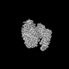 46711MC M: map data used to model this data C: citing same article ( |
|---|---|
| Similar structure data |
-Links
 PDBj
PDBj


- Assembly
| Deposited unit | 
|
|---|---|
| 1 |
|
-Components
-Ribonucleoside-diphosphate reductase 1 subunit ... , 2 types, 4 molecules ABCD
| #1: Protein | Mass: 85877.086 Da / Num. of mol.: 2 Source method: isolated from a genetically manipulated source Source: (gene. exp.) References: UniProt: P00452, ribonucleoside-diphosphate reductase #2: Protein | Mass: 43558.055 Da / Num. of mol.: 2 Source method: isolated from a genetically manipulated source Source: (gene. exp.) References: UniProt: P69924, ribonucleoside-diphosphate reductase |
|---|
-Non-polymers , 7 types, 638 molecules 


| #3: Chemical |  Mass: 491.182 Da / Num. of mol.: 2 / Source method: obtained synthetically / Formula: C10H16N5O12P3 / Feature type: SUBJECT OF INVESTIGATION Mass: 491.182 Da / Num. of mol.: 2 / Source method: obtained synthetically / Formula: C10H16N5O12P3 / Feature type: SUBJECT OF INVESTIGATION#4: Chemical | ChemComp-MG /  Mass: 24.305 Da / Num. of mol.: 4 / Source method: obtained synthetically / Formula: Mg Mass: 24.305 Da / Num. of mol.: 4 / Source method: obtained synthetically / Formula: Mg#5: Chemical | ChemComp-ATP /  Mass: 507.181 Da / Num. of mol.: 4 / Source method: obtained synthetically / Formula: C10H16N5O13P3 / Feature type: SUBJECT OF INVESTIGATION / Comment: ATP, energy-carrying molecule*YM Mass: 507.181 Da / Num. of mol.: 4 / Source method: obtained synthetically / Formula: C10H16N5O13P3 / Feature type: SUBJECT OF INVESTIGATION / Comment: ATP, energy-carrying molecule*YM#6: Chemical | ChemComp-UNL / | Mass: 214.110 Da / Num. of mol.: 1 / Source method: obtained synthetically / Feature type: SUBJECT OF INVESTIGATION #7: Chemical | ChemComp-A1A3L / | Mass: 402.192 Da / Num. of mol.: 1 / Source method: obtained synthetically / Formula: C9H16N4O10P2 / Feature type: SUBJECT OF INVESTIGATION #8: Chemical |  Mass: 127.689 Da / Num. of mol.: 2 / Source method: obtained synthetically / Formula: Fe2O / Feature type: SUBJECT OF INVESTIGATION Mass: 127.689 Da / Num. of mol.: 2 / Source method: obtained synthetically / Formula: Fe2O / Feature type: SUBJECT OF INVESTIGATION#9: Water | ChemComp-HOH / | Mass: 18.015 Da / Num. of mol.: 624 / Source method: isolated from a natural source / Formula: H2O |
|---|
-Details
| Has ligand of interest | Y |
|---|---|
| Has protein modification | Y |
-Experimental details
-Experiment
| Experiment | Method: ELECTRON MICROSCOPY |
|---|---|
| EM experiment | Aggregation state: PARTICLE / 3D reconstruction method: single particle reconstruction |
- Sample preparation
| Component | Name: Active state of class Ia ribonucleotide reductase trapped with mechanism-based inhibitor N3CDP Type: COMPLEX / Entity ID: #1-#2 / Source: RECOMBINANT |
|---|---|
| Molecular weight | Value: 0.26069 MDa / Experimental value: NO |
| Source (natural) | Organism: |
| Source (recombinant) | Organism: |
| Buffer solution | pH: 7.6 |
| Specimen | Embedding applied: NO / Shadowing applied: NO / Staining applied: NO / Vitrification applied: YES |
| Vitrification | Cryogen name: ETHANE |
- Electron microscopy imaging
| Experimental equipment |  Model: Titan Krios / Image courtesy: FEI Company |
|---|---|
| Microscopy | Model: FEI TITAN KRIOS |
| Electron gun | Electron source:  FIELD EMISSION GUN / Accelerating voltage: 300 kV / Illumination mode: OTHER FIELD EMISSION GUN / Accelerating voltage: 300 kV / Illumination mode: OTHER |
| Electron lens | Mode: BRIGHT FIELD / Nominal defocus max: 2300 nm / Nominal defocus min: 1000 nm |
| Image recording | Electron dose: 50.946 e/Å2 / Film or detector model: GATAN K2 SUMMIT (4k x 4k) |
- Processing
| EM software | Name: PHENIX / Version: 1.20.1_4487: / Category: model refinement | ||||||||||||||||||||||||
|---|---|---|---|---|---|---|---|---|---|---|---|---|---|---|---|---|---|---|---|---|---|---|---|---|---|
| CTF correction | Details: CTF correction using RELION's implementation of CTFFIND Type: NONE | ||||||||||||||||||||||||
| Symmetry | Point symmetry: C1 (asymmetric) | ||||||||||||||||||||||||
| 3D reconstruction | Resolution: 2.6 Å / Resolution method: FSC 0.143 CUT-OFF / Num. of particles: 440549 / Symmetry type: POINT | ||||||||||||||||||||||||
| Refine LS restraints |
|
