 Movie
Movie Controller


+ Open data

- Basic information
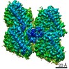
| Entry | Database: EMDB / ID: EMD-4643 | |||||||||
|---|---|---|---|---|---|---|---|---|---|---|
| Title | HsCKK (human CAMSAP1) decorated 13pf taxol-GDP microtubule | |||||||||
 Map data Map data | HsCKK-13pf microtubule symmetrised reconstruction (1x asymmetric unit, local resolution filtered) | |||||||||
 Sample Sample |
| |||||||||
 Keywords Keywords | Microtubule CAMSAP Calmodulin-regulated spectrum-associated proteins CKK Cryo-EM Cryo-Electron Microscopy / STRUCTURAL PROTEIN | |||||||||
| Function / homology |  Function and homology information Function and homology informationmicrotubule minus-end binding / Cilium Assembly / odontoblast differentiation / Post-chaperonin tubulin folding pathway / cytoskeleton-dependent intracellular transport / Carboxyterminal post-translational modifications of tubulin / Microtubule-dependent trafficking of connexons from Golgi to the plasma membrane / Sealing of the nuclear envelope (NE) by ESCRT-III / Intraflagellar transport / Formation of tubulin folding intermediates by CCT/TriC ...microtubule minus-end binding / Cilium Assembly / odontoblast differentiation / Post-chaperonin tubulin folding pathway / cytoskeleton-dependent intracellular transport / Carboxyterminal post-translational modifications of tubulin / Microtubule-dependent trafficking of connexons from Golgi to the plasma membrane / Sealing of the nuclear envelope (NE) by ESCRT-III / Intraflagellar transport / Formation of tubulin folding intermediates by CCT/TriC / regulation of cell morphogenesis / Gap junction assembly / Kinesins / Assembly and cell surface presentation of NMDA receptors / COPI-independent Golgi-to-ER retrograde traffic / GTPase activating protein binding / natural killer cell mediated cytotoxicity / COPI-dependent Golgi-to-ER retrograde traffic / spectrin binding / nuclear envelope lumen / intercellular bridge / regulation of synapse organization / Recycling pathway of L1 / regulation of microtubule polymerization / RHOH GTPase cycle / MHC class I protein binding / cellular response to interleukin-4 / microtubule-based process / RHO GTPases activate IQGAPs / Hedgehog 'off' state / COPI-mediated anterograde transport / cytoplasmic microtubule / Activation of AMPK downstream of NMDARs / ciliary tip / cytoskeleton organization / Loss of Nlp from mitotic centrosomes / Loss of proteins required for interphase microtubule organization from the centrosome / Recruitment of mitotic centrosome proteins and complexes / MHC class II antigen presentation / Recruitment of NuMA to mitotic centrosomes / Anchoring of the basal body to the plasma membrane / HSP90 chaperone cycle for steroid hormone receptors (SHR) in the presence of ligand / Mitotic Prometaphase / EML4 and NUDC in mitotic spindle formation / AURKA Activation by TPX2 / Resolution of Sister Chromatid Cohesion / sperm end piece / sperm principal piece / Translocation of SLC2A4 (GLUT4) to the plasma membrane / RHO GTPases Activate Formins / PKR-mediated signaling / microtubule cytoskeleton organization / mitotic spindle / structural constituent of cytoskeleton / neuron projection development / HCMV Early Events / cytoplasmic ribonucleoprotein granule / Aggrephagy / azurophil granule lumen / The role of GTSE1 in G2/M progression after G2 checkpoint / Separation of Sister Chromatids / microtubule cytoskeleton / Regulation of PLK1 Activity at G2/M Transition / mitotic cell cycle / double-stranded RNA binding / cell body / cilium / microtubule binding / Potential therapeutics for SARS / microtubule / Hydrolases; Acting on acid anhydrides; Acting on GTP to facilitate cellular and subcellular movement / cytoskeleton / calmodulin binding / membrane raft / protein domain specific binding / cell division / GTPase activity / ubiquitin protein ligase binding / Neutrophil degranulation / GTP binding / protein-containing complex binding / structural molecule activity / protein-containing complex / extracellular exosome / extracellular region / metal ion binding / nucleus / cytosol / cytoplasm Similarity search - Function | |||||||||
| Biological species |  Homo sapiens (human) Homo sapiens (human) | |||||||||
| Method | single particle reconstruction / cryo EM / Resolution: 3.7 Å | |||||||||
 Authors Authors | Atherton JM / Luo Y | |||||||||
| Funding support |  United Kingdom, United Kingdom,  Switzerland, 2 items Switzerland, 2 items
| |||||||||
 Citation Citation | Journal: Nat Commun / Year: 2019 Title: Structural determinants of microtubule minus end preference in CAMSAP CKK domains. Authors: Joseph Atherton / Yanzhang Luo / Shengqi Xiang / Chao Yang / Ankit Rai / Kai Jiang / Marcel Stangier / Annapurna Vemu / Alexander D Cook / Su Wang / Antonina Roll-Mecak / Michel O Steinmetz ...Authors: Joseph Atherton / Yanzhang Luo / Shengqi Xiang / Chao Yang / Ankit Rai / Kai Jiang / Marcel Stangier / Annapurna Vemu / Alexander D Cook / Su Wang / Antonina Roll-Mecak / Michel O Steinmetz / Anna Akhmanova / Marc Baldus / Carolyn A Moores /    Abstract: CAMSAP/Patronins regulate microtubule minus-end dynamics. Their end specificity is mediated by their CKK domains, which we proposed recognise specific tubulin conformations found at minus ends. To ...CAMSAP/Patronins regulate microtubule minus-end dynamics. Their end specificity is mediated by their CKK domains, which we proposed recognise specific tubulin conformations found at minus ends. To critically test this idea, we compared the human CAMSAP1 CKK domain (HsCKK) with a CKK domain from Naegleria gruberi (NgCKK), which lacks minus-end specificity. Here we report near-atomic cryo-electron microscopy structures of HsCKK- and NgCKK-microtubule complexes, which show that these CKK domains share the same protein fold, bind at the intradimer interprotofilament tubulin junction, but exhibit different footprints on microtubules. NMR experiments show that both HsCKK and NgCKK are remarkably rigid. However, whereas NgCKK binding does not alter the microtubule architecture, HsCKK remodels its microtubule interaction site and changes the underlying polymer structure because the tubulin lattice conformation is not optimal for its binding. Thus, in contrast to many MAPs, the HsCKK domain can differentiate subtly specific tubulin conformations to enable microtubule minus-end recognition. | |||||||||
| History |
|
- Structure visualization
| Movie |
Movie viewer |
|---|---|
| Structure viewer | EM map: SurfViewMolmilJmol/JSmol |
| Supplemental images |

- Downloads & links
-EMDB archive
| Map data | emd_4643.map.gz | 647.5 KB | EMDB map data format | |
|---|---|---|---|---|
| Header (meta data) | emd-4643-v30.xmlemd-4643.xml | 17.6 KB 17.6 KB | Display Display | EMDB header |
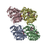
| Images | 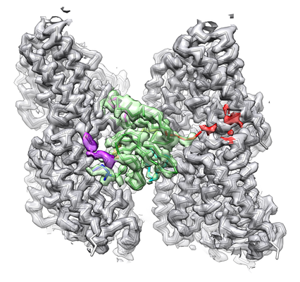 emd_4643.png emd_4643.png | 233.1 KB | ||
| Filedesc metadata | emd-4643.cif.gz | 6.8 KB | ||
| Others | emd_4643_additional.map.gz | 170.5 MB | ||
| Archive directory |  http://ftp.pdbj.org/pub/emdb/structures/EMD-4643ftp://ftp.pdbj.org/pub/emdb/structures/EMD-4643 http://ftp.pdbj.org/pub/emdb/structures/EMD-4643ftp://ftp.pdbj.org/pub/emdb/structures/EMD-4643 | HTTPS FTP |
-Related structure data
| Related structure data |  6qusMC  4644C  4650C  4654C 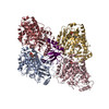 6quyC 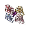 6qveC 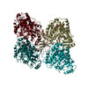 6qvjC M: atomic model generated by this map C: citing same article ( |
|---|---|
| Similar structure data | |
| EM raw data | EMPIAR-10795 (Title: A microtubule RELION-based pipeline for cryo-EM image processing Data size: 1.6 TB Data #1: Movies of microtubules decorated with CAMSAP1-CKK domain [micrographs - multiframe]) |

-Links
| EMDB pages | EMDB (EBI/PDBe) / EMDataResource |
|---|---|
| Related items in Molecule of the Month |


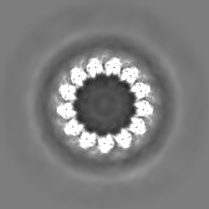
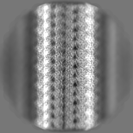
-Map
| File | Download / File: emd_4643.map.gz / Format: CCP4 / Size: 307.5 MB / Type: IMAGE STORED AS FLOATING POINT NUMBER (4 BYTES) | ||||||||||||||||||||||||||||||||||||||||||||||||||||||||||||||||||||
|---|---|---|---|---|---|---|---|---|---|---|---|---|---|---|---|---|---|---|---|---|---|---|---|---|---|---|---|---|---|---|---|---|---|---|---|---|---|---|---|---|---|---|---|---|---|---|---|---|---|---|---|---|---|---|---|---|---|---|---|---|---|---|---|---|---|---|---|---|---|
| Annotation | HsCKK-13pf microtubule symmetrised reconstruction (1x asymmetric unit, local resolution filtered) | ||||||||||||||||||||||||||||||||||||||||||||||||||||||||||||||||||||
| Projections & slices | Image control
Images are generated by Spider. generated in cubic-lattice coordinate | ||||||||||||||||||||||||||||||||||||||||||||||||||||||||||||||||||||
| Voxel size | X=Y=Z: 1.39 Å | ||||||||||||||||||||||||||||||||||||||||||||||||||||||||||||||||||||
| Density |
| ||||||||||||||||||||||||||||||||||||||||||||||||||||||||||||||||||||
| Symmetry | Space group: 1 | ||||||||||||||||||||||||||||||||||||||||||||||||||||||||||||||||||||
| Details | EMDB XML:
CCP4 map header:
| ||||||||||||||||||||||||||||||||||||||||||||||||||||||||||||||||||||
 Z (Sec.)
 Y (Row.)
 X (Col.)


-Supplemental data
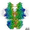
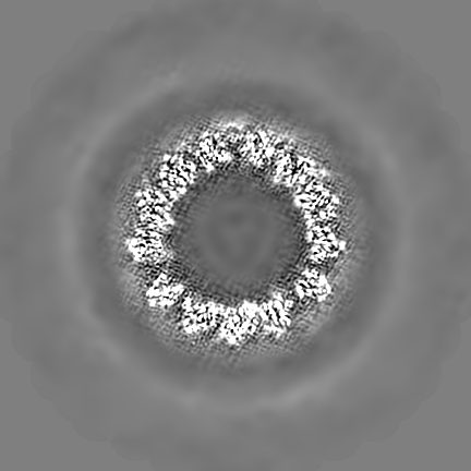
-Additional map: HsCKK-13pf microtubule C1 reconstruction (full map, local resolution...
| File | emd_4643_additional.map | ||||||||||||
|---|---|---|---|---|---|---|---|---|---|---|---|---|---|
| Annotation | HsCKK-13pf microtubule C1 reconstruction (full map, local resolution filtered) | ||||||||||||
| Projections & Slices |
| ||||||||||||
| Density Histograms |


- Sample components
+Entire : HsCKK (human CAMSAP1) decorated 13pf taxol-GDP microtubule
+Supramolecule #1: HsCKK (human CAMSAP1) decorated 13pf taxol-GDP microtubule
+Supramolecule #2: tubulin
+Supramolecule #3: Calmodulin-regulated spectrin-associated protein 1
+Macromolecule #1: Tubulin alpha-1B chain
+Macromolecule #2: Calmodulin-regulated spectrin-associated protein 1

+Macromolecule #3: Tubulin beta chain
+Macromolecule #4: GUANOSINE-5'-TRIPHOSPHATE

+Macromolecule #5: MAGNESIUM ION
+Macromolecule #6: GUANOSINE-5'-DIPHOSPHATE

+Macromolecule #7: TAXOL

-Experimental details
-Structure determination
| Method | cryo EM |
|---|---|
 Processing Processing | single particle reconstruction |
| Aggregation state | filament |
-Sample preparation
| Buffer | pH: 6.8 / Details: BRB20 |
|---|---|
| Vitrification | Cryogen name: ETHANE |
- Electron microscopy
| Microscope | FEI POLARA 300 |
|---|---|
| Image recording | Film or detector model: GATAN K2 SUMMIT (4k x 4k) / Detector mode: COUNTING / Average electron dose: 42.0 e/Å2 / Details: dose weighted images used in final reconstructions |
| Electron beam | Acceleration voltage: 300 kV / Electron source:  FIELD EMISSION GUN FIELD EMISSION GUN |
| Electron optics | Illumination mode: FLOOD BEAM / Imaging mode: BRIGHT FIELD |
| Experimental equipment |  Model: Tecnai Polara / Image courtesy: FEI Company |
-Image processing
| Startup model | Type of model: PDB ENTRY |
|---|---|
| Final reconstruction | Resolution.type: BY AUTHOR / Resolution: 3.7 Å / Resolution method: FSC 0.143 CUT-OFF / Number images used: 36342 |
| Initial angle assignment | Type: MAXIMUM LIKELIHOOD |
| Final angle assignment | Type: MAXIMUM LIKELIHOOD / Software - Name: RELION |
-Atomic model buiding 1
| Refinement | Space: REAL / Protocol: OTHER |
|---|---|
| Output model | PDB-6qus: |
