 ムービー
ムービー コントローラー


+ データを開く

- 基本情報
基本情報
| 登録情報 | データベース: EMDB / ID: EMD-3217 | |||||||||
|---|---|---|---|---|---|---|---|---|---|---|
| タイトル | In situ sub-tomogram average of the host-contact Chlamydia trachomatis type III secretion system | |||||||||
 マップデータ マップデータ | Sub-tomogram average of Chlamydia trachomatis type III secretion system (host-contact) | |||||||||
 試料 試料 |
| |||||||||
 キーワード キーワード | injectisome / type III secretion / T3SS | |||||||||
| 生物種 |   Chlamydia trachomatis (トラコーマクラミジア) Chlamydia trachomatis (トラコーマクラミジア) | |||||||||
| 手法 | サブトモグラム平均法 / クライオ電子顕微鏡法 / 解像度: 38.0 Å | |||||||||
 データ登録者 データ登録者 | Nans A / Kudryashev M / Saibil HR / Hayward RD | |||||||||
 引用 引用 | ジャーナル: Nat Commun / 年: 2015 タイトル: Structure of a bacterial type III secretion system in contact with a host membrane in situ. 著者: Andrea Nans / Mikhail Kudryashev / Helen R Saibil / Richard D Hayward /   要旨: Many bacterial pathogens of animals and plants use a conserved type III secretion system (T3SS) to inject virulence effector proteins directly into eukaryotic cells to subvert host functions. Contact ...Many bacterial pathogens of animals and plants use a conserved type III secretion system (T3SS) to inject virulence effector proteins directly into eukaryotic cells to subvert host functions. Contact with host membranes is critical for T3SS activation, yet little is known about T3SS architecture in this state or the conformational changes that drive effector translocation. Here we use cryo-electron tomography and sub-tomogram averaging to derive the intact structure of the primordial Chlamydia trachomatis T3SS in the presence and absence of host membrane contact. Comparison of the averaged structures demonstrates a marked compaction of the basal body (4 nm) occurs when the needle tip contacts the host cell membrane. This compaction is coupled to a stabilization of the cytosolic sorting platform-ATPase. Our findings reveal the first structure of a bacterial T3SS from a major human pathogen engaged with a eukaryotic host, and reveal striking 'pump-action' conformational changes that underpin effector injection. | |||||||||
| 履歴 |
|
- 構造の表示
構造の表示
| ムービー |
 ムービービューア ムービービューア |
|---|---|
| 構造ビューア | EMマップ: SurfViewMolmilJmol/JSmol |
| 添付画像 |

- ダウンロードとリンク
-EMDBアーカイブ
| マップデータ | emd_3217.map.gz | 40.7 MB | EMDBマップデータ形式 | |
|---|---|---|---|---|
| ヘッダ (付随情報) | emd-3217-v30.xmlemd-3217.xml | 8.4 KB 8.4 KB | 表示 表示 | EMDBヘッダ |
| 画像 | EMD-3217_snapshot2_500.tif | 978.8 KB | ||
| その他 | emd_3217_additional_1.map.gz | 129.1 MB | ||
| アーカイブディレクトリ |  http://ftp.pdbj.org/pub/emdb/structures/EMD-3217ftp://ftp.pdbj.org/pub/emdb/structures/EMD-3217 http://ftp.pdbj.org/pub/emdb/structures/EMD-3217ftp://ftp.pdbj.org/pub/emdb/structures/EMD-3217 | HTTPS FTP |
-検証レポート
| 文書・要旨 | emd_3217_validation.pdf.gz | 196.4 KB | 表示 | EMDB検証レポート |
|---|---|---|---|---|
| 文書・詳細版 | emd_3217_full_validation.pdf.gz | 195.5 KB | 表示 | |
| XML形式データ | emd_3217_validation.xml.gz | 6.2 KB | 表示 | |
| アーカイブディレクトリ | https://ftp.pdbj.org/pub/emdb/validation_reports/EMD-3217ftp://ftp.pdbj.org/pub/emdb/validation_reports/EMD-3217 | HTTPS FTP |
-関連構造データ
| 関連構造データ |  3216C C: 同じ文献を引用 ( |
|---|---|
| 類似構造データ | |
| 電子顕微鏡画像生データ | EMPIAR-10048 (タイトル: Cryo-electron tomogram of Chlamydia trachomatis with type III secretion system in contact with HeLa cell Data size: 13.6 Data #1: Aligned multi-frame micrographs that comprise a tilt series of Chlamydia trachomatis type III secretion systems in contact with a HeLa cell [class averages]) |
-リンク
| EMDBのページ | EMDB (EBI/PDBe) / EMDataResource |
|---|
-マップ
| ファイル | ダウンロード / ファイル: emd_3217.map.gz / 形式: CCP4 / 大きさ: 62.5 MB / タイプ: IMAGE STORED AS FLOATING POINT NUMBER (4 BYTES) | ||||||||||||||||||||||||||||||||||||||||||||||||||||||||||||||||||||
|---|---|---|---|---|---|---|---|---|---|---|---|---|---|---|---|---|---|---|---|---|---|---|---|---|---|---|---|---|---|---|---|---|---|---|---|---|---|---|---|---|---|---|---|---|---|---|---|---|---|---|---|---|---|---|---|---|---|---|---|---|---|---|---|---|---|---|---|---|---|
| 注釈 | Sub-tomogram average of Chlamydia trachomatis type III secretion system (host-contact) | ||||||||||||||||||||||||||||||||||||||||||||||||||||||||||||||||||||
| ボクセルのサイズ | X=Y=Z: 5.4 Å | ||||||||||||||||||||||||||||||||||||||||||||||||||||||||||||||||||||
| 密度 |
| ||||||||||||||||||||||||||||||||||||||||||||||||||||||||||||||||||||
| 対称性 | 空間群: 1 | ||||||||||||||||||||||||||||||||||||||||||||||||||||||||||||||||||||
| 詳細 | EMDB XML:
CCP4マップ ヘッダ情報:
| ||||||||||||||||||||||||||||||||||||||||||||||||||||||||||||||||||||
-添付データ
-添付マップデータ: emd 3217 additional 1.map
| ファイル | emd_3217_additional_1.map | ||||||||||||
|---|---|---|---|---|---|---|---|---|---|---|---|---|---|
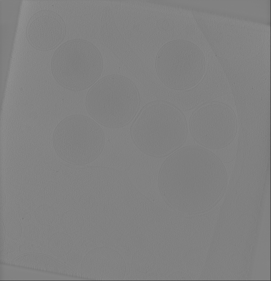
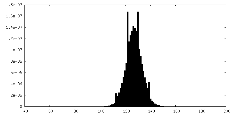
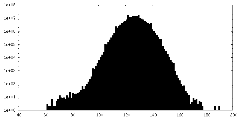
| 投影像・断面図 |
| ||||||||||||
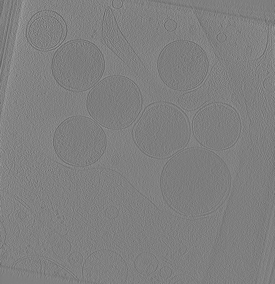
| 密度ヒストグラム |
 Z
Z Y
Y X
X


- 試料の構成要素
-全体 : Chlamydia trachomatis type III secretion system (host-contact)
| 全体 | 名称: Chlamydia trachomatis type III secretion system (host-contact) |
|---|---|
| 要素 |
|
-超分子 #1000: Chlamydia trachomatis type III secretion system (host-contact)
| 超分子 | 名称: Chlamydia trachomatis type III secretion system (host-contact) タイプ: sample / ID: 1000 / Number unique components: 1 |
|---|
-超分子 #1: Chlamydia trachomatis type III secretion system
| 超分子 | 名称: Chlamydia trachomatis type III secretion system / タイプ: organelle_or_cellular_component / ID: 1 詳細: Type III secretion systems were imaged in contact with U2OS or HeLa cells grown directly on EM grids. 組換発現: No |
|---|---|
| 由来(天然) | 生物種: Chlamydia trachomatis (トラコーマクラミジア) |
-実験情報
-構造解析
| 手法 | クライオ電子顕微鏡法 |
|---|---|
 解析 解析 | サブトモグラム平均法 |
| 試料の集合状態 | cell |
-試料調製
| グリッド | 詳細: 200 mesh gold Quantifoil 3.5/1 |
|---|---|
| 凍結 | 凍結剤: ETHANE / チャンバー内湿度: 100 % / 装置: FEI VITROBOT MARK IV / 手法: Blot for 6-10 seconds before plunging. |
- 電子顕微鏡法
| 顕微鏡 | FEI POLARA 300 |
|---|---|
| 特殊光学系 | エネルギーフィルター - 名称: Gatan Quantum エネルギーフィルター - エネルギー下限: 0.0 eV エネルギーフィルター - エネルギー上限: 20.0 eV |
| 日付 | 2014年11月10日 |
| 撮影 | カテゴリ: CCD フィルム・検出器のモデル: GATAN K2 SUMMIT (4k x 4k) 平均電子線量: 55 e/Å2 |
| 電子線 | 加速電圧: 300 kV / 電子線源:  FIELD EMISSION GUN FIELD EMISSION GUN |
| 電子光学系 | 照射モード: FLOOD BEAM / 撮影モード: BRIGHT FIELD / Cs: 2.3 mm / 最大 デフォーカス(公称値): -10.0 µm / 倍率(公称値): 41000 |
| 試料ステージ | 試料ホルダーモデル: SIDE ENTRY, EUCENTRIC / Tilt series - Axis1 - Min angle: -45 ° / Tilt series - Axis1 - Max angle: 60 ° |
| 実験機器 |  モデル: Tecnai Polara / 画像提供: FEI Company |
-画像解析
| 詳細 | Sub-tomograms were selected in IMOD and cropped in Dynamo. Alignment and averaging was carried out in Dynamo. |
|---|---|
| 最終 再構成 | 想定した対称性 - 点群: C12 (12回回転対称) / 解像度のタイプ: BY AUTHOR / 解像度: 38.0 Å / 解像度の算出法: OTHER / ソフトウェア - 名称: IMOD, Dynamo / 使用したサブトモグラム数: 196 |
| CTF補正 | 詳細: Each micrograph was phase-flipped in IMOD using the measured defocus |
