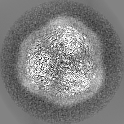
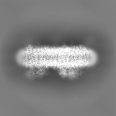
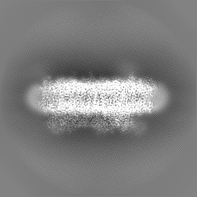
- EMDB-31605: Cryo-EM structure of cyanobacterial photosystem I in the presence... -
+
Open data
ID or keywords:
Loading...
-
Basic information
Entry
Database: EMDB / ID: EMD-31605
Title
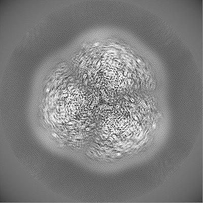
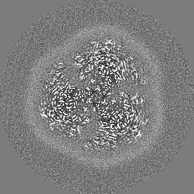
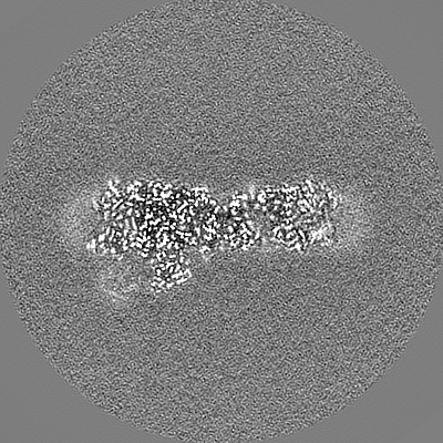
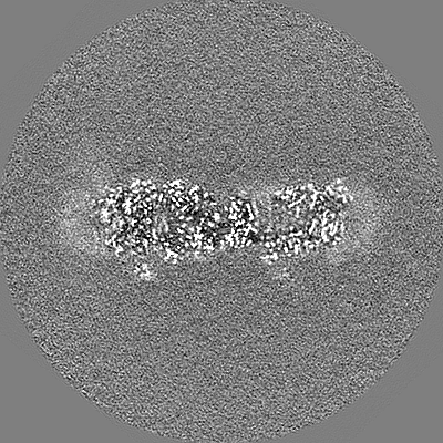
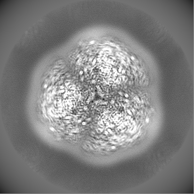
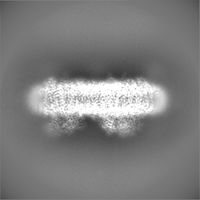
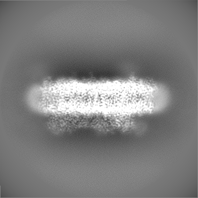
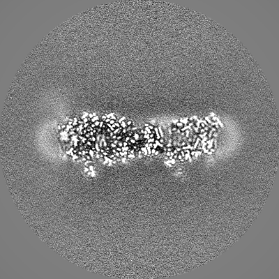
Cryo-EM structure of cyanobacterial photosystem I in the presence of ferredoxin and cytochrome c6
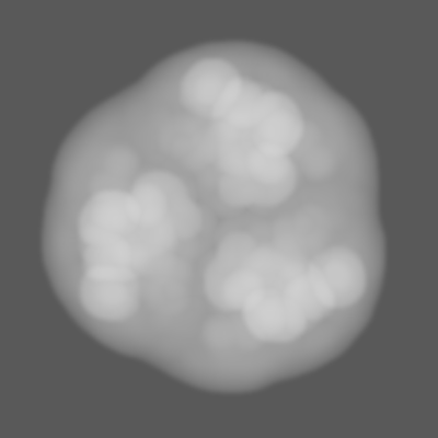
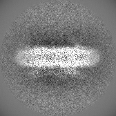
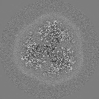
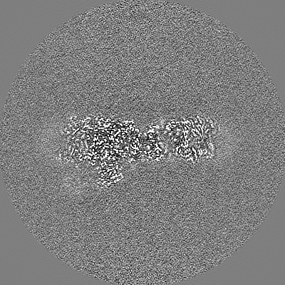
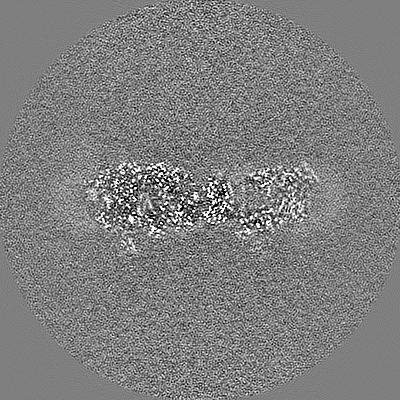
Map data
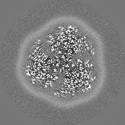
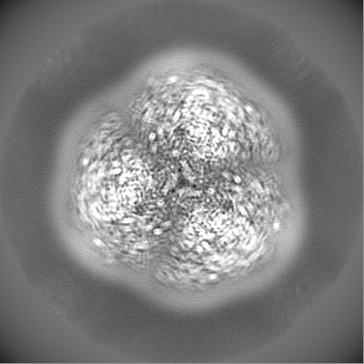
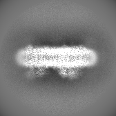
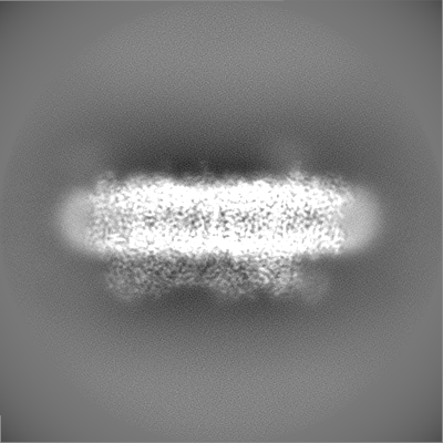
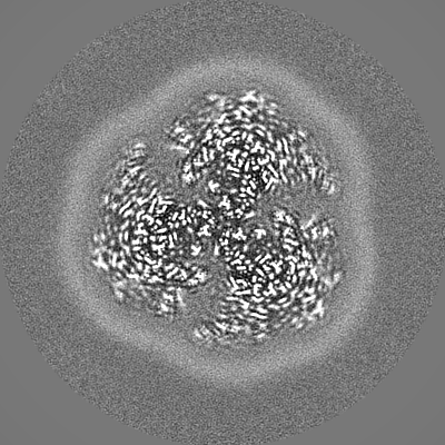
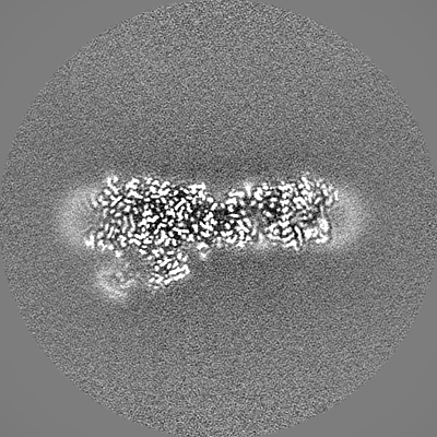
3D refined map in C3 symmetry, which was used for model building and refinement.
Sample
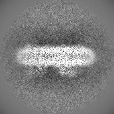
Complex: Cryo-EM map of cyanobacterial photosystem I in the presence of ferredoxin and cytochrome c6
Complex: Photosystem I trimer
Protein or peptide: x 10 types
Complex: Ga-Substituted Ferredoxin
Protein or peptide: x 3 types
Ligand: x 11 types
Keywords
Photosystem I / Ferredoxin / Cytochrome c6 / PHOTOSYNTHESIS
Function / homology
Function and homology information
photosystem I reaction center / photosystem I / photosynthetic electron transport in photosystem I / photosystem I / plasma membrane-derived thylakoid membrane / chlorophyll binding / photosynthesis / electron transport chain / 2 iron, 2 sulfur cluster binding / 4 iron, 4 sulfur cluster binding ...photosystem I reaction center / photosystem I / photosynthetic electron transport in photosystem I / photosystem I / plasma membrane-derived thylakoid membrane / chlorophyll binding / photosynthesis / electron transport chain / 2 iron, 2 sulfur cluster binding / 4 iron, 4 sulfur cluster binding / electron transfer activity / oxidoreductase activity / magnesium ion binding / metal ion binding Similarity search - Function
Photosystem I PsaX / Photosystem I PsaX superfamily / PsaX family / Photosystem I reaction center subunit PsaK / Ferredoxin [2Fe-2S], plant / Photosystem I reaction centre subunit PsaK / Photosystem I reaction centre subunit PsaK superfamily / Photosystem I psaG and psaK proteins signature. / Photosystem I PsaM, reaction centre superfamily / Photosystem I PsaM, reaction centre ...Photosystem I PsaX / Photosystem I PsaX superfamily / PsaX family / Photosystem I reaction center subunit PsaK / Ferredoxin [2Fe-2S], plant / Photosystem I reaction centre subunit PsaK / Photosystem I reaction centre subunit PsaK superfamily / Photosystem I psaG and psaK proteins signature. / Photosystem I PsaM, reaction centre superfamily / Photosystem I PsaM, reaction centre / Photosystem I protein M (PsaM) / Photosystem I reaction center subunit V/PsaK / Photosystem I psaG / psaK / Photosystem I PsaL, reaction centre subunit XI / Photosystem I, reaction centre subunit XI / Photosystem I PsaL, reaction centre subunit XI superfamily / Photosystem I reaction centre subunit XI / Photosystem I reaction centre subunit VIII / Photosystem I reaction centre subunit VIII / Photosystem I reaction centre subunit VIII superfamily / Photosystem I PsaF, reaction centre subunit III / Photosystem I PsaF, reaction centre subunit III superfamily / Photosystem I reaction centre subunit III / Photosystem I PsaD / Photosystem I, reaction centre subunit PsaD superfamily / PsaD / Photosystem I PsaE, reaction centre subunit IV / Photosystem I reaction centre subunit IV / PsaE / Photosystem I PsaJ, reaction centre subunit IX superfamily / Photosystem I PsaJ, reaction centre subunit IX / Photosystem I reaction centre subunit IX / PsaJ / Photosystem I PsaA / Photosystem I protein PsaC / Photosystem I PsaB / Photosystem I PsaA/PsaB, conserved site / Photosystem I psaA and psaB proteins signature. / : / Photosystem I PsaA/PsaB / Photosystem I PsaA/PsaB superfamily / Photosystem I psaA/psaB protein / 2Fe-2S ferredoxin, iron-sulphur binding site / 2Fe-2S ferredoxin-type iron-sulfur binding region signature. / Electron transport accessory-like domain superfamily / 2Fe-2S iron-sulfur cluster binding domain / Beta-grasp domain superfamily / 4Fe-4S dicluster domain / 2Fe-2S ferredoxin-type iron-sulfur binding domain profile. / 2Fe-2S ferredoxin-type iron-sulfur binding domain / 2Fe-2S ferredoxin-like superfamily / 4Fe-4S ferredoxin, iron-sulphur binding, conserved site / 4Fe-4S ferredoxin-type iron-sulfur binding region signature. / 4Fe-4S ferredoxin-type iron-sulfur binding domain profile. / 4Fe-4S ferredoxin-type, iron-sulphur binding domain Similarity search - Domain/homology
Ferredoxin-1 / Photosystem I reaction center subunit III / Photosystem I reaction center subunit XII / Photosystem I P700 chlorophyll a apoprotein A1 / Photosystem I P700 chlorophyll a apoprotein A2 / Photosystem I iron-sulfur center / Photosystem I reaction center subunit II / Photosystem I reaction center subunit IV / Photosystem I reaction center subunit PsaK / Photosystem I reaction center subunit VIII ...Ferredoxin-1 / Photosystem I reaction center subunit III / Photosystem I reaction center subunit XII / Photosystem I P700 chlorophyll a apoprotein A1 / Photosystem I P700 chlorophyll a apoprotein A2 / Photosystem I iron-sulfur center / Photosystem I reaction center subunit II / Photosystem I reaction center subunit IV / Photosystem I reaction center subunit PsaK / Photosystem I reaction center subunit VIII / Photosystem I reaction center subunit IX / Photosystem I reaction center subunit XI / Photosystem I 4.8K protein Similarity search - Component
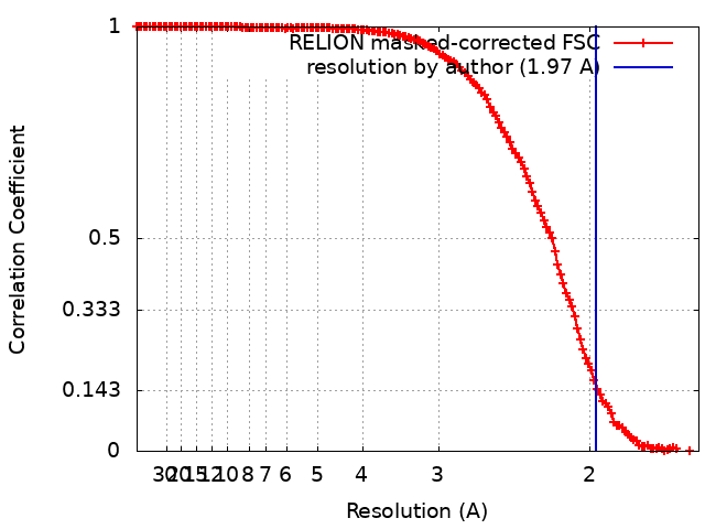
Journal: Commun Biol / Year: 2022 Title: Structure of cyanobacterial photosystem I complexed with ferredoxin at 1.97 Å resolution. Authors: Jiannan Li / Noriyuki Hamaoka / Fumiaki Makino / Akihiro Kawamoto / Yuxi Lin / Matthias Rögner / Marc M Nowaczyk / Young-Ho Lee / Keiichi Namba / Christoph Gerle / Genji Kurisu / Abstract: Photosystem I (PSI) is a light driven electron pump transferring electrons from Cytochrome c (Cyt c) to Ferredoxin (Fd). An understanding of this electron transfer process is hampered by a paucity of ...Photosystem I (PSI) is a light driven electron pump transferring electrons from Cytochrome c (Cyt c) to Ferredoxin (Fd). An understanding of this electron transfer process is hampered by a paucity of structural detail concerning PSI:Fd interface and the possible binding sites of Cyt c. Here we describe the high resolution cryo-EM structure of Thermosynechococcus elongatus BP-1 PSI in complex with Fd and a loosely bound Cyt c. Side chain interactions at the PSI:Fd interface including bridging water molecules are visualized in detail. The structure explains the properties of mutants of PsaE and PsaC that affect kinetics of Fd binding and suggests a molecular switch for the dissociation of Fd upon reduction. Calorimetry-based thermodynamic analyses confirms a single binding site for Fd and demonstrates that PSI:Fd complexation is purely driven by entropy. A possible reaction cycle for the efficient transfer of electrons from Cyt c to Fd via PSI is proposed.
Entire : Cryo-EM map of cyanobacterial photosystem I in the presence of fe...
Entire
Name: Cryo-EM map of cyanobacterial photosystem I in the presence of ferredoxin and cytochrome c6
Components
Complex: Cryo-EM map of cyanobacterial photosystem I in the presence of ferredoxin and cytochrome c6
Complex: Photosystem I trimer
Protein or peptide: Photosystem I P700 chlorophyll a apoprotein A1
Protein or peptide: Photosystem I P700 chlorophyll a apoprotein A2
Protein or peptide: Photosystem I iron-sulfur center
Protein or peptide: Photosystem I reaction center subunit II
Protein or peptide: Photosystem I reaction center subunit IV
Protein or peptide: Photosystem I reaction center subunit VIII
Protein or peptide: Photosystem I reaction center subunit IX
Protein or peptide: Photosystem I reaction center subunit PsaK
Protein or peptide: Photosystem I reaction center subunit XI
Protein or peptide: Photosystem I reaction center subunit XII
Complex: Ga-Substituted Ferredoxin
Protein or peptide: Photosystem I reaction center subunit III
Protein or peptide: Ferredoxin-1
Protein or peptide: Photosystem I 4.8K protein
Ligand: CHLOROPHYLL A ISOMER
Ligand: CHLOROPHYLL A
Ligand: PHYLLOQUINONE
Ligand: IRON/SULFUR CLUSTER
Ligand: BETA-CAROTENE
Ligand: 1,2-DIPALMITOYL-PHOSPHATIDYL-GLYCEROLE
Ligand: UNKNOWN LIGAND
Ligand: DIGALACTOSYL DIACYL GLYCEROL (DGDG)
Ligand: CALCIUM ION
Ligand: [2Ga-2S] cluster
Ligand: water
+
Supramolecule #1: Cryo-EM map of cyanobacterial photosystem I in the presence of fe...
Supramolecule
Name: Cryo-EM map of cyanobacterial photosystem I in the presence of ferredoxin and cytochrome c6 type: complex / ID: 1 / Parent: 0 / Macromolecule list: #1-#13 Details: Cytochrome C6 was not be built in model because of limited resolution
In the structure databanks used in Yorodumi, some data are registered as the other names, "COVID-19 virus" and "2019-nCoV". Here are the details of the virus and the list of structure data.
Jan 31, 2019. EMDB accession codes are about to change! (news from PDBe EMDB page)
EMDB accession codes are about to change! (news from PDBe EMDB page)
The allocation of 4 digits for EMDB accession codes will soon come to an end. Whilst these codes will remain in use, new EMDB accession codes will include an additional digit and will expand incrementally as the available range of codes is exhausted. The current 4-digit format prefixed with “EMD-” (i.e. EMD-XXXX) will advance to a 5-digit format (i.e. EMD-XXXXX), and so on. It is currently estimated that the 4-digit codes will be depleted around Spring 2019, at which point the 5-digit format will come into force.
The EM Navigator/Yorodumi systems omit the EMD- prefix.
Related info.:Q: What is EMD? / ID/Accession-code notation in Yorodumi/EM Navigator
Yorodumi is a browser for structure data from EMDB, PDB, SASBDB, etc.
This page is also the successor to EM Navigator detail page, and also detail information page/front-end page for Omokage search.
The word "yorodu" (or yorozu) is an old Japanese word meaning "ten thousand". "mi" (miru) is to see.
Related info.:EMDB / PDB / SASBDB / Comparison of 3 databanks / Yorodumi Search / Aug 31, 2016. New EM Navigator & Yorodumi / Yorodumi Papers / Jmol/JSmol / Function and homology information / Changes in new EM Navigator and Yorodumi
 Movie
Movie Controller

 Yorodumi
 Open data
 Basic information
 Map data
 Sample
 Keywords
 Function and homology information
 Thermosynechococcus elongatus BP-1 (bacteria) /
  Authors
 Citation


 Structure visualization
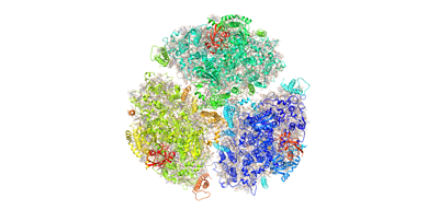
 Downloads & links
 emd_31605.png
 http://ftp.pdbj.org/pub/emdb/structures/EMD-31605
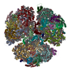


 Z (Sec.)
 Y (Row.)
 X (Col.)


 Sample components


 Processing
 Electron microscopy
 FIELD EMISSION GUN