[English] 日本語
 Yorodumi
- PDB-7fix: Cryo-EM structure of cyanobacterial photosystem I in the presence... -
+ Open data
- Basic information
| Entry | Database: PDB / ID: 7fix | ||||||
|---|---|---|---|---|---|---|---|
| Title | Cryo-EM structure of cyanobacterial photosystem I in the presence of ferredoxin and cytochrome c6 | ||||||
 Components Components |
| ||||||
 Keywords Keywords | PHOTOSYNTHESIS / Photosystem I / Ferredoxin / Cytochrome c6 | ||||||
| Function / homology |  Function and homology information Function and homology informationphotosystem I reaction center / photosystem I / photosynthetic electron transport in photosystem I / photosystem I / plasma membrane-derived thylakoid membrane / chlorophyll binding / photosynthesis / electron transport chain / 2 iron, 2 sulfur cluster binding / 4 iron, 4 sulfur cluster binding ...photosystem I reaction center / photosystem I / photosynthetic electron transport in photosystem I / photosystem I / plasma membrane-derived thylakoid membrane / chlorophyll binding / photosynthesis / electron transport chain / 2 iron, 2 sulfur cluster binding / 4 iron, 4 sulfur cluster binding / oxidoreductase activity / electron transfer activity / magnesium ion binding / metal ion binding / membrane Similarity search - Function | ||||||
| Biological species |   Thermosynechococcus vestitus BP-1 (bacteria) Thermosynechococcus vestitus BP-1 (bacteria) | ||||||
| Method | ELECTRON MICROSCOPY / single particle reconstruction / cryo EM / Resolution: 1.97 Å | ||||||
 Authors Authors | Li, J. / Kurisu, G. | ||||||
| Funding support | 1items
| ||||||
 Citation Citation |  Journal: Commun Biol / Year: 2022 Journal: Commun Biol / Year: 2022Title: Structure of cyanobacterial photosystem I complexed with ferredoxin at 1.97 Å resolution. Authors: Jiannan Li / Noriyuki Hamaoka / Fumiaki Makino / Akihiro Kawamoto / Yuxi Lin / Matthias Rögner / Marc M Nowaczyk / Young-Ho Lee / Keiichi Namba / Christoph Gerle / Genji Kurisu /    Abstract: Photosystem I (PSI) is a light driven electron pump transferring electrons from Cytochrome c (Cyt c) to Ferredoxin (Fd). An understanding of this electron transfer process is hampered by a paucity of ...Photosystem I (PSI) is a light driven electron pump transferring electrons from Cytochrome c (Cyt c) to Ferredoxin (Fd). An understanding of this electron transfer process is hampered by a paucity of structural detail concerning PSI:Fd interface and the possible binding sites of Cyt c. Here we describe the high resolution cryo-EM structure of Thermosynechococcus elongatus BP-1 PSI in complex with Fd and a loosely bound Cyt c. Side chain interactions at the PSI:Fd interface including bridging water molecules are visualized in detail. The structure explains the properties of mutants of PsaE and PsaC that affect kinetics of Fd binding and suggests a molecular switch for the dissociation of Fd upon reduction. Calorimetry-based thermodynamic analyses confirms a single binding site for Fd and demonstrates that PSI:Fd complexation is purely driven by entropy. A possible reaction cycle for the efficient transfer of electrons from Cyt c to Fd via PSI is proposed. | ||||||
| History |
|
- Structure visualization
| Structure viewer | Molecule:  Molmil Molmil Jmol/JSmol Jmol/JSmol |
|---|
- Downloads & links
- Download
| PDBx/mmCIF format |  7fix.cif.gz 7fix.cif.gz | 1.6 MB | Display |  PDBx/mmCIF format PDBx/mmCIF format |
|---|---|---|---|---|
| PDB format |  pdb7fix.ent.gz pdb7fix.ent.gz | Display |  PDB format PDB format | |
| PDBx/mmJSON format |  7fix.json.gz 7fix.json.gz | Tree view |  PDBx/mmJSON format PDBx/mmJSON format | |
| Others |  Other downloads Other downloads |
-Validation report
| Arichive directory |  https://data.pdbj.org/pub/pdb/validation_reports/fi/7fix https://data.pdbj.org/pub/pdb/validation_reports/fi/7fix ftp://data.pdbj.org/pub/pdb/validation_reports/fi/7fix ftp://data.pdbj.org/pub/pdb/validation_reports/fi/7fix | HTTPS FTP |
|---|
-Related structure data
| Related structure data |  31605MC M: map data used to model this data C: citing same article ( |
|---|---|
| Similar structure data | Similarity search - Function & homology  F&H Search F&H Search |
- Links
Links
- Assembly
| Deposited unit | 
|
|---|---|
| 1 |
|
- Components
-Photosystem I ... , 12 types, 36 molecules A1A2A3B1B2B3C1C2C3D1D2D3E1E2E3F1F2F3I1I2I3J1J2J3K1K2K3L1L2L3...
| #1: Protein | Mass: 83267.773 Da / Num. of mol.: 3 / Source method: isolated from a natural source Source: (natural)   Thermosynechococcus vestitus BP-1 (bacteria) Thermosynechococcus vestitus BP-1 (bacteria)Strain: BP-1 / References: UniProt: P0A405, photosystem I #2: Protein | Mass: 83123.648 Da / Num. of mol.: 3 / Source method: isolated from a natural source Source: (natural)   Thermosynechococcus vestitus BP-1 (bacteria) Thermosynechococcus vestitus BP-1 (bacteria)Strain: BP-1 / References: UniProt: P0A407, photosystem I #3: Protein | Mass: 8809.207 Da / Num. of mol.: 3 / Source method: isolated from a natural source Source: (natural)   Thermosynechococcus vestitus BP-1 (bacteria) Thermosynechococcus vestitus BP-1 (bacteria)Strain: BP-1 / References: UniProt: P0A415, photosystem I #4: Protein | Mass: 15389.494 Da / Num. of mol.: 3 / Source method: isolated from a natural source Source: (natural)   Thermosynechococcus vestitus BP-1 (bacteria) Thermosynechococcus vestitus BP-1 (bacteria)Strain: BP-1 / References: UniProt: P0A420 #5: Protein | Mass: 8399.485 Da / Num. of mol.: 3 / Source method: isolated from a natural source Source: (natural)   Thermosynechococcus vestitus BP-1 (bacteria) Thermosynechococcus vestitus BP-1 (bacteria)Strain: BP-1 / References: UniProt: P0A423 #6: Protein | Mass: 19098.062 Da / Num. of mol.: 3 Source method: isolated from a genetically manipulated source Source: (gene. exp.)   Thermosynechococcus vestitus BP-1 (bacteria) Thermosynechococcus vestitus BP-1 (bacteria)Strain: BP-1 / Gene: psaF, tlr2411 Production host:   Thermosynechococcus vestitus BP-1 (bacteria) Thermosynechococcus vestitus BP-1 (bacteria)Strain (production host): BP-1 / References: UniProt: P0A401 #7: Protein/peptide | Mass: 4297.234 Da / Num. of mol.: 3 / Source method: isolated from a natural source Source: (natural)   Thermosynechococcus vestitus BP-1 (bacteria) Thermosynechococcus vestitus BP-1 (bacteria)Strain: BP-1 / References: UniProt: P0A427 #8: Protein/peptide | Mass: 4770.698 Da / Num. of mol.: 3 / Source method: isolated from a natural source Source: (natural)   Thermosynechococcus vestitus BP-1 (bacteria) Thermosynechococcus vestitus BP-1 (bacteria)Strain: BP-1 / References: UniProt: P0A429 #9: Protein | Mass: 8483.983 Da / Num. of mol.: 3 / Source method: isolated from a natural source Source: (natural)   Thermosynechococcus vestitus BP-1 (bacteria) Thermosynechococcus vestitus BP-1 (bacteria)Strain: BP-1 / References: UniProt: P0A425 #10: Protein | Mass: 16261.685 Da / Num. of mol.: 3 / Source method: isolated from a natural source Source: (natural)   Thermosynechococcus vestitus BP-1 (bacteria) Thermosynechococcus vestitus BP-1 (bacteria)Strain: BP-1 / References: UniProt: Q8DGB4 #11: Protein/peptide | Mass: 3426.115 Da / Num. of mol.: 3 / Source method: isolated from a natural source Source: (natural)   Thermosynechococcus vestitus BP-1 (bacteria) Thermosynechococcus vestitus BP-1 (bacteria)Strain: BP-1 / References: UniProt: P0A403 #13: Protein/peptide | Mass: 4424.317 Da / Num. of mol.: 3 Source method: isolated from a genetically manipulated source Source: (gene. exp.)   Thermosynechococcus vestitus BP-1 (bacteria) Thermosynechococcus vestitus BP-1 (bacteria)Strain: BP-1 / Gene: tsr0813 Production host:   Thermosynechococcus vestitus BP-1 (bacteria) Thermosynechococcus vestitus BP-1 (bacteria)References: UniProt: Q8DKP6 |
|---|
-Protein / Sugars , 2 types, 6 molecules R1R2R3

| #12: Protein | Mass: 10853.959 Da / Num. of mol.: 3 Source method: isolated from a genetically manipulated source Source: (gene. exp.)   Thermosynechococcus vestitus BP-1 (bacteria) Thermosynechococcus vestitus BP-1 (bacteria)Strain: BP-1 / Gene: petF1, petF / Production host:  #21: Sugar | |
|---|
-Non-polymers , 10 types, 789 molecules 

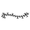


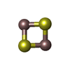


| #14: Chemical | | #15: Chemical | ChemComp-CLA / #16: Chemical | ChemComp-PQN / #17: Chemical | ChemComp-SF4 / #18: Chemical | ChemComp-BCR / #19: Chemical | ChemComp-LHG / #20: Chemical | ChemComp-UNL / Mass: 40.078 Da / Num. of mol.: 51 / Source method: obtained synthetically / Feature type: SUBJECT OF INVESTIGATION #22: Chemical | #23: Chemical | #24: Water | ChemComp-HOH / | |
|---|
-Details
| Has ligand of interest | Y |
|---|---|
| Has protein modification | Y |
-Experimental details
-Experiment
| Experiment | Method: ELECTRON MICROSCOPY |
|---|---|
| EM experiment | Aggregation state: PARTICLE / 3D reconstruction method: single particle reconstruction |
- Sample preparation
| Component |
| ||||||||||||||||||||||||||||
|---|---|---|---|---|---|---|---|---|---|---|---|---|---|---|---|---|---|---|---|---|---|---|---|---|---|---|---|---|---|
| Molecular weight |
| ||||||||||||||||||||||||||||
| Source (natural) |
| ||||||||||||||||||||||||||||
| Source (recombinant) | Organism:  | ||||||||||||||||||||||||||||
| Buffer solution | pH: 8 | ||||||||||||||||||||||||||||
| Specimen | Conc.: 30 mg/ml / Embedding applied: NO / Shadowing applied: NO / Staining applied: NO / Vitrification applied: YES | ||||||||||||||||||||||||||||
| Specimen support | Grid material: COPPER / Grid mesh size: 300 divisions/in. / Grid type: Quantifoil R1.2/1.3 | ||||||||||||||||||||||||||||
| Vitrification | Instrument: FEI VITROBOT MARK IV / Cryogen name: ETHANE / Humidity: 100 % / Chamber temperature: 277 K |
- Electron microscopy imaging
| Microscopy | Model: JEOL CRYO ARM 300 |
|---|---|
| Electron gun | Electron source:  FIELD EMISSION GUN / Accelerating voltage: 300 kV / Illumination mode: FLOOD BEAM FIELD EMISSION GUN / Accelerating voltage: 300 kV / Illumination mode: FLOOD BEAM |
| Electron lens | Mode: BRIGHT FIELD / Nominal defocus max: 1500 nm / Nominal defocus min: 500 nm / Cs: 2.7 mm |
| Specimen holder | Cryogen: NITROGEN |
| Image recording | Electron dose: 48 e/Å2 / Film or detector model: GATAN K3 BIOQUANTUM (6k x 4k) |
- Processing
| Software |
| ||||||||||||||||||||||||
|---|---|---|---|---|---|---|---|---|---|---|---|---|---|---|---|---|---|---|---|---|---|---|---|---|---|
| CTF correction | Type: PHASE FLIPPING AND AMPLITUDE CORRECTION | ||||||||||||||||||||||||
| Symmetry | Point symmetry: C3 (3 fold cyclic) | ||||||||||||||||||||||||
| 3D reconstruction | Resolution: 1.97 Å / Resolution method: FSC 0.143 CUT-OFF / Num. of particles: 207142 / Symmetry type: POINT | ||||||||||||||||||||||||
| Refinement | Cross valid method: NONE | ||||||||||||||||||||||||
| Refine LS restraints |
|
 Movie
Movie Controller


 PDBj
PDBj


