- EMDB-28711: CryoEM reconstruction of membrane-bound, left-handed CHMP1B+IST1 ... -
+
Open data
ID or keywords:
Loading...
-
Basic information
Entry
Database: EMDB / ID: EMD-28711
Title
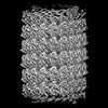
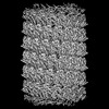
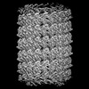
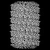
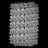
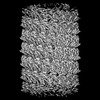
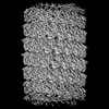
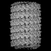
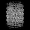
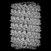
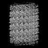
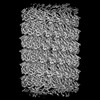
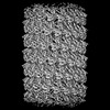
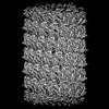
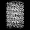
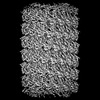
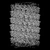
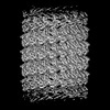
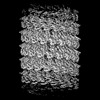
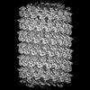
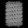
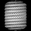
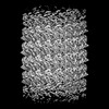
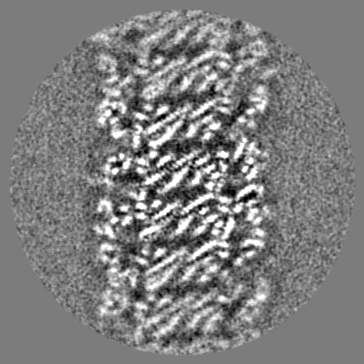
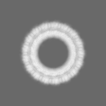
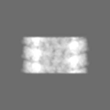
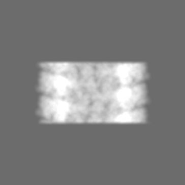
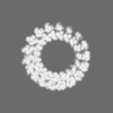
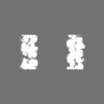
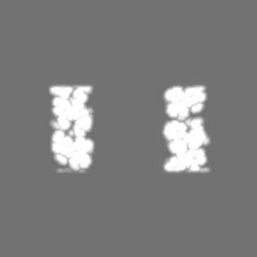
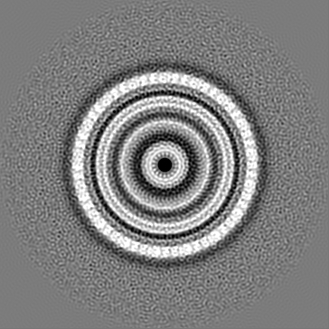
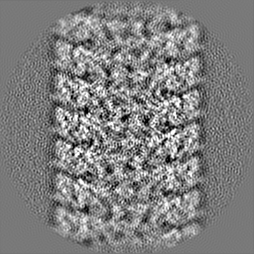
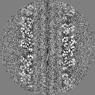
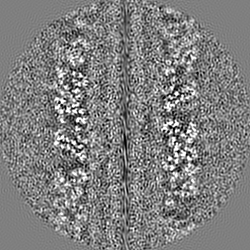
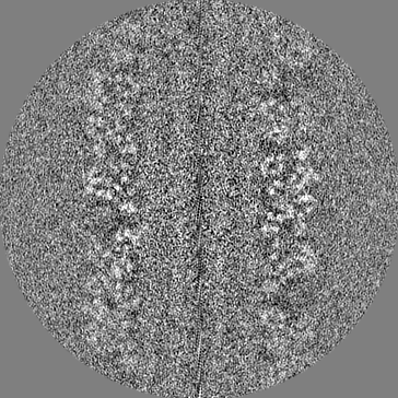
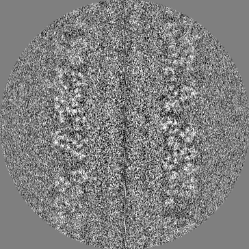
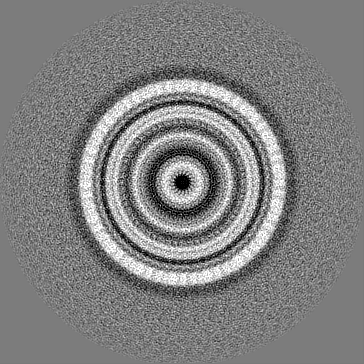
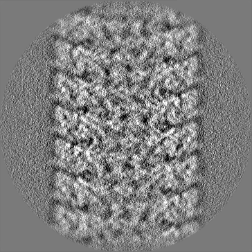
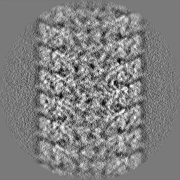
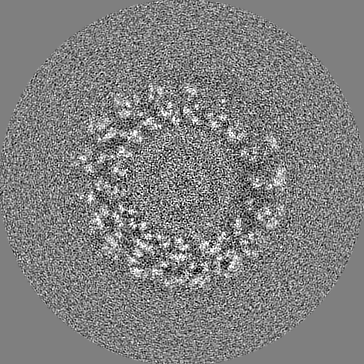
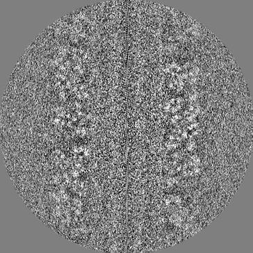
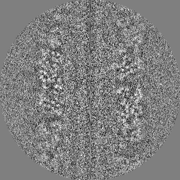
CryoEM reconstruction of membrane-bound, left-handed CHMP1B+IST1 filament with 30% cholesterol
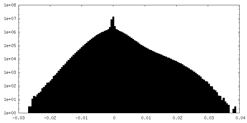
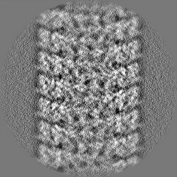
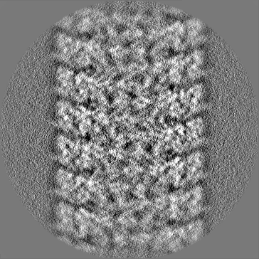
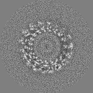
Map data
Left-handed, 17-subunit-per-turn, membrane-bound CHMP1B/IST1 with 30% cholesterol.
Sample
Complex: membrane-bound, left-handed CHMP1B+IST1 filament with 30% cholesterol
Protein or peptide: CHMP1B
Protein or peptide: IST1
Keywords
ESCRT / brominated / lipid / membrane / MEMBRANE PROTEIN
Function / homology
Function and homology information
MIT domain binding / multivesicular body-lysosome fusion / amphisome membrane / vesicle fusion with vacuole / ESCRT III complex disassembly / late endosome to lysosome transport / ESCRT III complex / kinetochore microtubule / endosome transport via multivesicular body sorting pathway / cytoskeleton-dependent cytokinesis ...MIT domain binding / multivesicular body-lysosome fusion / amphisome membrane / vesicle fusion with vacuole / ESCRT III complex disassembly / late endosome to lysosome transport / ESCRT III complex / kinetochore microtubule / endosome transport via multivesicular body sorting pathway / cytoskeleton-dependent cytokinesis / collateral sprouting / nuclear membrane reassembly / membrane coat / multivesicular body sorting pathway / positive regulation of collateral sprouting / Sealing of the nuclear envelope (NE) by ESCRT-III / midbody abscission / regulation of centrosome duplication / membrane fission / late endosome to vacuole transport / multivesicular body assembly / multivesicular body membrane / ubiquitin-dependent protein catabolic process via the multivesicular body sorting pathway / plasma membrane repair / Flemming body / regulation of mitotic spindle assembly / mitotic metaphase chromosome alignment / nucleus organization / viral budding via host ESCRT complex / endoplasmic reticulum-Golgi intermediate compartment / positive regulation of proteolysis / autophagosome membrane / autophagosome maturation / nuclear pore / multivesicular body / viral budding from plasma membrane / establishment of protein localization / kinetochore / autophagy / azurophil granule lumen / intracellular protein localization / nuclear envelope / protein transport / midbody / endosome membrane / cadherin binding / protein domain specific binding / cell division / lysosomal membrane / Neutrophil degranulation / centrosome / chromatin / protein-containing complex binding / extracellular exosome / extracellular region / nucleoplasm / identical protein binding / plasma membrane / cytosol Similarity search - Function
Vacuolar protein sorting-associated protein Ist1 / Vacuolar protein sorting-associated protein IST1-like / Regulator of Vps4 activity in the MVB pathway / Snf7 family / Snf7 Similarity search - Domain/homology
National Institutes of Health/National Institute of General Medical Sciences (NIH/NIGMS)
GM117593
United States
National Institutes of Health/National Institute of General Medical Sciences (NIH/NIGMS)
GM137109
United States
National Institutes of Health/National Institute of General Medical Sciences (NIH/NIGMS)
GM082545
United States
National Institutes of Health/National Institute of General Medical Sciences (NIH/NIGMS)
GM110772
United States
National Institutes of Health/National Institute of General Medical Sciences (NIH/NIGMS)
GM089740
United States
Howard Hughes Medical Institute (HHMI)
United States
Chan Zuckerberg Initiative
United States
Citation
Journal: Nat Struct Mol Biol / Year: 2020 Title: Membrane constriction and thinning by sequential ESCRT-III polymerization. Authors: Henry C Nguyen / Nathaniel Talledge / John McCullough / Abhimanyu Sharma / Frank R Moss / Janet H Iwasa / Michael D Vershinin / Wesley I Sundquist / Adam Frost / Abstract: The endosomal sorting complexes required for transport (ESCRTs) mediate diverse membrane remodeling events. These typically require ESCRT-III proteins to stabilize negatively curved membranes; ...The endosomal sorting complexes required for transport (ESCRTs) mediate diverse membrane remodeling events. These typically require ESCRT-III proteins to stabilize negatively curved membranes; however, recent work has indicated that certain ESCRT-IIIs also participate in positive-curvature membrane-shaping reactions. ESCRT-IIIs polymerize into membrane-binding filaments, but the structural basis for negative versus positive membrane remodeling by these proteins remains poorly understood. To learn how certain ESCRT-IIIs shape positively curved membranes, we determined structures of human membrane-bound CHMP1B-only, membrane-bound CHMP1B + IST1, and IST1-only filaments by cryo-EM. Our structures show how CHMP1B first polymerizes into a single-stranded helical filament, shaping membranes into moderate-curvature tubules. Subsequently, IST1 assembles a second strand on CHMP1B, further constricting the membrane tube and reducing its diameter nearly to the fission point. Each step of constriction thins the underlying bilayer, lowering the barrier to membrane fission. Our structures reveal how a two-component, sequential polymerization mechanism drives membrane tubulation, constriction and bilayer thinning.
In the structure databanks used in Yorodumi, some data are registered as the other names, "COVID-19 virus" and "2019-nCoV". Here are the details of the virus and the list of structure data.
Jan 31, 2019. EMDB accession codes are about to change! (news from PDBe EMDB page)
EMDB accession codes are about to change! (news from PDBe EMDB page)
The allocation of 4 digits for EMDB accession codes will soon come to an end. Whilst these codes will remain in use, new EMDB accession codes will include an additional digit and will expand incrementally as the available range of codes is exhausted. The current 4-digit format prefixed with “EMD-” (i.e. EMD-XXXX) will advance to a 5-digit format (i.e. EMD-XXXXX), and so on. It is currently estimated that the 4-digit codes will be depleted around Spring 2019, at which point the 5-digit format will come into force.
The EM Navigator/Yorodumi systems omit the EMD- prefix.
Related info.:Q: What is EMD? / ID/Accession-code notation in Yorodumi/EM Navigator
Yorodumi is a browser for structure data from EMDB, PDB, SASBDB, etc.
This page is also the successor to EM Navigator detail page, and also detail information page/front-end page for Omokage search.
The word "yorodu" (or yorozu) is an old Japanese word meaning "ten thousand". "mi" (miru) is to see.
Related info.:EMDB / PDB / SASBDB / Comparison of 3 databanks / Yorodumi Search / Aug 31, 2016. New EM Navigator & Yorodumi / Yorodumi Papers / Jmol/JSmol / Function and homology information / Changes in new EM Navigator and Yorodumi
 Movie
Movie Controller

 Yorodumi
 Open data
 Basic information
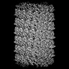
 Map data
 Sample
 Keywords
 Function and homology information
 Homo sapiens (human)
 Authors
 United States, 7 items
  Citation
 Structure visualization
 Downloads & links
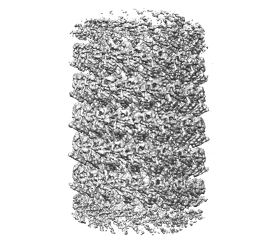 emd_28711.png
 http://ftp.pdbj.org/pub/emdb/structures/EMD-28711
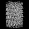


 Z (Sec.)
 Y (Row.)
 X (Col.)


 Sample components
 Processing
 Electron microscopy
 FIELD EMISSION GUN

