National Institutes of Health/National Institute of General Medical Sciences (NIH/NIGMS)
GM143805
United States
National Institutes of Health/National Institute of General Medical Sciences (NIH/NIGMS)
GM139616
United States
National Institutes of Health/National Institute of General Medical Sciences (NIH/NIGMS)
GM119032
United States
Citation
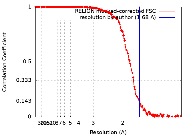
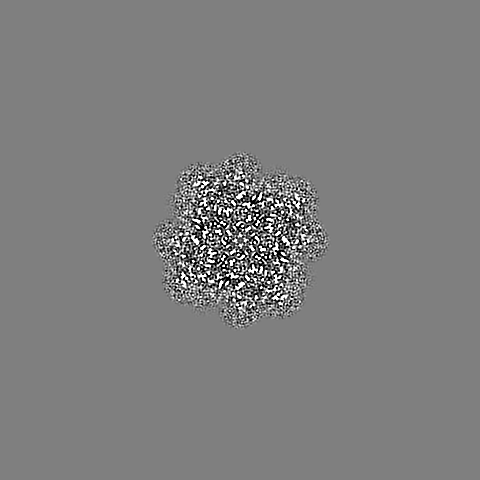
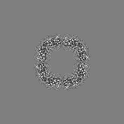
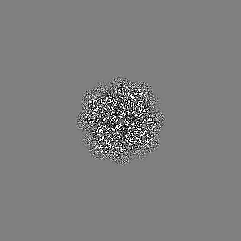
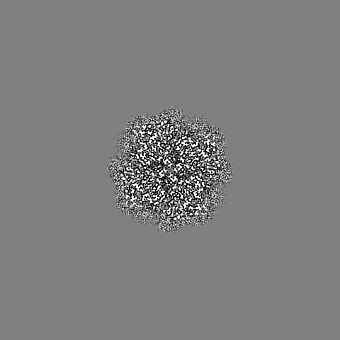
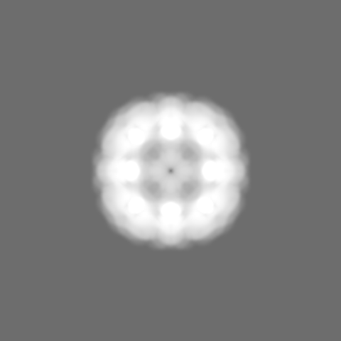
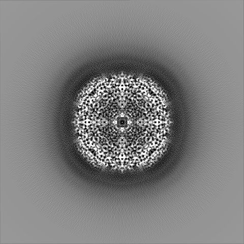
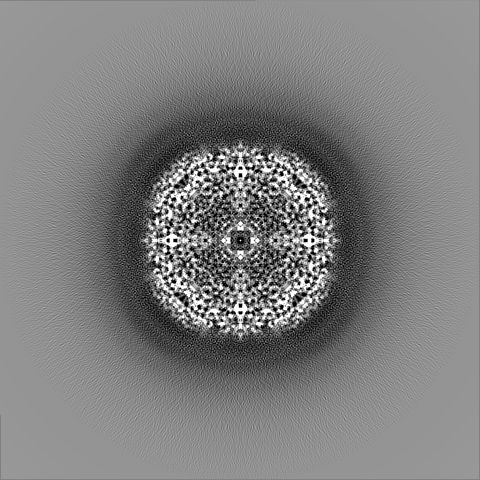
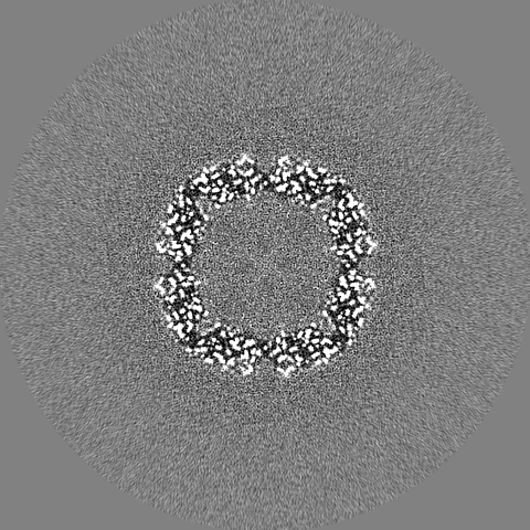
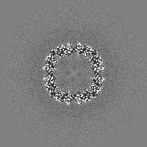
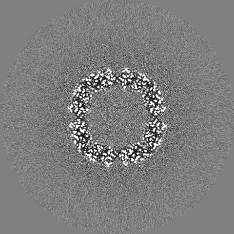
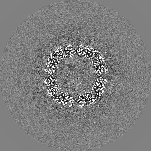
Journal: J Struct Biol X / Year: 2023 Title: Characterizing the resolution and throughput of the Apollo direct electron detector. Authors: Ruizhi Peng / Xiaofeng Fu / Joshua H Mendez / Peter S Randolph / Benjamin E Bammes / Scott M Stagg / Abstract: Advances in electron detection have been essential to the success of high-resolution cryo-EM structure determination. A new generation of direct electron detector called the Apollo, has been ...Advances in electron detection have been essential to the success of high-resolution cryo-EM structure determination. A new generation of direct electron detector called the Apollo, has been developed by Direct Electron. The Apollo uses a novel event-based MAPS detector custom designed for ultra-fast electron counting. We have evaluated this new camera, finding that it delivers high detective quantum efficiency (DQE) and low coincidence loss, enabling high-quality electron counting data acquisition at up to nearly 80 input electrons per pixel per second. We further characterized the performance of Apollo for single particle cryo-EM on real biological samples. Using mouse apoferritin, Apollo yielded better than 1.9 Å resolution reconstructions at all three tested dose rates from a half-day data collection session each. With longer collection time and improved specimen preparation, mouse apoferritin was reconstructed to 1.66 Å resolution. Applied to a more challenging small protein aldolase, we obtained a 2.24 Å resolution reconstruction. The high quality of the map indicates that the Apollo has sufficiently high DQE to reconstruct smaller proteins and complexes with high-fidelity. Our results demonstrate that the Apollo camera performs well across a broad range of dose rates and is capable of capturing high quality data that produce high-resolution reconstructions for large and small single particle samples.
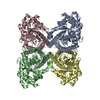
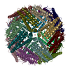
Protein or peptide: Ferritin heavy chain, N-terminally processed
Ligand: FE (III) ION
Ligand: water
-
Supramolecule #1: Mouse apoferritin heavy chain
Supramolecule
Name: Mouse apoferritin heavy chain / type: complex / ID: 1 / Parent: 0 / Macromolecule list: #1 Details: Ferritin is a universal intracellular protein that stores iron and releases it in a controlled fashion.
Source (natural)
Organism: Mus musculus (house mouse)
Molecular weight
Theoretical: 506 KDa
-
Macromolecule #1: Ferritin heavy chain, N-terminally processed
Macromolecule
Name: Ferritin heavy chain, N-terminally processed / type: protein_or_peptide / ID: 1 / Number of copies: 24 / Enantiomer: LEVO
Cryogen name: ETHANE / Chamber humidity: 95 % / Chamber temperature: 277.15 K / Instrument: FEI VITROBOT MARK IV
-
Electron microscopy
Microscope
FEI TITAN KRIOS
Image recording
Film or detector model: DIRECT ELECTRON APOLLO (4k x 4k) / Digitization - Dimensions - Width: 8192 pixel / Digitization - Dimensions - Height: 8192 pixel / Number grids imaged: 1 / Number real images: 972 / Average exposure time: 1.264 sec. / Average electron dose: 60.0 e/Å2
Electron beam
Acceleration voltage: 300 kV / Electron source: FIELD EMISSION GUN
In the structure databanks used in Yorodumi, some data are registered as the other names, "COVID-19 virus" and "2019-nCoV". Here are the details of the virus and the list of structure data.
Jan 31, 2019. EMDB accession codes are about to change! (news from PDBe EMDB page)
EMDB accession codes are about to change! (news from PDBe EMDB page)
The allocation of 4 digits for EMDB accession codes will soon come to an end. Whilst these codes will remain in use, new EMDB accession codes will include an additional digit and will expand incrementally as the available range of codes is exhausted. The current 4-digit format prefixed with “EMD-” (i.e. EMD-XXXX) will advance to a 5-digit format (i.e. EMD-XXXXX), and so on. It is currently estimated that the 4-digit codes will be depleted around Spring 2019, at which point the 5-digit format will come into force.
The EM Navigator/Yorodumi systems omit the EMD- prefix.
Related info.:Q: What is EMD? / ID/Accession-code notation in Yorodumi/EM Navigator
Yorodumi is a browser for structure data from EMDB, PDB, SASBDB, etc.
This page is also the successor to EM Navigator detail page, and also detail information page/front-end page for Omokage search.
The word "yorodu" (or yorozu) is an old Japanese word meaning "ten thousand". "mi" (miru) is to see.
Related info.:EMDB / PDB / SASBDB / Comparison of 3 databanks / Yorodumi Search / Aug 31, 2016. New EM Navigator & Yorodumi / Yorodumi Papers / Jmol/JSmol / Function and homology information / Changes in new EM Navigator and Yorodumi
 Movie
Movie Controller

 Yorodumi
 Open data
 Basic information
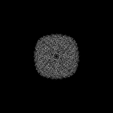
 Map data
 Sample
 Keywords
 Function and homology information
 Authors
 United States, 3 items
  Citation
 Structure visualization
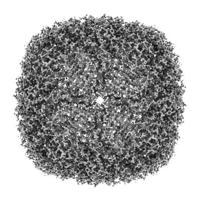
 Downloads & links
 emd_28269.png
 http://ftp.pdbj.org/pub/emdb/structures/EMD-28269


 Z (Sec.)
 Y (Row.)
 X (Col.)


 Sample components

 Processing
 Electron microscopy
 FIELD EMISSION GUN