 Movie
Movie Controller

[English] 日本語
 Yorodumi
- PDB-8ehg: Rabbit muscle aldolase determined using single-particle cryo-EM w... -
+ Open data

- Basic information
| Entry | Database: PDB / ID: 8ehg | ||||||||||||
|---|---|---|---|---|---|---|---|---|---|---|---|---|---|
| Title | Rabbit muscle aldolase determined using single-particle cryo-EM with Apollo camera. | ||||||||||||
 Components Components | Fructose-bisphosphate aldolase A | ||||||||||||
 Keywords Keywords | SUGAR BINDING PROTEIN / glycolysis | ||||||||||||
| Function / homology |  Function and homology information Function and homology informationnegative regulation of Arp2/3 complex-mediated actin nucleation / fructose-bisphosphate aldolase / fructose-bisphosphate aldolase activity / M band / I band / glycolytic process / protein homotetramerization / positive regulation of cell migration Similarity search - Function | ||||||||||||
| Biological species |  | ||||||||||||
| Method | ELECTRON MICROSCOPY / single particle reconstruction / cryo EM / Resolution: 2.24 Å | ||||||||||||
 Authors Authors | Peng, R. / Fu, X. / Mendez, J.H. / Randolph, P.H. / Bammes, B. / Stagg, S.M. | ||||||||||||
| Funding support |  United States, 3items United States, 3items
| ||||||||||||
 Citation Citation | Journal: J Struct Biol X / Year: 2023 Title: Characterizing the resolution and throughput of the Apollo direct electron detector. Authors: Ruizhi Peng / Xiaofeng Fu / Joshua H Mendez / Peter S Randolph / Benjamin E Bammes / Scott M Stagg / Abstract: Advances in electron detection have been essential to the success of high-resolution cryo-EM structure determination. A new generation of direct electron detector called the Apollo, has been ...Advances in electron detection have been essential to the success of high-resolution cryo-EM structure determination. A new generation of direct electron detector called the Apollo, has been developed by Direct Electron. The Apollo uses a novel event-based MAPS detector custom designed for ultra-fast electron counting. We have evaluated this new camera, finding that it delivers high detective quantum efficiency (DQE) and low coincidence loss, enabling high-quality electron counting data acquisition at up to nearly 80 input electrons per pixel per second. We further characterized the performance of Apollo for single particle cryo-EM on real biological samples. Using mouse apoferritin, Apollo yielded better than 1.9 Å resolution reconstructions at all three tested dose rates from a half-day data collection session each. With longer collection time and improved specimen preparation, mouse apoferritin was reconstructed to 1.66 Å resolution. Applied to a more challenging small protein aldolase, we obtained a 2.24 Å resolution reconstruction. The high quality of the map indicates that the Apollo has sufficiently high DQE to reconstruct smaller proteins and complexes with high-fidelity. Our results demonstrate that the Apollo camera performs well across a broad range of dose rates and is capable of capturing high quality data that produce high-resolution reconstructions for large and small single particle samples. | ||||||||||||
| History |
|
- Structure visualization
| Structure viewer | Molecule: MolmilJmol/JSmol |
|---|
- Downloads & links
-Download
| PDBx/mmCIF format | 8ehg.cif.gz | 312.6 KB | Display | PDBx/mmCIF format |
|---|---|---|---|---|
| PDB format | pdb8ehg.ent.gz | 203.6 KB | Display | PDB format |
| PDBx/mmJSON format | 8ehg.json.gz | Tree view | PDBx/mmJSON format | |
| Others |  Other downloads Other downloads |
-Validation report
| Arichive directory | https://data.pdbj.org/pub/pdb/validation_reports/eh/8ehgftp://data.pdbj.org/pub/pdb/validation_reports/eh/8ehg | HTTPS FTP |
|---|
-Related structure data
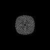
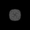
| Related structure data |  28147MC 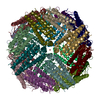 8emqC 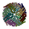 8en7C M: map data used to model this data C: citing same article ( |
|---|---|
| Similar structure data |
-Links
 PDBj
PDBj
- Assembly
| Deposited unit | 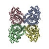
| ||||||||||||||||||||||||||||||||||||||||||||||||||||||||||||||||||
|---|---|---|---|---|---|---|---|---|---|---|---|---|---|---|---|---|---|---|---|---|---|---|---|---|---|---|---|---|---|---|---|---|---|---|---|---|---|---|---|---|---|---|---|---|---|---|---|---|---|---|---|---|---|---|---|---|---|---|---|---|---|---|---|---|---|---|---|
| 1 |
| ||||||||||||||||||||||||||||||||||||||||||||||||||||||||||||||||||
| Noncrystallographic symmetry (NCS) | NCS domain:
NCS domain segments:
NCS oper:
|
-Components
| #1: Protein | Mass: 39394.875 Da / Num. of mol.: 4 / Source method: isolated from a natural source / Source: (natural) |
|---|
-Experimental details
-Experiment
| Experiment | Method: ELECTRON MICROSCOPY |
|---|---|
| EM experiment | Aggregation state: PARTICLE / 3D reconstruction method: single particle reconstruction |
- Sample preparation
| Component | Name: Aldolase from rabbit muscle / Type: COMPLEX Details: Plays a key role in glycolysis and gluconeogenesis. In addition, may also function as scaffolding protein. Entity ID: all / Source: NATURAL | ||||||||||||||||||||
|---|---|---|---|---|---|---|---|---|---|---|---|---|---|---|---|---|---|---|---|---|---|
| Molecular weight | Value: 14921 MDa / Experimental value: NO | ||||||||||||||||||||
| Source (natural) | Organism: | ||||||||||||||||||||
| Buffer solution | pH: 7.5 / Details: DTT are added freshly before use. | ||||||||||||||||||||
| Buffer component |
| ||||||||||||||||||||
| Specimen | Conc.: 4 mg/ml / Embedding applied: NO / Shadowing applied: NO / Staining applied: NO / Vitrification applied: YES | ||||||||||||||||||||
| Specimen support | Grid material: GOLD / Grid mesh size: 300 divisions/in. / Grid type: UltrAuFoil R1.2/1.3 | ||||||||||||||||||||
| Vitrification | Instrument: FEI VITROBOT MARK IV / Cryogen name: ETHANE / Humidity: 95 % / Chamber temperature: 277.15 K |
- Electron microscopy imaging
| Experimental equipment |  Model: Titan Krios / Image courtesy: FEI Company |
|---|---|
| Microscopy | Model: FEI TITAN KRIOS |
| Electron gun | Electron source:  FIELD EMISSION GUN / Accelerating voltage: 300 kV / Illumination mode: FLOOD BEAM FIELD EMISSION GUN / Accelerating voltage: 300 kV / Illumination mode: FLOOD BEAM |
| Electron lens | Mode: BRIGHT FIELD / Nominal magnification: 75000 X / Calibrated magnification: 72621 X / Nominal defocus max: 1500 nm / Nominal defocus min: 800 nm / Cs: 2.7 mm / C2 aperture diameter: 80 µm / Alignment procedure: ZEMLIN TABLEAU |
| Specimen holder | Cryogen: NITROGEN / Specimen holder model: FEI TITAN KRIOS AUTOGRID HOLDER |
| Image recording | Average exposure time: 1.347 sec. / Electron dose: 60 e/Å2 / Film or detector model: OTHER / Num. of grids imaged: 1 / Num. of real images: 8682 Details: Images were collected using Direct Electron Apollo camera at a fixed movie frame rate of 60 frames/second. |
| Image scans | Width: 8192 / Height: 8192 |
- Processing
| Software |
| ||||||||||||||||||||||||||||||||||||||||
|---|---|---|---|---|---|---|---|---|---|---|---|---|---|---|---|---|---|---|---|---|---|---|---|---|---|---|---|---|---|---|---|---|---|---|---|---|---|---|---|---|---|
| EM software |
| ||||||||||||||||||||||||||||||||||||||||
| Image processing | Details: Direct Electron Apollo | ||||||||||||||||||||||||||||||||||||||||
| CTF correction | Type: PHASE FLIPPING AND AMPLITUDE CORRECTION | ||||||||||||||||||||||||||||||||||||||||
| Particle selection | Num. of particles selected: 3805094 Details: particles were picked using templates projected from EMD-21023. | ||||||||||||||||||||||||||||||||||||||||
| Symmetry | Point symmetry: D2 (2x2 fold dihedral) | ||||||||||||||||||||||||||||||||||||||||
| 3D reconstruction | Resolution: 2.24 Å / Resolution method: FSC 0.143 CUT-OFF / Num. of particles: 722778 / Algorithm: BACK PROJECTION / Num. of class averages: 1 / Symmetry type: POINT | ||||||||||||||||||||||||||||||||||||||||
| Refinement | Cross valid method: NONE Stereochemistry target values: GeoStd + Monomer Library + CDL v1.2 | ||||||||||||||||||||||||||||||||||||||||
| Displacement parameters | Biso mean: 8.14 Å2 | ||||||||||||||||||||||||||||||||||||||||
| Refine LS restraints |
| ||||||||||||||||||||||||||||||||||||||||
| Refine LS restraints NCS |
|
