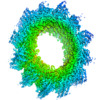
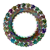
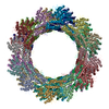
+ データを開く
- 基本情報
基本情報
| 登録情報 | データベース: EMDB / ID: EMD-2699 | |||||||||
|---|---|---|---|---|---|---|---|---|---|---|
| タイトル | VipA/VipB, contractile sheath of the type VI secretion system | |||||||||
 マップデータ マップデータ | Structure of the native helical assembly isolated from Vibrio cholerae | |||||||||
 試料 試料 |
| |||||||||
 キーワード キーワード | VipA / VipB / Vibrio / T6SS / cryo-EM / sheath | |||||||||
| 機能・相同性 | Type VI secretion system TssC-like / TssC1, N-terminal / TssC1, C-terminal / EvpB/VC_A0108, tail sheath N-terminal domain / EvpB/VC_A0108, tail sheath gpW/gp25-like domain / Type VI secretion system sheath protein TssB1 / Type VI secretion system, VipA, VC_A0107 or Hcp2 / Type VI secretion system contractile sheath large subunit / Type VI secretion system contractile sheath small subunit 機能・相同性情報 機能・相同性情報 | |||||||||
| 生物種 |  Vibrio cholerae O1 biovar El Tor str. N16961 (コレラ菌) Vibrio cholerae O1 biovar El Tor str. N16961 (コレラ菌) | |||||||||
| 手法 | らせん対称体再構成法 / クライオ電子顕微鏡法 / 解像度: 3.5 Å | |||||||||
 データ登録者 データ登録者 | Kudryashev M / Wang R / Brackmann M / Scherer S / Maier T / DiMaio F / Baker D / Stahlberg H / Egelman EH / Basler M | |||||||||
 引用 引用 |  ジャーナル: Cell / 年: 2015 ジャーナル: Cell / 年: 2015タイトル: Structure of the type VI secretion system contractile sheath. 著者: Mikhail Kudryashev / Ray Yu-Ruei Wang / Maximilian Brackmann / Sebastian Scherer / Timm Maier / David Baker / Frank DiMaio / Henning Stahlberg / Edward H Egelman / Marek Basler /   要旨: Bacteria use rapid contraction of a long sheath of the type VI secretion system (T6SS) to deliver effectors into a target cell. Here, we present an atomic-resolution structure of a native contracted ...Bacteria use rapid contraction of a long sheath of the type VI secretion system (T6SS) to deliver effectors into a target cell. Here, we present an atomic-resolution structure of a native contracted Vibrio cholerae sheath determined by cryo-electron microscopy. The sheath subunits, composed of tightly interacting proteins VipA and VipB, assemble into a six-start helix. The helix is stabilized by a core domain assembled from four β strands donated by one VipA and two VipB molecules. The fold of inner and middle layers is conserved between T6SS and phage sheaths. However, the structure of the outer layer is distinct and suggests a mechanism of interaction of the bacterial sheath with an accessory ATPase, ClpV, that facilitates multiple rounds of effector delivery. Our results provide a mechanistic insight into assembly of contractile nanomachines that bacteria and phages use to translocate macromolecules across membranes. | |||||||||
| 履歴 |
|
- 構造の表示
構造の表示
| ムービー |
 ムービービューア ムービービューア |
|---|---|
| 構造ビューア | EMマップ:  SurfView SurfView Molmil Molmil Jmol/JSmol Jmol/JSmol |
| 添付画像 |
- ダウンロードとリンク
-EMDBアーカイブ
| マップデータ |  emd_2699.map.gz emd_2699.map.gz | 72.4 MB |  EMDBマップデータ形式 EMDBマップデータ形式 | |
|---|---|---|---|---|
| ヘッダ (付随情報) |  emd-2699-v30.xml emd-2699-v30.xml emd-2699.xml emd-2699.xml | 11.4 KB 11.4 KB | 表示 表示 |  EMDBヘッダ EMDBヘッダ |
| 画像 |  emd_2699.png emd_2699.png | 328.3 KB | ||
| アーカイブディレクトリ |  http://ftp.pdbj.org/pub/emdb/structures/EMD-2699 http://ftp.pdbj.org/pub/emdb/structures/EMD-2699 ftp://ftp.pdbj.org/pub/emdb/structures/EMD-2699 ftp://ftp.pdbj.org/pub/emdb/structures/EMD-2699 | HTTPS FTP |
-検証レポート
| 文書・要旨 |  emd_2699_validation.pdf.gz emd_2699_validation.pdf.gz | 370.2 KB | 表示 |  EMDB検証レポート EMDB検証レポート |
|---|---|---|---|---|
| 文書・詳細版 |  emd_2699_full_validation.pdf.gz emd_2699_full_validation.pdf.gz | 369.8 KB | 表示 | |
| XML形式データ |  emd_2699_validation.xml.gz emd_2699_validation.xml.gz | 4.5 KB | 表示 | |
| アーカイブディレクトリ |  https://ftp.pdbj.org/pub/emdb/validation_reports/EMD-2699 https://ftp.pdbj.org/pub/emdb/validation_reports/EMD-2699 ftp://ftp.pdbj.org/pub/emdb/validation_reports/EMD-2699 ftp://ftp.pdbj.org/pub/emdb/validation_reports/EMD-2699 | HTTPS FTP |
-関連構造データ
| 関連構造データ |  3j9gMC M: このマップから作成された原子モデル C: 同じ文献を引用 ( |
|---|---|
| 類似構造データ | |
| 電子顕微鏡画像生データ |  EMPIAR-10019 (タイトル: VipA/VipB, sheath of the bacterial type IV secretion system, micrographs for helical reconstruction taken on a K2 detector EMPIAR-10019 (タイトル: VipA/VipB, sheath of the bacterial type IV secretion system, micrographs for helical reconstruction taken on a K2 detectorData size: 4.1 Data #1: Frame-averaged micrographs [micrographs - single frame]) |
- リンク
リンク
| EMDBのページ |  EMDB (EBI/PDBe) / EMDB (EBI/PDBe) /  EMDataResource EMDataResource |
|---|
- マップ
マップ
| ファイル |  ダウンロード / ファイル: emd_2699.map.gz / 形式: CCP4 / 大きさ: 268.2 MB / タイプ: IMAGE STORED AS FLOATING POINT NUMBER (4 BYTES) ダウンロード / ファイル: emd_2699.map.gz / 形式: CCP4 / 大きさ: 268.2 MB / タイプ: IMAGE STORED AS FLOATING POINT NUMBER (4 BYTES) | ||||||||||||||||||||||||||||||||||||||||||||||||||||||||||||||||||||
|---|---|---|---|---|---|---|---|---|---|---|---|---|---|---|---|---|---|---|---|---|---|---|---|---|---|---|---|---|---|---|---|---|---|---|---|---|---|---|---|---|---|---|---|---|---|---|---|---|---|---|---|---|---|---|---|---|---|---|---|---|---|---|---|---|---|---|---|---|---|
| 注釈 | Structure of the native helical assembly isolated from Vibrio cholerae | ||||||||||||||||||||||||||||||||||||||||||||||||||||||||||||||||||||
| 投影像・断面図 | 画像のコントロール
画像は Spider により作成 これらの図は立方格子座標系で作成されたものです | ||||||||||||||||||||||||||||||||||||||||||||||||||||||||||||||||||||
| ボクセルのサイズ | X=Y=Z: 0.5 Å | ||||||||||||||||||||||||||||||||||||||||||||||||||||||||||||||||||||
| 密度 |
| ||||||||||||||||||||||||||||||||||||||||||||||||||||||||||||||||||||
| 対称性 | 空間群: 1 | ||||||||||||||||||||||||||||||||||||||||||||||||||||||||||||||||||||
| 詳細 | EMDB XML:
CCP4マップ ヘッダ情報:
| ||||||||||||||||||||||||||||||||||||||||||||||||||||||||||||||||||||
-添付データ
- 試料の構成要素
-全体 : Native VipA/B sheath purified from Vibrio cholerae
| 全体 | 名称: Native VipA/B sheath purified from Vibrio cholerae |
|---|---|
| 要素 |
|
-超分子 #1000: Native VipA/B sheath purified from Vibrio cholerae
| 超分子 | 名称: Native VipA/B sheath purified from Vibrio cholerae / タイプ: sample / ID: 1000 集合状態: heterodimer of VipA and VipB assembled in a helix Number unique components: 2 |
|---|
-分子 #1: VipA
| 分子 | 名称: VipA / タイプ: protein_or_peptide / ID: 1 / 集合状態: helix built of heterodimers / 組換発現: No / データベース: NCBI |
|---|---|
| 由来(天然) | 生物種:  Vibrio cholerae O1 biovar El Tor str. N16961 (コレラ菌) Vibrio cholerae O1 biovar El Tor str. N16961 (コレラ菌)株: N16961 / 細胞中の位置: cytoplasm |
| 分子量 | 理論値: 74 KDa |
| 配列 | UniProtKB: Type VI secretion system contractile sheath large subunit |
-分子 #2: VipB
| 分子 | 名称: VipB / タイプ: protein_or_peptide / ID: 2 / 集合状態: helix built of heterodimers / 組換発現: No / データベース: NCBI |
|---|---|
| 由来(天然) | 生物種:  Vibrio cholerae O1 biovar El Tor str. N16961 (コレラ菌) Vibrio cholerae O1 biovar El Tor str. N16961 (コレラ菌)株: N16961 / 細胞中の位置: cytoplasm |
| 分子量 | 理論値: 74 KDa |
| 配列 | UniProtKB: Type VI secretion system contractile sheath large subunit |
-実験情報
-構造解析
| 手法 | クライオ電子顕微鏡法 |
|---|---|
 解析 解析 | らせん対称体再構成法 |
| 試料の集合状態 | helical array |
- 試料調製
試料調製
| 濃度 | 0.1 mg/mL |
|---|---|
| 緩衝液 | pH: 7.4 / 詳細: PBS |
| グリッド | 詳細: Quantifoil holey carbon grids |
| 凍結 | 凍結剤: ETHANE / 装置: FEI VITROBOT MARK IV / 手法: Blot for 1.5 seconds before plunging |
| 詳細 | native polymer |
- 電子顕微鏡法
| 顕微鏡 | FEI TITAN KRIOS |
|---|---|
| アライメント法 | Legacy - 非点収差: Was corrected manually at high magnification Legacy - Electron beam tilt params: 0 |
| 日付 | 2013年11月15日 |
| 撮影 | カテゴリ: CCD / フィルム・検出器のモデル: GATAN K2 (4k x 4k) / 実像数: 72 / 平均電子線量: 30 e/Å2 詳細: Images were collected in dose fractionation mode and further aligned by Li and Cheng's algorithm in real time using the 2dx_automator |
| 電子線 | 加速電圧: 300 kV / 電子線源:  FIELD EMISSION GUN FIELD EMISSION GUN |
| 電子光学系 | 倍率(補正後): 29000 / 照射モード: FLOOD BEAM / 撮影モード: BRIGHT FIELD / Cs: 2.7 mm / 最大 デフォーカス(公称値): 2.0 µm / 最小 デフォーカス(公称値): 0.4 µm / 倍率(公称値): 29000 |
| 試料ステージ | 試料ホルダーモデル: FEI TITAN KRIOS AUTOGRID HOLDER Tilt angle min: -5 / Tilt angle max: 5 |
| 実験機器 |  モデル: Titan Krios / 画像提供: FEI Company |
- 画像解析
画像解析
| 詳細 | We used Iterative Helical Real Space Reconstruction methodology(IHRSR) |
|---|---|
| 最終 再構成 | 想定した対称性 - らせんパラメータ - Δz: 21.8 Å 想定した対称性 - らせんパラメータ - ΔΦ: 29.4 ° 想定した対称性 - らせんパラメータ - 軸対称性: C6 (6回回転対称) アルゴリズム: OTHER / 解像度のタイプ: BY AUTHOR / 解像度: 3.5 Å / 解像度の算出法: OTHER ソフトウェア - 名称: 2dx_automator, Ctffind, Helixboxer, IHRSR/Spider 詳細: Resolution was detected by Resmap, it varied from 3.2 A in well ordered parts till 5 A outside of the helix. |
| CTF補正 | 詳細: Ctffind for each micrograph |
 ムービー
ムービー コントローラー


 Z (Sec.)
 Y (Row.)
 X (Col.)


