Journal: J Biol Chem / Year: 2023 Title: The nematode α-catenin ortholog, HMP1, has an extended α-helix when bound to actin filaments. Authors: Erumbi S Rangarajan / Emmanuel W Smith / Tina Izard / Abstract: The regulation of cell-cell junctions during epidermal morphogenesis ensures tissue integrity, a process regulated by α-catenin. This cytoskeletal protein connects the cadherin complex to ...The regulation of cell-cell junctions during epidermal morphogenesis ensures tissue integrity, a process regulated by α-catenin. This cytoskeletal protein connects the cadherin complex to filamentous actin at cell-cell junctions. The cadherin-catenin complex plays key roles in cell physiology, organism development, and disease. While mutagenesis of Caenorhabditis elegans cadherin and catenin shows that these proteins are key for embryonic morphogenesis, we know surprisingly little about their structure and attachment to the cytoskeleton. In contrast to mammalian α-catenin that functions as a dimer or monomer, the α-catenin ortholog from C. elegans, HMP1 for humpback, is a monomer. Our cryogenic electron microscopy (cryoEM) structure of HMP1/α-catenin reveals that the amino- and carboxy-terminal domains of HMP1/α-catenin are disordered and not in contact with the remaining HMP1/α-catenin middle domain. Since the carboxy-terminal HMP1/α-catenin domain is the F-actin-binding domain (FABD), this interdomain constellation suggests that HMP1/α-catenin is constitutively active, which we confirm biochemically. Our perhaps most surprising finding, given the high sequence similarity between the mammalian and nematode proteins, is our cryoEM structure of HMP1/α-catenin bound to F-actin. Unlike the structure of mammalian α-catenin bound to F-actin, binding to F-actin seems to allosterically convert a loop region of the HMP1/α-catenin FABD to extend an HMP1/α-catenin FABD α-helix. We use cryoEM and bundling assays to show for the first time how the FABD of HMP1/α-catenin bundles actin in the absence of force. Collectively, our data advance our understanding of α-catenin regulation of cell-cell contacts and additionally aid our understanding of the evolution of multicellularity in metazoans.
In the structure databanks used in Yorodumi, some data are registered as the other names, "COVID-19 virus" and "2019-nCoV". Here are the details of the virus and the list of structure data.
Jan 31, 2019. EMDB accession codes are about to change! (news from PDBe EMDB page)
EMDB accession codes are about to change! (news from PDBe EMDB page)
The allocation of 4 digits for EMDB accession codes will soon come to an end. Whilst these codes will remain in use, new EMDB accession codes will include an additional digit and will expand incrementally as the available range of codes is exhausted. The current 4-digit format prefixed with “EMD-” (i.e. EMD-XXXX) will advance to a 5-digit format (i.e. EMD-XXXXX), and so on. It is currently estimated that the 4-digit codes will be depleted around Spring 2019, at which point the 5-digit format will come into force.
The EM Navigator/Yorodumi systems omit the EMD- prefix.
Related info.:Q: What is EMD? / ID/Accession-code notation in Yorodumi/EM Navigator
Yorodumi is a browser for structure data from EMDB, PDB, SASBDB, etc.
This page is also the successor to EM Navigator detail page, and also detail information page/front-end page for Omokage search.
The word "yorodu" (or yorozu) is an old Japanese word meaning "ten thousand". "mi" (miru) is to see.
Related info.:EMDB / PDB / SASBDB / Comparison of 3 databanks / Yorodumi Search / Aug 31, 2016. New EM Navigator & Yorodumi / Yorodumi Papers / Jmol/JSmol / Function and homology information / Changes in new EM Navigator and Yorodumi
 Movie
Movie Controller

 Yorodumi
 Open data
 Basic information
 Map data
 Sample
 Function and homology information

 Authors
 United States, 1 items
  Citation
 Structure visualization
 Downloads & links
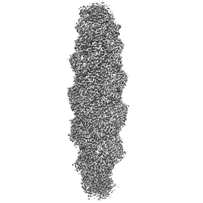 emd_26805.png
 http://ftp.pdbj.org/pub/emdb/structures/EMD-26805


 Z (Sec.)
 Y (Row.)
 X (Col.)


 Sample components

 Processing
 Electron microscopy
 FIELD EMISSION GUN
