- EMDB-26060: Cryo-em structure of human prothrombin:prothrombinase at 4.1 Angs... -
+
Open data
ID or keywords:
Loading...
-
Basic information
Entry
Database: EMDB / ID: EMD-26060
Title
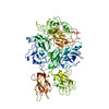
Cryo-em structure of human prothrombin:prothrombinase at 4.1 Angstrom resolution
Map data
prothrombin:prothrombinase
Sample
Complex: prothrombin prothrombinase
Complex: Prothrombin (E.C.3.4.21.5), Factor X light chain, Activated factor Xa heavy chain
Protein or peptide: Prothrombin
Protein or peptide: Factor X light chain
Protein or peptide: Activated factor Xa heavy chain
Complex: Coagulation factor Va
Protein or peptide: Coagulation factor Va
Protein or peptide: Coagulation factor Va
Keywords
prothrombin prothrombinase / BLOOD CLOTTING
Function / homology
Function and homology information
response to vitamin K / coagulation factor Xa / platelet alpha granule / Cargo concentration in the ER / COPII-coated ER to Golgi transport vesicle / Defective factor IX causes thrombophilia / Defective cofactor function of FVIIIa variant / Defective F9 variant does not activate FX / : / COPII-mediated vesicle transport ...response to vitamin K / coagulation factor Xa / platelet alpha granule / Cargo concentration in the ER / COPII-coated ER to Golgi transport vesicle / Defective factor IX causes thrombophilia / Defective cofactor function of FVIIIa variant / Defective F9 variant does not activate FX / : / COPII-mediated vesicle transport / blood circulation / : / thrombospondin receptor activity / thrombin / thrombin-activated receptor signaling pathway / ligand-gated ion channel signaling pathway / Defective factor XII causes hereditary angioedema / negative regulation of astrocyte differentiation / regulation of blood coagulation / neutrophil-mediated killing of gram-negative bacterium / positive regulation of phospholipase C-activating G protein-coupled receptor signaling pathway / Defective F8 cleavage by thrombin / Platelet Aggregation (Plug Formation) / positive regulation of collagen biosynthetic process / negative regulation of platelet activation / negative regulation of blood coagulation / negative regulation of fibrinolysis / positive regulation of blood coagulation / blood coagulation, fibrin clot formation / positive regulation of TOR signaling / Transport of gamma-carboxylated protein precursors from the endoplasmic reticulum to the Golgi apparatus / : / Gamma-carboxylation of protein precursors / Removal of aminoterminal propeptides from gamma-carboxylated proteins / regulation of cytosolic calcium ion concentration / fibrinolysis / : / negative regulation of proteolysis / endoplasmic reticulum-Golgi intermediate compartment membrane / negative regulation of cytokine production involved in inflammatory response / platelet alpha granule lumen / Regulation of Complement cascade / positive regulation of release of sequestered calcium ion into cytosol / acute-phase response / Cell surface interactions at the vascular wall / Peptide ligand-binding receptors / positive regulation of receptor signaling pathway via JAK-STAT / growth factor activity / Post-translational protein phosphorylation / lipopolysaccharide binding / response to wounding / positive regulation of protein localization to nucleus / platelet activation / phospholipid binding / Golgi lumen / Regulation of Insulin-like Growth Factor (IGF) transport and uptake by Insulin-like Growth Factor Binding Proteins (IGFBPs) / positive regulation of reactive oxygen species metabolic process / positive regulation of insulin secretion / blood coagulation / Platelet degranulation / regulation of cell shape / heparin binding / antimicrobial humoral immune response mediated by antimicrobial peptide / extracellular vesicle / Thrombin signalling through proteinase activated receptors (PARs) / positive regulation of cell growth / blood microparticle / G alpha (q) signalling events / positive regulation of phosphatidylinositol 3-kinase/protein kinase B signal transduction / cell surface receptor signaling pathway / positive regulation of cell migration / endoplasmic reticulum lumen / copper ion binding / receptor ligand activity / serine-type endopeptidase activity / external side of plasma membrane / signaling receptor binding / positive regulation of cell population proliferation / calcium ion binding / proteolysis / : / extracellular exosome / extracellular region / membrane / plasma membrane Similarity search - Function
National Institutes of Health/National Heart, Lung, and Blood Institute (NIH/NHLBI)
United States
Citation
Journal: Blood / Year: 2022 Title: Cryo-EM structure of the prothrombin-prothrombinase complex. Authors: Eliza A Ruben / Brock Summers / Michael J Rau / James A J Fitzpatrick / Enrico Di Cera / Abstract: The intrinsic and extrinsic pathways of the coagulation cascade converge to a common step where the prothrombinase complex, comprising the enzyme factor Xa (fXa), the cofactor fVa, Ca2+ and ...The intrinsic and extrinsic pathways of the coagulation cascade converge to a common step where the prothrombinase complex, comprising the enzyme factor Xa (fXa), the cofactor fVa, Ca2+ and phospholipids, activates the zymogen prothrombin to the protease thrombin. The reaction entails cleavage at 2 sites, R271 and R320, generating the intermediates prethrombin 2 and meizothrombin, respectively. The molecular basis of these interactions that are central to hemostasis remains elusive. We solved 2 cryogenic electron microscopy (cryo-EM) structures of the fVa-fXa complex, 1 free on nanodiscs at 5.3-Å resolution and the other bound to prothrombin at near atomic 4.1-Å resolution. In the prothrombin-fVa-fXa complex, the Gla domains of fXa and prothrombin align on a plane with the C1 and C2 domains of fVa for interaction with membranes. Prothrombin and fXa emerge from this plane in curved conformations that bring their protease domains in contact with each other against the A2 domain of fVa. The 672ESTVMATRKMHDRLEPEDEE691 segment of the A2 domain closes on the protease domain of fXa like a lid to fix orientation of the active site. The 696YDYQNRL702 segment binds to prothrombin and establishes the pathway of activation by sequestering R271 against D697 and directing R320 toward the active site of fXa. The cryo-EM structure provides a molecular view of prothrombin activation along the meizothrombin pathway and suggests a mechanism for cleavage at the alternative R271 site. The findings advance our basic knowledge of a key step of coagulation and bear broad relevance to other interactions in the blood.
In the structure databanks used in Yorodumi, some data are registered as the other names, "COVID-19 virus" and "2019-nCoV". Here are the details of the virus and the list of structure data.
Jan 31, 2019. EMDB accession codes are about to change! (news from PDBe EMDB page)
EMDB accession codes are about to change! (news from PDBe EMDB page)
The allocation of 4 digits for EMDB accession codes will soon come to an end. Whilst these codes will remain in use, new EMDB accession codes will include an additional digit and will expand incrementally as the available range of codes is exhausted. The current 4-digit format prefixed with “EMD-” (i.e. EMD-XXXX) will advance to a 5-digit format (i.e. EMD-XXXXX), and so on. It is currently estimated that the 4-digit codes will be depleted around Spring 2019, at which point the 5-digit format will come into force.
The EM Navigator/Yorodumi systems omit the EMD- prefix.
Related info.:Q: What is EMD? / ID/Accession-code notation in Yorodumi/EM Navigator
Yorodumi is a browser for structure data from EMDB, PDB, SASBDB, etc.
This page is also the successor to EM Navigator detail page, and also detail information page/front-end page for Omokage search.
The word "yorodu" (or yorozu) is an old Japanese word meaning "ten thousand". "mi" (miru) is to see.
Related info.:EMDB / PDB / SASBDB / Comparison of 3 databanks / Yorodumi Search / Aug 31, 2016. New EM Navigator & Yorodumi / Yorodumi Papers / Jmol/JSmol / Function and homology information / Changes in new EM Navigator and Yorodumi
 Movie
Movie Controller

 Yorodumi
 Open data
 Basic information
 Map data
 Sample
 Keywords
 Function and homology information
 Homo sapiens (human)
 Authors
 United States, 1 items
  Citation
 Structure visualization
 Downloads & links
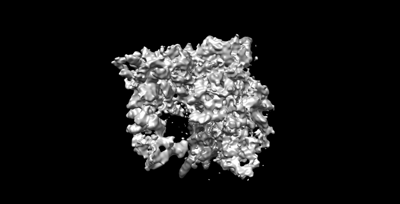 emd_26060.png
 http://ftp.pdbj.org/pub/emdb/structures/EMD-26060


 Z (Sec.)
 Y (Row.)
 X (Col.)


 Sample components
 Mesocricetus auratus (golden hamster)
 Processing
 Electron microscopy
 FIELD EMISSION GUN