[English] 日本語
 Yorodumi
- EMDB-2526: The electron crystallography structure of the cAMP-bound potassiu... -
+ Open data
- Basic information
| Entry | Database: EMDB / ID: EMD-2526 | |||||||||
|---|---|---|---|---|---|---|---|---|---|---|
| Title | The electron crystallography structure of the cAMP-bound potassium channel MloK1 | |||||||||
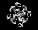
 Map data Map data | Tetrameric MloK1 in the presence of cAMP | |||||||||
 Sample Sample |
| |||||||||
 Keywords Keywords | Electron crystallography / 2dx / voltage gated potassium channel / CNBD / 2D crystal | |||||||||
| Function / homology |  Function and homology information Function and homology informationtransport / potassium channel activity / membrane => GO:0016020 / cAMP binding / monoatomic ion transport / potassium ion transmembrane transport / potassium ion transport / nucleotide binding / protein-containing complex binding / identical protein binding ...transport / potassium channel activity / membrane => GO:0016020 / cAMP binding / monoatomic ion transport / potassium ion transmembrane transport / potassium ion transport / nucleotide binding / protein-containing complex binding / identical protein binding / membrane / plasma membrane Similarity search - Function | |||||||||
| Biological species |  Mesorhizobium loti (bacteria) Mesorhizobium loti (bacteria) | |||||||||
| Method | electron crystallography / cryo EM / Resolution: 7.0 Å | |||||||||
 Authors Authors | Kowal J / Chami M / Baumgartner P / Arheit M / Chiu P-L / Rangl M / Scheuring S / Schroeder GF / Nimigean CM / Stahlberg H | |||||||||
 Citation Citation |  Journal: Nat Commun / Year: 2014 Journal: Nat Commun / Year: 2014Title: Ligand-induced structural changes in the cyclic nucleotide-modulated potassium channel MloK1. Authors: Julia Kowal / Mohamed Chami / Paul Baumgartner / Marcel Arheit / Po-Lin Chiu / Martina Rangl / Simon Scheuring / Gunnar F Schröder / Crina M Nimigean / Henning Stahlberg /     Abstract: Cyclic nucleotide-modulated ion channels are important for signal transduction and pacemaking in eukaryotes. The molecular determinants of ligand gating in these channels are still unknown, mainly ...Cyclic nucleotide-modulated ion channels are important for signal transduction and pacemaking in eukaryotes. The molecular determinants of ligand gating in these channels are still unknown, mainly because of a lack of direct structural information. Here we report ligand-induced conformational changes in full-length MloK1, a cyclic nucleotide-modulated potassium channel from the bacterium Mesorhizobium loti, analysed by electron crystallography and atomic force microscopy. Upon cAMP binding, the cyclic nucleotide-binding domains move vertically towards the membrane, and directly contact the S1-S4 voltage sensor domains. This is accompanied by a significant shift and tilt of the voltage sensor domain helices. In both states, the inner pore-lining helices are in an 'open' conformation. We propose a mechanism in which ligand binding can favour pore opening via a direct interaction between the cyclic nucleotide-binding domains and voltage sensors. This offers a simple mechanistic hypothesis for the coupling between ligand gating and voltage sensing in eukaryotic HCN channels. | |||||||||
| History |
|
- Structure visualization
| Movie |
 Movie viewer Movie viewer |
|---|---|
| Structure viewer | EM map:  SurfView SurfView Molmil Molmil Jmol/JSmol Jmol/JSmol |
| Supplemental images |
- Downloads & links
-EMDB archive
| Map data |  emd_2526.map.gz emd_2526.map.gz | 4.2 MB |  EMDB map data format EMDB map data format | |
|---|---|---|---|---|
| Header (meta data) |  emd-2526-v30.xml emd-2526-v30.xml emd-2526.xml emd-2526.xml | 13.1 KB 13.1 KB | Display Display |  EMDB header EMDB header |
| Images |  EMD-2526.png EMD-2526.png | 107.2 KB | ||
| Archive directory |  http://ftp.pdbj.org/pub/emdb/structures/EMD-2526 http://ftp.pdbj.org/pub/emdb/structures/EMD-2526 ftp://ftp.pdbj.org/pub/emdb/structures/EMD-2526 ftp://ftp.pdbj.org/pub/emdb/structures/EMD-2526 | HTTPS FTP |
-Validation report
| Summary document |  emd_2526_validation.pdf.gz emd_2526_validation.pdf.gz | 223.6 KB | Display |  EMDB validaton report EMDB validaton report |
|---|---|---|---|---|
| Full document |  emd_2526_full_validation.pdf.gz emd_2526_full_validation.pdf.gz | 222.7 KB | Display | |
| Data in XML |  emd_2526_validation.xml.gz emd_2526_validation.xml.gz | 5.3 KB | Display | |
| Arichive directory |  https://ftp.pdbj.org/pub/emdb/validation_reports/EMD-2526 https://ftp.pdbj.org/pub/emdb/validation_reports/EMD-2526 ftp://ftp.pdbj.org/pub/emdb/validation_reports/EMD-2526 ftp://ftp.pdbj.org/pub/emdb/validation_reports/EMD-2526 | HTTPS FTP |
-Related structure data
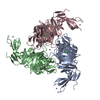
| Related structure data |  4chvMC  2527C  4chwC M: atomic model generated by this map C: citing same article ( |
|---|---|
| Similar structure data | |
| EM raw data |  EMPIAR-10006 (Title: 2D crystal images of the potassium channel MloK1 with and without cAMP ligand EMPIAR-10006 (Title: 2D crystal images of the potassium channel MloK1 with and without cAMP ligandData size: 11.6 Data #1: Potassium channel MloK1 with cAMP ligand [micrographs - single frame] Data #2: Potassium channel MloK1 without cAMP ligand [micrographs - single frame]) |
- Links
Links
| EMDB pages |  EMDB (EBI/PDBe) / EMDB (EBI/PDBe) /  EMDataResource EMDataResource |
|---|---|
| Related items in Molecule of the Month |
- Map
Map
| File |  Download / File: emd_2526.map.gz / Format: CCP4 / Size: 4.7 MB / Type: IMAGE STORED AS FLOATING POINT NUMBER (4 BYTES) Download / File: emd_2526.map.gz / Format: CCP4 / Size: 4.7 MB / Type: IMAGE STORED AS FLOATING POINT NUMBER (4 BYTES) | ||||||||||||||||||||||||||||||||||||||||||||||||||||||||||||||||||||
|---|---|---|---|---|---|---|---|---|---|---|---|---|---|---|---|---|---|---|---|---|---|---|---|---|---|---|---|---|---|---|---|---|---|---|---|---|---|---|---|---|---|---|---|---|---|---|---|---|---|---|---|---|---|---|---|---|---|---|---|---|---|---|---|---|---|---|---|---|---|
| Annotation | Tetrameric MloK1 in the presence of cAMP | ||||||||||||||||||||||||||||||||||||||||||||||||||||||||||||||||||||
| Projections & slices | Image control
Images are generated by Spider. | ||||||||||||||||||||||||||||||||||||||||||||||||||||||||||||||||||||
| Voxel size | X=Y=Z: 0.975 Å | ||||||||||||||||||||||||||||||||||||||||||||||||||||||||||||||||||||
| Density |
| ||||||||||||||||||||||||||||||||||||||||||||||||||||||||||||||||||||
| Symmetry | Space group: 1 | ||||||||||||||||||||||||||||||||||||||||||||||||||||||||||||||||||||
| Details | EMDB XML:
CCP4 map header:
| ||||||||||||||||||||||||||||||||||||||||||||||||||||||||||||||||||||
-Supplemental data
- Sample components
-Entire : MloK1 with cAMP
| Entire | Name: MloK1 with cAMP |
|---|---|
| Components |
|
-Supramolecule #1000: MloK1 with cAMP
| Supramolecule | Name: MloK1 with cAMP / type: sample / ID: 1000 / Oligomeric state: tetramer / Number unique components: 1 |
|---|---|
| Molecular weight | Theoretical: 148 KDa Method: Calculated from sequence for the tetrameric assembly. Lipids are not included. |
-Macromolecule #1: MloK1
| Macromolecule | Name: MloK1 / type: protein_or_peptide / ID: 1 / Name.synonym: MlotiK1 / Details: cAMP present in buffer / Number of copies: 4 / Oligomeric state: Tetramer / Recombinant expression: Yes |
|---|---|
| Source (natural) | Organism:  Mesorhizobium loti (bacteria) / synonym: Rhizobium / Location in cell: Membrane Mesorhizobium loti (bacteria) / synonym: Rhizobium / Location in cell: Membrane |
| Molecular weight | Theoretical: 148 KDa |
| Recombinant expression | Organism:  |
| Sequence | UniProtKB: Cyclic nucleotide-gated potassium channel mll3241 GO: transport, monoatomic ion transport, potassium ion transport, potassium ion transmembrane transport, nucleotide binding, potassium channel activity, cAMP binding, plasma membrane, membrane, membrane => GO:0016020 InterPro: Potassium channel domain, Cyclic nucleotide-binding domain superfamily, Cyclic nucleotide-binding, conserved site, Cyclic nucleotide-binding domain, INTERPRO: IPR003091, 1-aminocyclopropane- ...InterPro: Potassium channel domain, Cyclic nucleotide-binding domain superfamily, Cyclic nucleotide-binding, conserved site, Cyclic nucleotide-binding domain, INTERPRO: IPR003091, 1-aminocyclopropane-1-carboxylate deaminase/D-cysteine desulfhydrase, RmlC-like jelly roll fold |
-Experimental details
-Structure determination
| Method | cryo EM |
|---|---|
 Processing Processing | electron crystallography |
| Aggregation state | 2D array |
- Sample preparation
| Concentration | 0.7 mg/mL |
|---|---|
| Buffer | pH: 7.6 Details: 20 mM KCl, 1 mM BaCl2, 1 mM EDTA, 20 mM Tris, 0.2 mM cAMP |
| Grid | Details: Holey carbon film (Quantifoil) covered with ultra thin carbon film. Vitrified in crystallization buffer solution by plunge freezing into ethane. |
| Vitrification | Cryogen name: ETHANE / Chamber humidity: 90 % / Chamber temperature: 120 K / Instrument: FEI VITROBOT MARK IV / Method: Blot for 3 seconds before plunging |
| Details | Dialysis |
| Crystal formation | Details: Dialysis |
- Electron microscopy
| Microscope | FEI/PHILIPS CM200FEG |
|---|---|
| Temperature | Min: 85 K / Average: 85 K |
| Date | Mar 1, 2012 |
| Image recording | Category: FILM / Film or detector model: KODAK SO-163 FILM / Digitization - Scanner: PRIMESCAN / Digitization - Sampling interval: 5 µm / Number real images: 78 / Average electron dose: 5 e/Å2 / Od range: 1.4 / Bits/pixel: 16 |
| Tilt angle min | 0 |
| Electron beam | Acceleration voltage: 200 kV / Electron source:  FIELD EMISSION GUN FIELD EMISSION GUN |
| Electron optics | Illumination mode: FLOOD BEAM / Imaging mode: BRIGHT FIELD / Cs: 2.0 mm / Nominal defocus max: 3.077 µm / Nominal defocus min: 0.655 µm / Nominal magnification: 50000 |
| Sample stage | Specimen holder model: GATAN LIQUID NITROGEN / Tilt angle max: 46 / Tilt series - Axis1 - Min angle: 0 ° / Tilt series - Axis1 - Max angle: 46 ° |
- Image processing
| Details | 2dx |
|---|---|
| Final reconstruction | Resolution.type: BY AUTHOR / Resolution: 7.0 Å / Resolution method: OTHER / Software - Name: 2dx / Details: Resolution was limited to 7 x 7 x 12 Angstroems |
| Crystal parameters | Unit cell - A: 131 Å / Unit cell - B: 131 Å / Unit cell - C: 400 Å / Unit cell - γ: 90.0 ° / Unit cell - α: 90.0 ° / Unit cell - β: 90.0 ° / Plane group: P 4 21 2 |
 Movie
Movie Controller


 Z (Sec.)
 Y (Row.)
 X (Col.)


