National Institutes of Health/National Institute Of Allergy and Infectious Diseases (NIH/NIAID)
R01AI087946
United States
National Institutes of Health/National Institute Of Allergy and Infectious Diseases (NIH/NIAID)
R01AI132818
United States
Citation
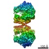
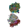
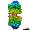
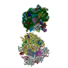
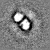
Journal: Cell / Year: 2021 Title: Interconnecting solvent quality, transcription, and chromosome folding in Escherichia coli. Authors: Yingjie Xiang / Ivan V Surovtsev / Yunjie Chang / Sander K Govers / Bradley R Parry / Jun Liu / Christine Jacobs-Wagner / Abstract: All cells fold their genomes, including bacterial cells, where the chromosome is compacted into a domain-organized meshwork called the nucleoid. How compaction and domain organization arise is not ...All cells fold their genomes, including bacterial cells, where the chromosome is compacted into a domain-organized meshwork called the nucleoid. How compaction and domain organization arise is not fully understood. Here, we describe a method to estimate the average mesh size of the nucleoid in Escherichia coli. Using nucleoid mesh size and DNA concentration estimates, we find that the cytoplasm behaves as a poor solvent for the chromosome when the cell is considered as a simple semidilute polymer solution. Monte Carlo simulations suggest that a poor solvent leads to chromosome compaction and DNA density heterogeneity (i.e., domain formation) at physiological DNA concentration. Fluorescence microscopy reveals that the heterogeneous DNA density negatively correlates with ribosome density within the nucleoid, consistent with cryoelectron tomography data. Drug experiments, together with past observations, suggest the hypothesis that RNAs contribute to the poor solvent effects, connecting chromosome compaction and domain formation to transcription and intracellular organization.
History
Deposition
Oct 20, 2020
-
Header (metadata) release
Jun 30, 2021
-
Map release
Jun 30, 2021
-
Update
Jan 11, 2023
-
Current status
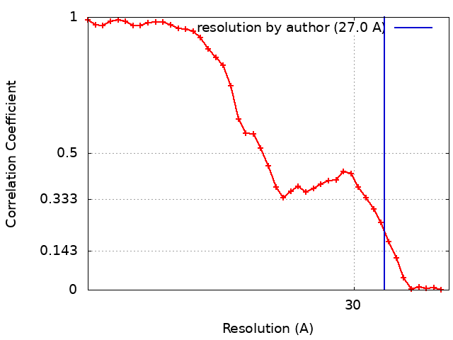
Jan 11, 2023
Processing site: RCSB / Status: Released
-
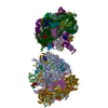
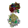
Structure visualization
Movie
Surface view with section colored by density value
EMPIAR-10589 (Title: cryo-FIB and cryo-ET study of ribosome and polysome structures in E. coli Data size: 9.7 Data #1: Unaligned tilt series of cryo-FIB milling E. coli cells [tilt series])
In the structure databanks used in Yorodumi, some data are registered as the other names, "COVID-19 virus" and "2019-nCoV". Here are the details of the virus and the list of structure data.
Jan 31, 2019. EMDB accession codes are about to change! (news from PDBe EMDB page)
EMDB accession codes are about to change! (news from PDBe EMDB page)
The allocation of 4 digits for EMDB accession codes will soon come to an end. Whilst these codes will remain in use, new EMDB accession codes will include an additional digit and will expand incrementally as the available range of codes is exhausted. The current 4-digit format prefixed with “EMD-” (i.e. EMD-XXXX) will advance to a 5-digit format (i.e. EMD-XXXXX), and so on. It is currently estimated that the 4-digit codes will be depleted around Spring 2019, at which point the 5-digit format will come into force.
The EM Navigator/Yorodumi systems omit the EMD- prefix.
Related info.:Q: What is EMD? / ID/Accession-code notation in Yorodumi/EM Navigator
Yorodumi is a browser for structure data from EMDB, PDB, SASBDB, etc.
This page is also the successor to EM Navigator detail page, and also detail information page/front-end page for Omokage search.
The word "yorodu" (or yorozu) is an old Japanese word meaning "ten thousand". "mi" (miru) is to see.
Related info.:EMDB / PDB / SASBDB / Comparison of 3 databanks / Yorodumi Search / Aug 31, 2016. New EM Navigator & Yorodumi / Yorodumi Papers / Jmol/JSmol / Function and homology information / Changes in new EM Navigator and Yorodumi
 Movie
Movie Controller


 Open data
 Basic information
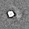
 Map data
 Sample
 Authors
 United States, 2 items
  Citation
 Structure visualization
 Movie viewer
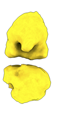
 Downloads & links
 emd_22878.png
 http://ftp.pdbj.org/pub/emdb/structures/EMD-22878


 Z (Sec.)
 Y (Row.)
 X (Col.)


 Sample components
 Processing
 Electron microscopy
 FIELD EMISSION GUN