 Movie
Movie Controller


+ Open data

- Basic information
| Entry | 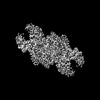 | |||||||||
|---|---|---|---|---|---|---|---|---|---|---|
| Title | Escherichia coli SduA complex | |||||||||
 Map data Map data | ||||||||||
 Sample Sample |
| |||||||||
 Keywords Keywords | SduA / Shedu / Prokaryotic immune system / IMMUNE SYSTEM | |||||||||
| Biological species |  | |||||||||
| Method | single particle reconstruction / cryo EM / Resolution: 2.95 Å | |||||||||
 Authors Authors | Loeff L / Jinek M / Asanovic I / Boneberg F / Pfleiderer MM / Ferdigg A / Ackle F / Martinez J | |||||||||
| Funding support | European Union, 2 items
| |||||||||
 Citation Citation | Journal: Cell / Year: 2025 Title: DNA end sensing and cleavage by the Shedu anti-phage defense system. Authors: Luuk Loeff / Alexander Walter / Gian Tizio Rosalen / Martin Jinek /  Abstract: The detection of molecular patterns associated with invading pathogens is a hallmark of innate immune systems. Prokaryotes deploy sophisticated host defense mechanisms in innate anti-phage immunity. ...The detection of molecular patterns associated with invading pathogens is a hallmark of innate immune systems. Prokaryotes deploy sophisticated host defense mechanisms in innate anti-phage immunity. Shedu is a single-component defense system comprising a putative nuclease SduA. Here, we report cryoelectron microscopy (cryo-EM) structures of apo- and double-stranded DNA (dsDNA)-bound tetrameric SduA assemblies, revealing that the N-terminal domains of SduA form a clamp that recognizes free DNA ends. End binding positions the DNA over the PD-(D/E)XK nuclease domain, resulting in dsDNA nicking at a fixed distance from the 5' end. The end-directed DNA nicking activity of Shedu prevents propagation of linear DNA in vivo. Finally, we show that phages escape Shedu immunity by suppressing their recombination-dependent DNA replication pathway. Taken together, these results define the antiviral mechanism of Shedu systems, underlining the paradigm that recognition of pathogen-specific nucleic acid structures is a conserved feature of innate immunity across all domains of life. | |||||||||
| History |
|
- Structure visualization
| Supplemental images |
|---|

- Downloads & links
-EMDB archive
| Map data | emd_17844.map.gz | 82 MB |  EMDB map data format EMDB map data format | |
|---|---|---|---|---|
| Header (meta data) | emd-17844-v30.xmlemd-17844.xml | 18.8 KB 18.8 KB | Display Display | EMDB header |
| FSC (resolution estimation) | emd_17844_fsc.xml | 11.5 KB | Display | FSC data file |
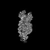
| Images |  emd_17844.png emd_17844.png | 482.3 KB | ||
| Masks | emd_17844_msk_1.map | 160.8 MB | Mask map | |
| Filedesc metadata | emd-17844.cif.gz | 6.2 KB | ||
| Others | emd_17844_additional_1.map.gzemd_17844_half_map_1.map.gzemd_17844_half_map_2.map.gz | 79.2 MB 148.8 MB 148.8 MB | ||
| Archive directory |  http://ftp.pdbj.org/pub/emdb/structures/EMD-17844ftp://ftp.pdbj.org/pub/emdb/structures/EMD-17844 http://ftp.pdbj.org/pub/emdb/structures/EMD-17844ftp://ftp.pdbj.org/pub/emdb/structures/EMD-17844 | HTTPS FTP |
-Related structure data
| Related structure data |  8ps4MC  8ps5C  8ps6C M: atomic model generated by this map C: citing same article ( |
|---|
-Links
| EMDB pages | EMDB (EBI/PDBe) / EMDataResource |
|---|
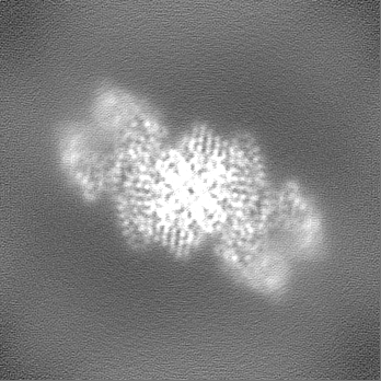
-Map
| File | Download / File: emd_17844.map.gz / Format: CCP4 / Size: 160.8 MB / Type: IMAGE STORED AS FLOATING POINT NUMBER (4 BYTES) | ||||||||||||||||||||||||||||||||||||
|---|---|---|---|---|---|---|---|---|---|---|---|---|---|---|---|---|---|---|---|---|---|---|---|---|---|---|---|---|---|---|---|---|---|---|---|---|---|
| Projections & slices | Image control
Images are generated by Spider. | ||||||||||||||||||||||||||||||||||||
| Voxel size | X=Y=Z: 0.65 Å | ||||||||||||||||||||||||||||||||||||
| Density |
| ||||||||||||||||||||||||||||||||||||
| Symmetry | Space group: 1 | ||||||||||||||||||||||||||||||||||||
| Details | EMDB XML:
|
 Z (Sec.)
 Y (Row.)
 X (Col.)


-Supplemental data
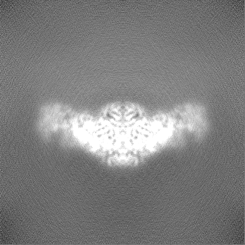
-Mask #1
| File | emd_17844_msk_1.map | ||||||||||||
|---|---|---|---|---|---|---|---|---|---|---|---|---|---|
| Projections & Slices |
| ||||||||||||
| Density Histograms |


-Additional map: #1
| File | emd_17844_additional_1.map | ||||||||||||
|---|---|---|---|---|---|---|---|---|---|---|---|---|---|
| Projections & Slices |
| ||||||||||||
| Density Histograms |


-Half map: #2
| File | emd_17844_half_map_1.map | ||||||||||||
|---|---|---|---|---|---|---|---|---|---|---|---|---|---|
| Projections & Slices |
| ||||||||||||
| Density Histograms |


-Half map: #1
| File | emd_17844_half_map_2.map | ||||||||||||
|---|---|---|---|---|---|---|---|---|---|---|---|---|---|
| Projections & Slices |
| ||||||||||||
| Density Histograms |


- Sample components
-Entire : Tetrameric SduA complex
| Entire | Name: Tetrameric SduA complex |
|---|---|
| Components |
|
-Supramolecule #1: Tetrameric SduA complex
| Supramolecule | Name: Tetrameric SduA complex / type: organelle_or_cellular_component / ID: 1 / Parent: 0 / Macromolecule list: #1 |
|---|---|
| Source (natural) | Organism: |
| Molecular weight | Theoretical: 194 KDa |
-Macromolecule #1: Shedu effector protein
| Macromolecule | Name: Shedu effector protein / type: protein_or_peptide / ID: 1 / Number of copies: 4 / Enantiomer: LEVO |
|---|---|
| Source (natural) | Organism: |
| Molecular weight | Theoretical: 47.465273 KDa |
| Recombinant expression | Organism: |
| Sequence | String: SNAMLQFSFV SNDVVMTYDG DSGEQIIWVW ESLNKFQTVC ISRIFNFQLQ DLRNPPSTVQ DFNDYEYSFN FGTLNNEYIT VPGRILSIN RDVLIHKSIK LERKVFASER NVSIFGRLSK LLDHTNPIII GGDKPEAIPK SVFQELQSKF PNTGELDRYA N ARVHAILA ...String: SNAMLQFSFV SNDVVMTYDG DSGEQIIWVW ESLNKFQTVC ISRIFNFQLQ DLRNPPSTVQ DFNDYEYSFN FGTLNNEYIT VPGRILSIN RDVLIHKSIK LERKVFASER NVSIFGRLSK LLDHTNPIII GGDKPEAIPK SVFQELQSKF PNTGELDRYA N ARVHAILA GYLDGMKDAR ERYEHYLNRK TVIRKTDKLD LEVLNKLEIE KYTLIRDIIQ DALNNKTNLS EDDWQSLMIP FI TLLFPKY IKVLEKVKIF DYYSNPSAKT NRFIDIALVD ANGNLDIIEV KKPFDDKILR KTPYRDNYIP TSELSGGIMQ AEK YIFHLS KWGVKGEKEL TNAYKNSLPA GMCIRISNPK AIIIVGRDQI ANGNMTDGQL LDFEIIKRKY ANMIDILTYD DLLR RLNNT IEALKG |
-Macromolecule #2: MAGNESIUM ION
| Macromolecule | Name: MAGNESIUM ION / type: ligand / ID: 2 / Number of copies: 2 / Formula: MG |
|---|---|
| Molecular weight | Theoretical: 24.305 Da |
-Experimental details
-Structure determination
| Method | cryo EM |
|---|---|
 Processing Processing | single particle reconstruction |
| Aggregation state | particle |
-Sample preparation
| Concentration | 2 mg/mL | ||||||||||||
|---|---|---|---|---|---|---|---|---|---|---|---|---|---|
| Buffer | pH: 8 Component:
Details: 20 mM Tris-HCl pH 8, 150 mM NaCl, 5 mM MgCl2 | ||||||||||||
| Grid | Model: Quantifoil R1.2/1.3 / Material: GOLD / Mesh: 200 | ||||||||||||
| Vitrification | Cryogen name: ETHANE / Chamber humidity: 85 % / Chamber temperature: 277.15 K / Instrument: FEI VITROBOT MARK IV | ||||||||||||
| Details | Samples was monodisperse |
- Electron microscopy
| Microscope | TFS KRIOS |
|---|---|
| Image recording | Film or detector model: GATAN K3 (6k x 4k) / Number real images: 5289 / Average electron dose: 66.529 e/Å2 |
| Electron beam | Acceleration voltage: 300 kV / Electron source:  FIELD EMISSION GUN FIELD EMISSION GUN |
| Electron optics | C2 aperture diameter: 50.0 µm / Calibrated magnification: 130000 / Illumination mode: FLOOD BEAM / Imaging mode: BRIGHT FIELD / Cs: 2.7 mm / Nominal defocus max: 2.4 µm / Nominal defocus min: 1.0 µm |
| Sample stage | Specimen holder model: FEI TITAN KRIOS AUTOGRID HOLDER / Cooling holder cryogen: NITROGEN |
| Experimental equipment |  Model: Titan Krios / Image courtesy: FEI Company |

