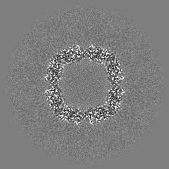
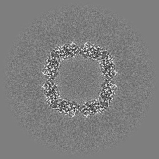
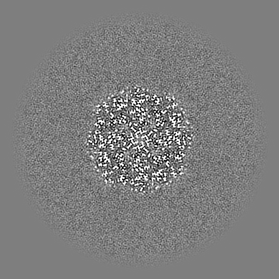
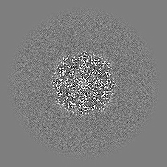
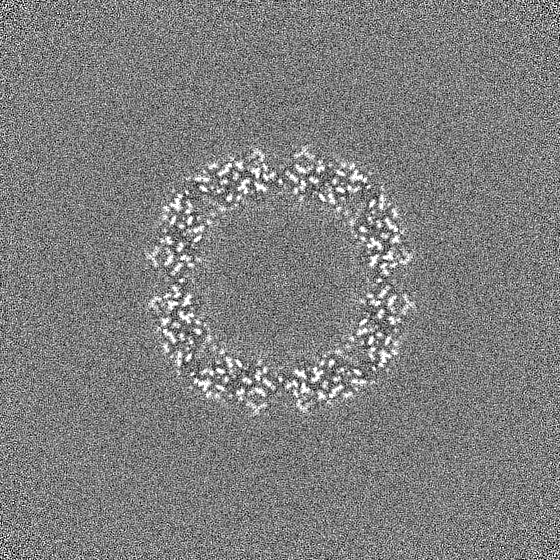
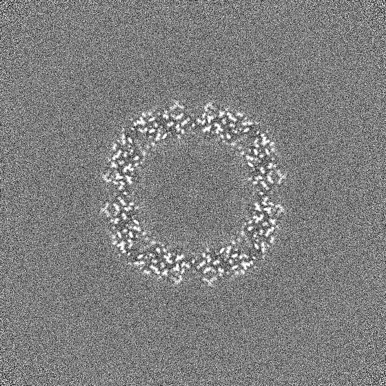
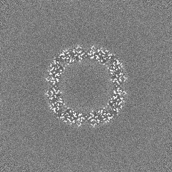
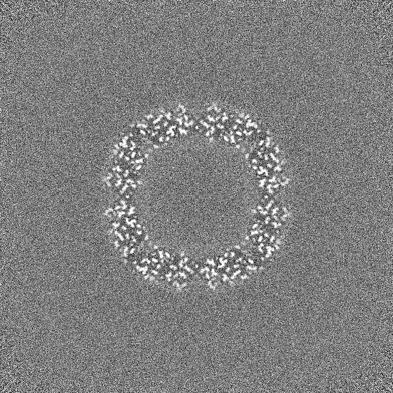
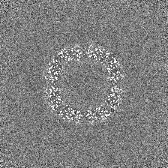
Journal: Acta Crystallogr D Struct Biol / Year: 2023 Title: Near-atomic resolution reconstructions from in situ revitrified cryo samples. Authors: Gabriele Bongiovanni / Oliver F Harder / Jonathan M Voss / Marcel Drabbels / Ulrich J Lorenz / Abstract: A microsecond time-resolved version of cryo-electron microscopy (cryo-EM) has recently been introduced to enable observation of the fast conformational motions of proteins. The technique involves ...A microsecond time-resolved version of cryo-electron microscopy (cryo-EM) has recently been introduced to enable observation of the fast conformational motions of proteins. The technique involves locally melting a cryo sample with a laser beam to allow the proteins to undergo dynamics in the liquid phase. When the laser is switched off, the sample cools within just a few microseconds and revitrifies, trapping particles in their transient configurations, in which they can subsequently be imaged. Two alternative implementations of the technique have previously been described, using either an optical microscope or performing revitrification experiments in situ. Here, it is shown that it is possible to obtain near-atomic resolution reconstructions from in situ revitrified cryo samples. Moreover, the resulting map is indistinguishable from that obtained from a conventional sample within the spatial resolution. Interestingly, it is observed that revitrification leads to a more homogeneous angular distribution of the particles, suggesting that revitrification may potentially be used to overcome issues of preferred particle orientation.
In the structure databanks used in Yorodumi, some data are registered as the other names, "COVID-19 virus" and "2019-nCoV". Here are the details of the virus and the list of structure data.
Jan 31, 2019. EMDB accession codes are about to change! (news from PDBe EMDB page)
EMDB accession codes are about to change! (news from PDBe EMDB page)
The allocation of 4 digits for EMDB accession codes will soon come to an end. Whilst these codes will remain in use, new EMDB accession codes will include an additional digit and will expand incrementally as the available range of codes is exhausted. The current 4-digit format prefixed with “EMD-” (i.e. EMD-XXXX) will advance to a 5-digit format (i.e. EMD-XXXXX), and so on. It is currently estimated that the 4-digit codes will be depleted around Spring 2019, at which point the 5-digit format will come into force.
The EM Navigator/Yorodumi systems omit the EMD- prefix.
Related info.:Q: What is EMD? / ID/Accession-code notation in Yorodumi/EM Navigator
Yorodumi is a browser for structure data from EMDB, PDB, SASBDB, etc.
This page is also the successor to EM Navigator detail page, and also detail information page/front-end page for Omokage search.
The word "yorodu" (or yorozu) is an old Japanese word meaning "ten thousand". "mi" (miru) is to see.
Related info.:EMDB / PDB / SASBDB / Comparison of 3 databanks / Yorodumi Search / Aug 31, 2016. New EM Navigator & Yorodumi / Yorodumi Papers / Jmol/JSmol / Function and homology information / Changes in new EM Navigator and Yorodumi
 Movie
Movie Controller


 Open data
 Basic information
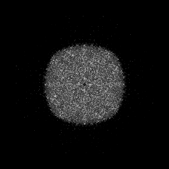
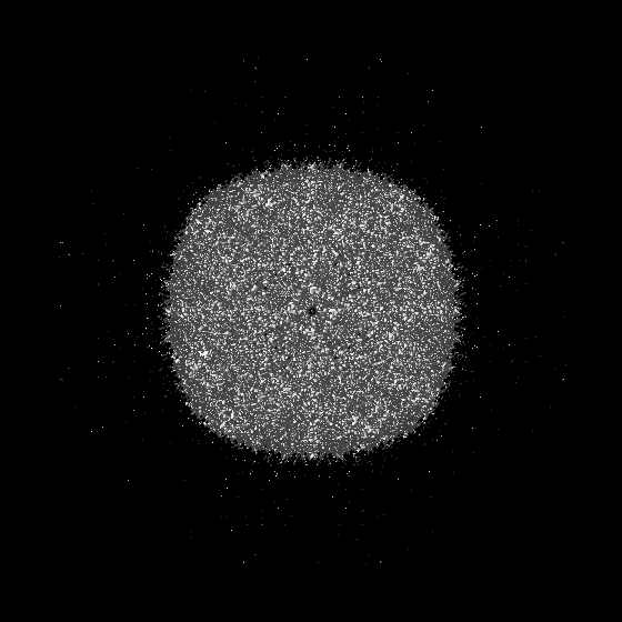
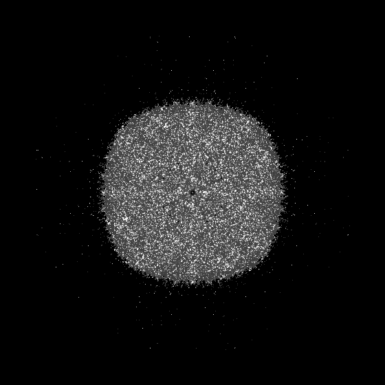
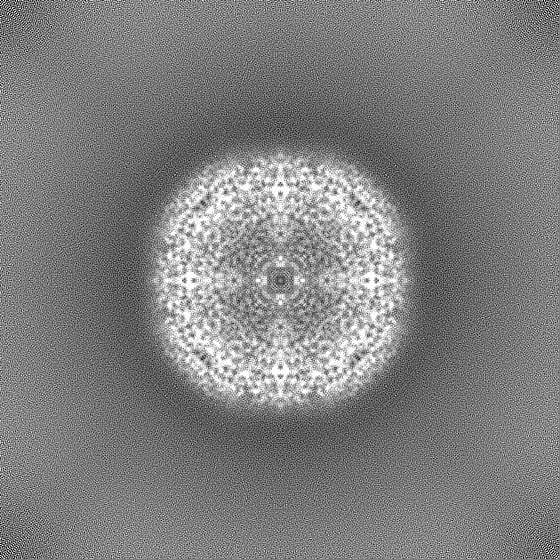
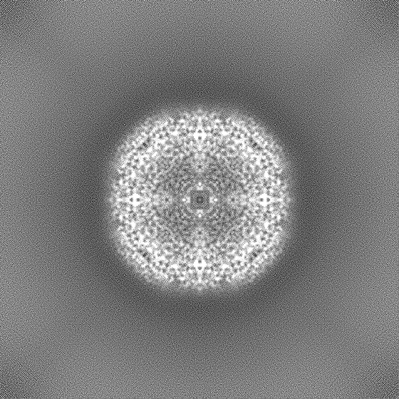
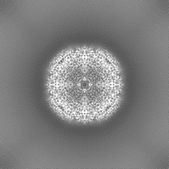
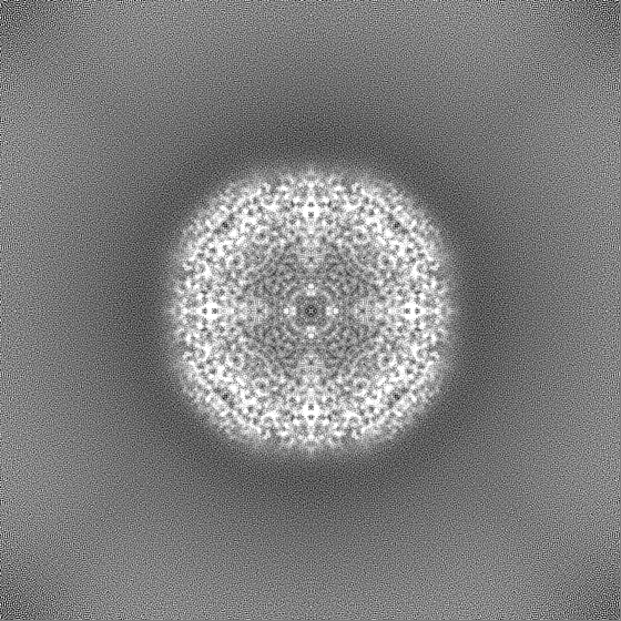
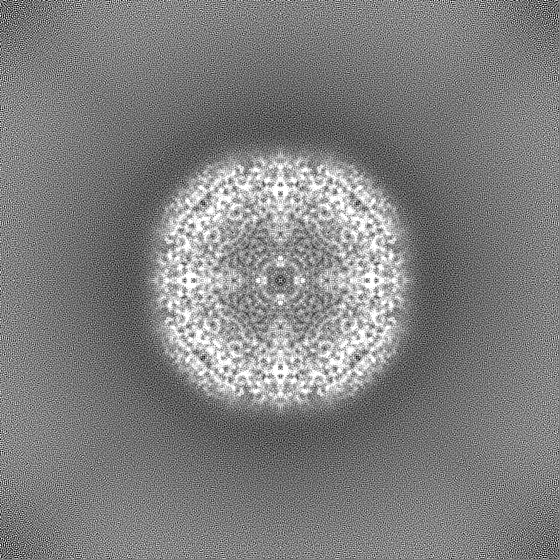
 Map data
 Sample
 Keywords
 Authors
 Citation
 Structure visualization
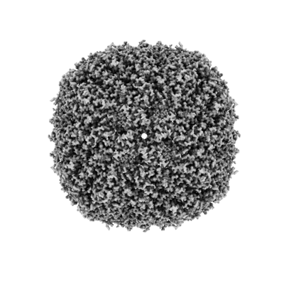
 Downloads & links
 EMDB map data format
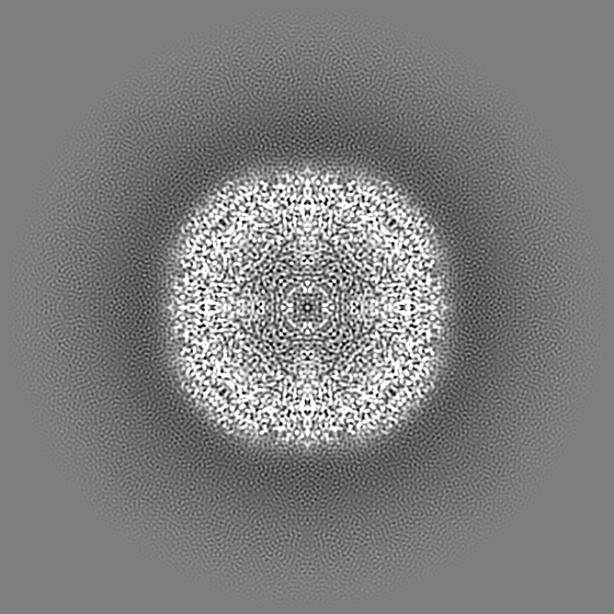
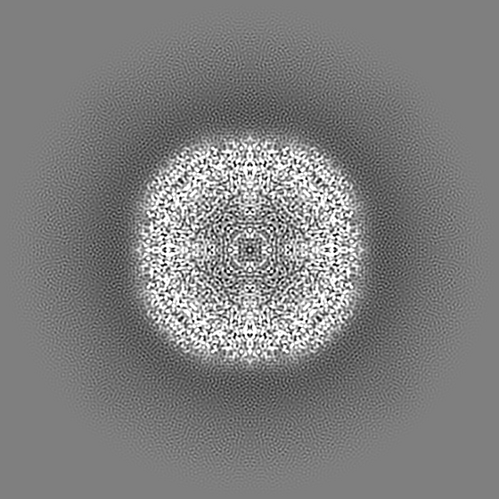
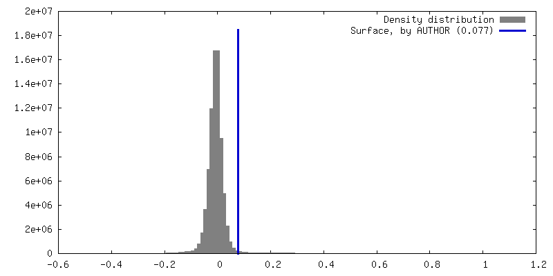
 emd_15715.png
 http://ftp.pdbj.org/pub/emdb/structures/EMD-15715
 Z (Sec.)
 Y (Row.)
 X (Col.)


 Sample components
 Processing
 Electron microscopy
 FIELD EMISSION GUN
