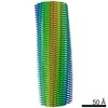
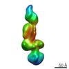
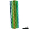
Journal: Nature / Year: 2022 Title: Structure of pathological TDP-43 filaments from ALS with FTLD. Authors: Diana Arseni / Masato Hasegawa / Alexey G Murzin / Fuyuki Kametani / Makoto Arai / Mari Yoshida / Benjamin Ryskeldi-Falcon / Abstract: The abnormal aggregation of TAR DNA-binding protein 43 kDa (TDP-43) in neurons and glia is the defining pathological hallmark of the neurodegenerative disease amyotrophic lateral sclerosis (ALS) ...The abnormal aggregation of TAR DNA-binding protein 43 kDa (TDP-43) in neurons and glia is the defining pathological hallmark of the neurodegenerative disease amyotrophic lateral sclerosis (ALS) and multiple forms of frontotemporal lobar degeneration (FTLD). It is also common in other diseases, including Alzheimer's and Parkinson's. No disease-modifying therapies exist for these conditions and early diagnosis is not possible. The structures of pathological TDP-43 aggregates are unknown. Here we used cryo-electron microscopy to determine the structures of aggregated TDP-43 in the frontal and motor cortices of an individual who had ALS with FTLD and from the frontal cortex of a second individual with the same diagnosis. An identical amyloid-like filament structure comprising a single protofilament was found in both brain regions and individuals. The ordered filament core spans residues 282-360 in the TDP-43 low-complexity domain and adopts a previously undescribed double-spiral-shaped fold, which shows no similarity to those of TDP-43 filaments formed in vitro. An abundance of glycine and neutral polar residues facilitates numerous turns and restricts β-strand length, which results in an absence of β-sheet stacking that is associated with cross-β amyloid structure. An uneven distribution of residues gives rise to structurally and chemically distinct surfaces that face external densities and suggest possible ligand-binding sites. This work enhances our understanding of the molecular pathogenesis of ALS and FTLD and informs the development of diagnostic and therapeutic agents that target aggregated TDP-43.
History
Deposition
Oct 8, 2021
-
Header (metadata) release
Dec 15, 2021
-
Map release
Dec 15, 2021
-
Update
Jul 17, 2024
-
Current status
Jul 17, 2024
Processing site: PDBe / Status: Released
-
Structure visualization
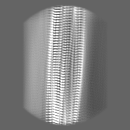
Movie
Surface view with section colored by density value
EMPIAR-10830 (Title: Structure of pathological TDP-43 filaments from ALS with FTLD (Individual 1, frontal cortex) Data size: 3.9 TB Data #1: Unaligned multiframe movies [micrographs - multiframe])
Entire : Pathological TDP-43 filaments extracted from the frontal cortex o...
Entire
Name: Pathological TDP-43 filaments extracted from the frontal cortex of an individual that succumbed to ALS with FTLD.
Components
Tissue: Pathological TDP-43 filaments extracted from the frontal cortex of an individual that succumbed to ALS with FTLD.
Protein or peptide: TAR DNA-binding protein 43
-
Supramolecule #1: Pathological TDP-43 filaments extracted from the frontal cortex o...
Supramolecule
Name: Pathological TDP-43 filaments extracted from the frontal cortex of an individual that succumbed to ALS with FTLD. type: tissue / ID: 1 / Parent: 0 / Macromolecule list: all
Source (natural)
Organism: Homo sapiens (human) / Tissue: Brain
-
Macromolecule #1: TAR DNA-binding protein 43
Macromolecule
Name: TAR DNA-binding protein 43 / type: protein_or_peptide / ID: 1 / Number of copies: 4 / Enantiomer: LEVO
In the structure databanks used in Yorodumi, some data are registered as the other names, "COVID-19 virus" and "2019-nCoV". Here are the details of the virus and the list of structure data.
Jan 31, 2019. EMDB accession codes are about to change! (news from PDBe EMDB page)
EMDB accession codes are about to change! (news from PDBe EMDB page)
The allocation of 4 digits for EMDB accession codes will soon come to an end. Whilst these codes will remain in use, new EMDB accession codes will include an additional digit and will expand incrementally as the available range of codes is exhausted. The current 4-digit format prefixed with “EMD-” (i.e. EMD-XXXX) will advance to a 5-digit format (i.e. EMD-XXXXX), and so on. It is currently estimated that the 4-digit codes will be depleted around Spring 2019, at which point the 5-digit format will come into force.
The EM Navigator/Yorodumi systems omit the EMD- prefix.
Related info.:Q: What is EMD? / ID/Accession-code notation in Yorodumi/EM Navigator
Yorodumi is a browser for structure data from EMDB, PDB, SASBDB, etc.
This page is also the successor to EM Navigator detail page, and also detail information page/front-end page for Omokage search.
The word "yorodu" (or yorozu) is an old Japanese word meaning "ten thousand". "mi" (miru) is to see.
Related info.:EMDB / PDB / SASBDB / Comparison of 3 databanks / Yorodumi Search / Aug 31, 2016. New EM Navigator & Yorodumi / Yorodumi Papers / Jmol/JSmol / Function and homology information / Changes in new EM Navigator and Yorodumi
 Movie
Movie Controller


 Open data
 Basic information
 Map data
 Sample
 Keywords
 Function and homology information
 Homo sapiens (human)
 Authors
 United Kingdom, 1 items
  Citation
 Structure visualization
 Downloads & links
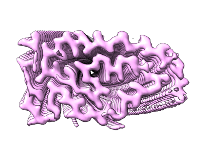 emd_13708.png
 http://ftp.pdbj.org/pub/emdb/structures/EMD-13708


 Z (Sec.)
 Y (Row.)
 X (Col.)


 Sample components
 Processing
 Electron microscopy
 FIELD EMISSION GUN

