 ムービー
ムービー コントローラー


+ データを開く

- 基本情報
基本情報
| 登録情報 | データベース: EMDB / ID: EMD-0995 | |||||||||
|---|---|---|---|---|---|---|---|---|---|---|
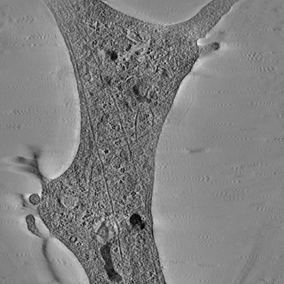
| タイトル | Ultra-high voltage electron microscope tomography using 700-nm-thick neurite section acquired at 15,000 magnification at an accelerating voltage of 1 MV | |||||||||
 マップデータ マップデータ | 700nm-thick neurite section of cultured PC12 cells (Magnification 15K) | |||||||||
 試料 試料 |
| |||||||||
| 生物種 |  | |||||||||
| 手法 | 電子線トモグラフィー法 / ネガティブ染色法 | |||||||||
 データ登録者 データ登録者 | Nishida T / Yoshimura R / Nishi R / Imoto Y / Endo Y | |||||||||
 引用 引用 | ジャーナル: J Microsc / 年: 2020 タイトル: Application of ultra-high voltage electron microscope tomography to 3D imaging of microtubules in neurites of cultured PC12 cells. 著者: T Nishida / R Yoshimura / R Nishi / Y Imoto / Y Endo /  要旨: Electron tomography methods using the conventional transmission electron microscope have been widely used to investigate the three-dimensional distribution patterns of various cellular structures ...Electron tomography methods using the conventional transmission electron microscope have been widely used to investigate the three-dimensional distribution patterns of various cellular structures including microtubules in neurites. Because the penetrating power of electrons depends on the section thickness and accelerating voltage, conventional TEM, having acceleration voltages up to 200 kV, is limited to sample thicknesses of 0.2 µm or less. In this paper, we show that the ultra-high voltage electron microscope (UHVEM), employing acceleration voltages of higher than 1000 kV (1 MV), allowed distinct reconstruction of the three-dimensional array of microtubules in a 0.7-µm-thick neurite section. The detailed structure of microtubules was more clearly reconstructed from a 0.7-µm-thick section at an accelerating voltage of 1 MV compared with a 1.0 µm section at 2 MV. Furthermore, the entire distribution of each microtubule in a neurite could be reconstructed from serial-section UHVEM tomography. Application of optimised UHVEM tomography will provide new insights, bridging the gap between the structure and function of widely-distributed cellular organelles such as microtubules for neurite outgrowth. LAY DESCRIPTION: An optimal 3D visualisation of microtubule cytoskeleton using ultra-high voltage electron microscopy tomography The ultra-high voltage electron microscope (UHVEM) is able to visualise a micrometre-thick specimen at nanoscale spatial resolution because of the high-energy electron beam penetrating such a specimen. In this study, we determined the optimal conditions necessary for microtubule cytoskeleton imaging within 0.7-µm-thick section using a combination with UHVEM and electron tomography method. Our approach provides excellent 3D information about the complex arrangement of the individual microtubule filaments that make up the microtubule network. | |||||||||
| 履歴 |
|
- 構造の表示
構造の表示
| ムービー |
 ムービービューア ムービービューア |
|---|---|
| 添付画像 |
- ダウンロードとリンク
-EMDBアーカイブ
| マップデータ | emd_0995.map.gz | 127.2 MB | EMDBマップデータ形式 | |
|---|---|---|---|---|
| ヘッダ (付随情報) | emd-0995-v30.xmlemd-0995.xml | 8.6 KB 8.6 KB | 表示 表示 | EMDBヘッダ |
| 画像 |  emd_0995.png emd_0995.png | 118.5 KB | ||
| アーカイブディレクトリ |  http://ftp.pdbj.org/pub/emdb/structures/EMD-0995ftp://ftp.pdbj.org/pub/emdb/structures/EMD-0995 http://ftp.pdbj.org/pub/emdb/structures/EMD-0995ftp://ftp.pdbj.org/pub/emdb/structures/EMD-0995 | HTTPS FTP |
-検証レポート
| 文書・要旨 | emd_0995_validation.pdf.gz | 78 KB | 表示 | EMDB検証レポート |
|---|---|---|---|---|
| 文書・詳細版 | emd_0995_full_validation.pdf.gz | 76.8 KB | 表示 | |
| XML形式データ | emd_0995_validation.xml.gz | 497 B | 表示 | |
| アーカイブディレクトリ | https://ftp.pdbj.org/pub/emdb/validation_reports/EMD-0995ftp://ftp.pdbj.org/pub/emdb/validation_reports/EMD-0995 | HTTPS FTP |
-関連構造データ
| 関連構造データ |  0987C  0994C  0996C C: 同じ文献を引用 ( |
|---|---|
| 電子顕微鏡画像生データ | EMPIAR-10355 (タイトル: Ultra-high voltage electron microscope tomography tilt series of 0.7-μm-thick neurite section acquired at 15,000× magnification at an accelerating voltage of 1 MV Data size: 120.3 MB Data #1: Ultra-high voltage electron microscope tomography tilt series of 0.7-μm-thick neurite section acquired at 15,000× magnification at an accelerating voltage of 1 MV [tilt series]) |
-リンク
| EMDBのページ | EMDB (EBI/PDBe) / EMDataResource |
|---|
-マップ
| ファイル | ダウンロード / ファイル: emd_0995.map.gz / 形式: CCP4 / 大きさ: 174.2 MB / タイプ: IMAGE STORED AS SIGNED INTEGER (2 BYTES) | ||||||||||||||||||||||||||||||||||||||||||||||||||||||||||||
|---|---|---|---|---|---|---|---|---|---|---|---|---|---|---|---|---|---|---|---|---|---|---|---|---|---|---|---|---|---|---|---|---|---|---|---|---|---|---|---|---|---|---|---|---|---|---|---|---|---|---|---|---|---|---|---|---|---|---|---|---|---|
| 注釈 | 700nm-thick neurite section of cultured PC12 cells (Magnification 15K) | ||||||||||||||||||||||||||||||||||||||||||||||||||||||||||||
| 投影像・断面図 | 画像のコントロール
画像は Spider により作成 これらの図は立方格子座標系で作成されたものです | ||||||||||||||||||||||||||||||||||||||||||||||||||||||||||||
| ボクセルのサイズ | X=Y=Z: 45.2 Å | ||||||||||||||||||||||||||||||||||||||||||||||||||||||||||||
| 密度 |
| ||||||||||||||||||||||||||||||||||||||||||||||||||||||||||||
| 対称性 | 空間群: 1 | ||||||||||||||||||||||||||||||||||||||||||||||||||||||||||||
| 詳細 | EMDB XML:
CCP4マップ ヘッダ情報:
| ||||||||||||||||||||||||||||||||||||||||||||||||||||||||||||
 Z (Sec.)
 Y (Row.)
 X (Col.)


-添付データ
- 試料の構成要素
-全体 : Microtubule distribution in neurite of differentiated PC12 cell
| 全体 | 名称: Microtubule distribution in neurite of differentiated PC12 cell |
|---|---|
| 要素 |
|
-超分子 #1: Microtubule distribution in neurite of differentiated PC12 cell
| 超分子 | 名称: Microtubule distribution in neurite of differentiated PC12 cell タイプ: organelle_or_cellular_component / ID: 1 / 親要素: 0 |
|---|---|
| 由来(天然) | 生物種: |
-実験情報
-構造解析
| 手法 | ネガティブ染色法 |
|---|---|
 解析 解析 | 電子線トモグラフィー法 |
| 試料の集合状態 | cell |
-試料調製
| 緩衝液 | pH: 7.3 |
|---|---|
| 染色 | タイプ: POSITIVE / 材質: Uranyl Acetate, Lead |
| 糖包埋 | 材質: resin |
| グリッド | 材質: COPPER / メッシュ: 50 / 支持フィルム - 材質: FORMVAR |
| 切片作成 | ウルトラミクロトーム - 装置: Reichert-Jung Ultracut E ウルトラミクロトーム - 温度: 300 K / ウルトラミクロトーム - 最終 厚さ: 700 nm |
| 位置合わせマーカー | Manufacturer: BBI Solutions / 直径: 20 nm |
- 電子顕微鏡法
| 顕微鏡 | HITACHI H3000 UHVEM |
|---|---|
| 撮影 | フィルム・検出器のモデル: OTHER / 平均電子線量: 13.0 e/Å2 |
| 電子線 | 加速電圧: 1000 kV / 電子線源: LAB6 |
| 電子光学系 | 照射モード: FLOOD BEAM / 撮影モード: BRIGHT FIELD / 倍率(公称値): 15000 |
| 試料ステージ | 試料ホルダーモデル: OTHER |
-画像解析
| 最終 再構成 | アルゴリズム: BACK PROJECTION / ソフトウェア - 名称:  IMOD / 使用した粒子像数: 67 IMOD / 使用した粒子像数: 67 |
|---|
