 Movie
Movie Controller
 Structure viewers
 About Yorodumi Papers


+Search query
-Structure paper
| Title | Application of ultra-high voltage electron microscope tomography to 3D imaging of microtubules in neurites of cultured PC12 cells. |
|---|---|
| Journal, issue, pages | J Microsc, Vol. 278, Issue 1, Page 42-48, Year 2020 |
| Publish date | Mar 18, 2020 |
 Authors Authors | T Nishida / R Yoshimura / R Nishi / Y Imoto / Y Endo /  |
| PubMed Abstract | Electron tomography methods using the conventional transmission electron microscope have been widely used to investigate the three-dimensional distribution patterns of various cellular structures ...Electron tomography methods using the conventional transmission electron microscope have been widely used to investigate the three-dimensional distribution patterns of various cellular structures including microtubules in neurites. Because the penetrating power of electrons depends on the section thickness and accelerating voltage, conventional TEM, having acceleration voltages up to 200 kV, is limited to sample thicknesses of 0.2 µm or less. In this paper, we show that the ultra-high voltage electron microscope (UHVEM), employing acceleration voltages of higher than 1000 kV (1 MV), allowed distinct reconstruction of the three-dimensional array of microtubules in a 0.7-µm-thick neurite section. The detailed structure of microtubules was more clearly reconstructed from a 0.7-µm-thick section at an accelerating voltage of 1 MV compared with a 1.0 µm section at 2 MV. Furthermore, the entire distribution of each microtubule in a neurite could be reconstructed from serial-section UHVEM tomography. Application of optimised UHVEM tomography will provide new insights, bridging the gap between the structure and function of widely-distributed cellular organelles such as microtubules for neurite outgrowth. LAY DESCRIPTION: An optimal 3D visualisation of microtubule cytoskeleton using ultra-high voltage electron microscopy tomography The ultra-high voltage electron microscope (UHVEM) is able to visualise a micrometre-thick specimen at nanoscale spatial resolution because of the high-energy electron beam penetrating such a specimen. In this study, we determined the optimal conditions necessary for microtubule cytoskeleton imaging within 0.7-µm-thick section using a combination with UHVEM and electron tomography method. Our approach provides excellent 3D information about the complex arrangement of the individual microtubule filaments that make up the microtubule network. |
 External links External links | J Microsc / PubMed:32133640 |
| Methods | EM (tomography) |
| Structure data | 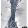 EMDB-0987: 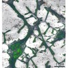 EMDB-0994: 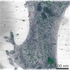 EMDB-0995: 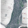 EMDB-0996: |
| Source |
|

