 Movie
Movie Controller


+ Open data

- Basic information
| Entry | Database: EMDB / ID: EMD-0732 | |||||||||
|---|---|---|---|---|---|---|---|---|---|---|
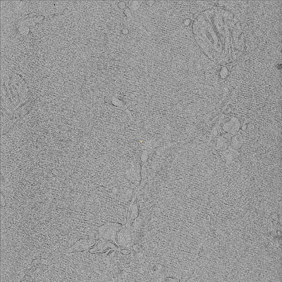
| Title | Cryo electron tomogram of cryo-lamella of rat skeleton muscle | |||||||||
 Map data Map data | ||||||||||
 Sample Sample |
| |||||||||
| Biological species |  | |||||||||
| Method | electron tomography | |||||||||
 Authors Authors | Zhang J / Zhang D / Sun L / Sun F | |||||||||
| Funding support |  China, 2 items China, 2 items
| |||||||||
 Citation Citation | Journal: J Struct Biol / Year: 2021 Title: VHUT-cryo-FIB, a method to fabricate frozen hydrated lamellae from tissue specimens for in situ cryo-electron tomography. Authors: Jianguo Zhang / Danyang Zhang / Lei Sun / Gang Ji / Xiaojun Huang / Tongxin Niu / Jiashu Xu / Chengying Ma / Yun Zhu / Ning Gao / Wei Xu / Fei Sun / Abstract: Cryo-electron tomography (cryo-ET) provides a promising approach to study intact structures of macromolecules in situ, but the efficient preparation of high-quality cryosections represents a ...Cryo-electron tomography (cryo-ET) provides a promising approach to study intact structures of macromolecules in situ, but the efficient preparation of high-quality cryosections represents a bottleneck. Although cryo-focused ion beam (cryo-FIB) milling has emerged for large and flat cryo-lamella preparation, its application to tissue specimens remains challenging. Here, we report an integrated workflow, VHUT-cryo-FIB, for efficiently preparing frozen hydrated tissue lamella that can be readily used in subsequent cryo-ET studies. The workflow includes vibratome slicing, high-pressure freezing, ultramicrotome cryo-trimming and cryo-FIB milling. Two strategies were developed for loading cryo-lamella via a side-entry cryo-holder or an FEI AutoGrid. The workflow was validated by using various tissue specimens, including rat skeletal muscle, rat liver and spinach leaf specimens, and in situ structures of ribosomes were obtained at nanometer resolution from the spinach and liver samples. | |||||||||
| History |
|
- Structure visualization
| Movie |
 Movie viewer Movie viewer |
|---|---|
| Supplemental images |
- Downloads & links
-EMDB archive
| Map data | emd_0732.map.gz | 648.8 MB | EMDB map data format | |
|---|---|---|---|---|
| Header (meta data) | emd-0732-v30.xmlemd-0732.xml | 13.1 KB 13.1 KB | Display Display | EMDB header |
| Images |  emd_0732.png emd_0732.png | 125.2 KB | ||
| Archive directory |  http://ftp.pdbj.org/pub/emdb/structures/EMD-0732ftp://ftp.pdbj.org/pub/emdb/structures/EMD-0732 http://ftp.pdbj.org/pub/emdb/structures/EMD-0732ftp://ftp.pdbj.org/pub/emdb/structures/EMD-0732 | HTTPS FTP |
-Related structure data
| Related structure data |  0733C  0734C C: citing same article ( |
|---|---|
| EM raw data | EMPIAR-10301 (Title: Cryo electron tomography of muscle tissue lamella from mice Data size: 1.3 Data #1: Unaligned tilt series of cryo lamella of rat skeleton muscle [tilt series]) |
-Links
| EMDB pages | EMDB (EBI/PDBe) / EMDataResource |
|---|
-Map
| File | Download / File: emd_0732.map.gz / Format: CCP4 / Size: 700 MB / Type: IMAGE STORED AS FLOATING POINT NUMBER (4 BYTES) | ||||||||||||||||||||||||||||||||||||||||||||||||||||||||||||
|---|---|---|---|---|---|---|---|---|---|---|---|---|---|---|---|---|---|---|---|---|---|---|---|---|---|---|---|---|---|---|---|---|---|---|---|---|---|---|---|---|---|---|---|---|---|---|---|---|---|---|---|---|---|---|---|---|---|---|---|---|---|
| Projections & slices | Image control
Images are generated by Spider. generated in cubic-lattice coordinate | ||||||||||||||||||||||||||||||||||||||||||||||||||||||||||||
| Voxel size | X=Y=Z: 26.1 Å | ||||||||||||||||||||||||||||||||||||||||||||||||||||||||||||
| Density |
| ||||||||||||||||||||||||||||||||||||||||||||||||||||||||||||
| Symmetry | Space group: 1 | ||||||||||||||||||||||||||||||||||||||||||||||||||||||||||||
| Details | EMDB XML:
CCP4 map header:
| ||||||||||||||||||||||||||||||||||||||||||||||||||||||||||||
 Z (Sec.)
 Y (Row.)
 X (Col.)


-Supplemental data
- Sample components
-Entire : cryo-lamella of rat skeleton muscle
| Entire | Name: cryo-lamella of rat skeleton muscle |
|---|---|
| Components |
|
-Supramolecule #1: cryo-lamella of rat skeleton muscle
| Supramolecule | Name: cryo-lamella of rat skeleton muscle / type: tissue / ID: 1 / Parent: 0 |
|---|---|
| Source (natural) | Organism: |
-Experimental details
-Structure determination
 Processing Processing | electron tomography |
|---|---|
| Aggregation state | tissue |
-Sample preparation
| Buffer | pH: 7 / Details: phosphate buffered saline (PBS) |
|---|---|
| Details | Mice were sacrificed and muscle was removed and cut into small bulks with a razor blade. Then a bulk of muscle was fixed on the specimen holder of VT1200S vibrating blade microtome for slicing. The thickness of each slice was set to 80-100 um. The generated muscle slice was cut into appropriate size for high pressure freezing. |
| High pressure freezing | Instrument: OTHER Details: Muscle tissue slice was put in the recess of the carrier and cryoprotectant 1-hexadecene was added to fill the surrounding area. Then a sapphire disk was loaded on top of the carrier before ...Details: Muscle tissue slice was put in the recess of the carrier and cryoprotectant 1-hexadecene was added to fill the surrounding area. Then a sapphire disk was loaded on top of the carrier before the whole composed sandwich was frozen.. The value given for _emd_high_pressure_freezing.instrument is HPF COMPACT 01. This is not in a list of allowed values {'BAL-TEC HPM 010', 'LEICA EM HPM100', 'OTHER', 'LEICA EM PACT2', 'LEICA EM PACT', 'EMS-002 RAPID IMMERSION FREEZER'} so OTHER is written into the XML file. |
| Cryo protectant | 1-hexadecene |
| Sectioning | Focused ion beam - Instrument: OTHER / Focused ion beam - Ion: OTHER / Focused ion beam - Voltage: 30 kV / Focused ion beam - Current: 0.08 nA / Focused ion beam - Duration: 3600 sec. / Focused ion beam - Temperature: 93 K / Focused ion beam - Initial thickness: 200 nm / Focused ion beam - Final thickness: 20 nm Focused ion beam - Details: Then the carrier was transfer with the cryo-transfer shuttle17 into the SEM chamber by using Quorum PP3000T cryotransfer system under -180 degree. To improve sample ...Focused ion beam - Details: Then the carrier was transfer with the cryo-transfer shuttle17 into the SEM chamber by using Quorum PP3000T cryotransfer system under -180 degree. To improve sample conductivity and reduce curtaining artifacts, the samples were deposited with organometallic platinum using the in situ gas injection system (GIS) operated at 5 seconds gas injection time before milling. During the cryo-FIB milling process, the milling angle is nearly in parallel with the carrier, and the milling was performed parallel from both sides of the sample platform to produce lamella. Rough milling is produced with the accelerating voltage of the ion beam at 30 kV, and current at 0.79 nA-0.43 nA. The initial milling width is about 20 um and depth is about 20 um. To facilitate tomography data collection, ice at the notch above lamella was removed to get a trapezoid-shaped milling pattern. After rough milling, one side of the lamella is jagged from the main platform. When the thickness of lamella reaches about 1 um the ion current is reduced to 0.23 nA or 80 pA until thickness finally reaching 150 to 250 nm.. The value given for _emd_sectioning_focused_ion_beam.instrument is Helios NanoLab 600i. This is not in a list of allowed values {'DB235', 'OTHER'} so OTHER is written into the XML file. |
- Electron microscopy
| Microscope | TFS TALOS F200C |
|---|---|
| Image recording | Film or detector model: FEI CETA (4k x 4k) / Digitization - Dimensions - Width: 4096 pixel / Digitization - Dimensions - Height: 4096 pixel / Digitization - Sampling interval: 14.0 µm / Number grids imaged: 1 / Number real images: 82 / Average exposure time: 1.0 sec. / Average electron dose: 3.0 e/Å2 |
| Electron beam | Acceleration voltage: 200 kV / Electron source:  FIELD EMISSION GUN FIELD EMISSION GUN |
| Electron optics | Calibrated defocus max: 10.0 µm / Calibrated defocus min: 8.0 µm / Illumination mode: FLOOD BEAM / Imaging mode: BRIGHT FIELD / Cs: 2.7 mm / Nominal magnification: 13500 |
| Sample stage | Specimen holder model: GATAN 910 MULTI-SPECIMEN SINGLE TILT CRYO TRANSFER HOLDER Cooling holder cryogen: NITROGEN |
| Experimental equipment |  Model: Tecnai F20 / Image courtesy: FEI Company |
-Image processing
| Final reconstruction | Algorithm: FOURIER SPACE / Software - Name: ICON (ver. 1.2.9) Details: ICON version 1.2.9 was used for the final reconstruction and the reconstruction algorithm is iterative compressed-sensing optimized NUFFT. Number images used: 43 |
|---|
