 Movie
Movie Controller

[English] 日本語
 Yorodumi
- ChemComp-VD3: (1S,3Z)-3-[(2E)-2-[(1R,3AR,7AS)-7A-METHYL-1-[(2R)-6-METHYLHEPTAN-... -
+ Open data

- Basic information
| Entry |  Database: PDB chemical components / ID: VD3 Database: PDB chemical components / ID: VD3 |
|---|---|
| Name | Name: ( Synonyms: VITAMIN D3 |
-Chemical information
| Composition |  | ||||||
|---|---|---|---|---|---|---|---|
| Others | Type: NON-POLYMER / PDB classification: HETAIN / Three letter code: VD3 | ||||||
| History |
| ||||||
 External links External links | UniChem / ChemSpider / ChEMBL / CompTox / LipidMaps / PubChem / SureChEMBL / Wikipedia search / Google search |
- Structure visualization
| Structure viewer | Molecule:  MolmilJmol/JSmol MolmilJmol/JSmol |
|---|
-Details
-SMILES
| ACDLabs 10.04 | | CACTVS 3.341 | OpenEye OEToolkits 1.5.0 | |
|---|
-SMILES CANONICAL
| CACTVS 3.341 | | OpenEye OEToolkits 1.5.0 | |
|---|
-InChI
| InChI 1.03 |
|---|
-InChIKey
| InChI 1.03 |
|---|
-SYSTEMATIC NAME
| ACDLabs 10.04 | (| OpenEye OEToolkits 1.5.0 | ( | |
|---|
-PDB entries
Showing all 6 items
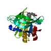
PDB-2gj5: 
Crystal structure of a secondary vitamin D3 binding site of milk beta-lactoglobulin
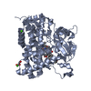
PDB-3a50:
Structure of cytochrome P450 Vdh mutant (Vdh-K1) obtained by directed evolution with bound vitamin D3
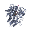
PDB-3c6g:
Crystal structure of CYP2R1 in complex with vitamin D3

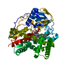
PDB-3vrm:
Structure of cytochrome P450 Vdh mutant T107A with bound vitamin D3
PDB-6t0g:
Crystal structure of CYP124 in complex with vitamin D3

PDB-8vxg:
The crystal structure of CYP125MRCA, an ancestrally reconstructed CYP125 enzyme
