 Movie
Movie Controller


+ Open data

- Basic information
| Entry |  Database: PDB chemical components / ID: RUU Database: PDB chemical components / ID: RUU |
|---|---|
| Name | Name: |
-Chemical information
| Composition |  | ||||||||||||||||
|---|---|---|---|---|---|---|---|---|---|---|---|---|---|---|---|---|---|
| Others |  4ns7 | ||||||||||||||||
| History |
| ||||||||||||||||
 External links External links | UniChem / ChemSpider / PubChem / SureChEMBL / Wikipedia search / Google search |
- Structure visualization
| Structure viewer | Molecule:  MolmilJmol/JSmol MolmilJmol/JSmol |
|---|
-Details
-SMILES
| ACDLabs 12.01 | | CACTVS 3.385 | OpenEye OEToolkits 1.7.6 | |
|---|
-SMILES CANONICAL
| CACTVS 3.385 | | OpenEye OEToolkits 1.7.6 | |
|---|
-InChI
| InChI 1.03 |
|---|
-InChIKey
| InChI 1.03 |
|---|
-SYSTEMATIC NAME
| ACDLabs 12.01 | | OpenEye OEToolkits 1.7.6 | ( | |
|---|
-CONDENSED IUPAC CARBOHYDRATE SYMBOL
| GMML 1.0 |
|---|
-COMMON NAME
| GMML 1.0 |
|---|
-IUPAC CARBOHYDRATE SYMBOL
| PDB-CARE 1.0 |
|---|
-PDB entries
Showing all 4 items

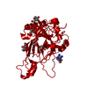
PDB-4q0q: 
Crystal structure of Acinetobacter sp. DL28 L-ribose isomerase in complex with L-ribulose
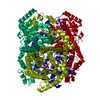
PDB-4q0v:
Crystal structure of Acinetobacter sp. DL28 L-ribose isomerase mutant E204Q in complex with L-ribulose
PDB-4qe1:
Room temperature X-ray structure of D-xylose isomerase in complex with two Cd2+ ions and L-ribulose
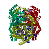
PDB-4qe5:
Room temperature X-ray structure of D-xylose isomerase in complex with two Mg2+ ions and L-ribulose
