 Movie
Movie Controller


+ Open data

- Basic information
| Entry |  Database: PDB chemical components / ID: PXP Database: PDB chemical components / ID: PXP |
|---|---|
| Name | Name: |
-Chemical information
| Composition |  | ||||
|---|---|---|---|---|---|
| Others | Type: NON-POLYMER / PDB classification: HETAIN / Three letter code: PXP / Model coordinates PDB-ID: 1HO4 | ||||
| History |
| ||||
 External links External links | UniChem / ChemSpider / Brenda / ChEBI / CompTox / DrugBank / HMDB / PubChem / SureChEMBL / Wikipedia search / Google search |
- Structure visualization
| Structure viewer | Molecule:  MolmilJmol/JSmol MolmilJmol/JSmol |
|---|
-Details
-SMILES
| ACDLabs 10.04 | | CACTVS 3.341 | OpenEye OEToolkits 1.5.0 | |
|---|
-SMILES CANONICAL
| CACTVS 3.341 | | OpenEye OEToolkits 1.5.0 | |
|---|
-InChI
| InChI 1.03 |
|---|
-InChIKey
| InChI 1.03 |
|---|
-SYSTEMATIC NAME
| ACDLabs 10.04 | [| OpenEye OEToolkits 1.5.0 | [ | |
|---|
-PDB entries
Showing all 5 items
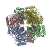
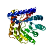
PDB-1ho4: 
CRYSTAL STRUCTURE OF PYRIDOXINE 5'-PHOSPHATE SYNTHASE IN COMPLEX WITH PYRIDOXINE 5'-PHOSPHATE AND INORGANIC PHOSPHATE
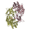
PDB-1szr:
A Dimer interface mutant of ornithine decarboxylase reveals structure of gem diamine intermediate

PDB-3f4n:
Crystal Structure of Pyridoxal Phosphate Biosynthetic Protein PdxJ from Yersinia pestis
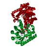
PDB-3o6d:
Pyridoxal phosphate biosynthetic protein PdxJ from Campylobacter jejuni in complex with pyridoxine-5'-phosphate
PDB-7ubq:
The crystal structure of the wild-type of E. coli YGGS in complex with PNP
